Foundations visual system
1/20
There's no tags or description
Looks like no tags are added yet.
Name | Mastery | Learn | Test | Matching | Spaced |
|---|
No study sessions yet.
21 Terms
CN 3, 4 and 6 _____ the _____
CN 3, 4 and 6 MOVE the EYES
CN 2 (OPTIC) is the nerve of
VISION
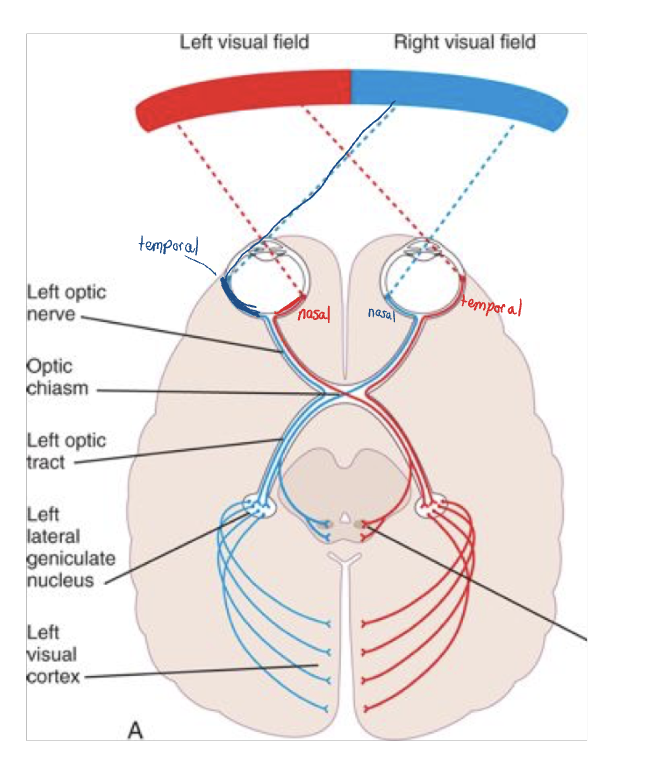
pathway of visual stimuli:
cornea → iris (controls amount of light by controlling the pupil) → lens (fine tunes the image) → retina → rods and cones → A focused image will land directly in the center of the macula → fovea
data congregates on the optic disc
Optic Nerve
All axons converge to form the
The fibers CROSS at a location known as the _____ _____
Then split into the R and L ______ ______
These axons continue and synapse in the ________
The LGN projects via the _______ ________ to the primary Visual Cortex (Area 17, 18 & 19)
All axons converge to form the Optic Nerve (CN 2)
The fibers CROSS at a location known as the OPTIC CHIASM
Then split into the R and L OPTIC TRACT
These axons continue and synapse in the THALAMUS
The LGN projects via the OPTIC RADIATIONS to the primary Visual Cortex (Area 17, 18 & 19)
Visual Pathway
Object in RIGHT visual field → Data is transmitted from the
Temporal fibers DO NOT
Nasal fibers
Therefore, the _____ hemisphere receives information about the _____ visual field (and vice versa)
Object in RIGHT visual field → Data is transmitted from the LEFT half of each retina (temporal portion of LEFT retinal, nasal portion of RIGHT retina)
Temporal fibers DO NOT CROSS → Transmit data to the LEFT Visual Cortex
Nasal fibers CROSS → Transmit data to the LEFT Visual Cortex
Therefore, the LEFT hemisphere receives information about the RIGHT visual field (and vice versa)
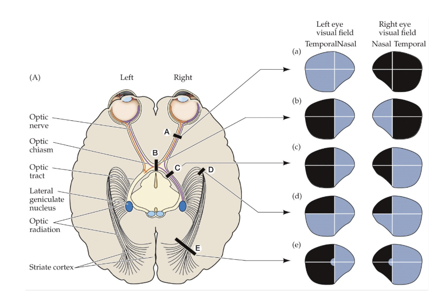
Lesions of the Visual Fields
A. Retinal Lesion:
B. Optic Nerve Lesion: Loss of vision in
C. Optic Chiasm Lesion
Loss of vision in
_______ _______
D. Optic Tract Lesion: ______ ______
Loss of vision on
Temporal loss of the _____ side
Nasal loss of the ______ side
A. Retinal Lesion: blind spot in one eye
B. Optic Nerve Lesion: Loss of vision in one eye
C. Optic Chiasm Lesion
Loss of vision in the temporal half each eye
Bitemporal hemianopia
D. Optic Tract Lesion: Homonymous hemianopia
Loss of vision on one half of each eye
Temporal loss of the SAME side
Nasal loss of the OPPOSITE side
Localization
Frontal
Parietal
Occipital
Temporal
Frontal
movement, higher order cognition, decision making and planning, language
Parietal
processing and integration of sensory input
Occipital
vision
Temporal
hearing, learning, and memory
Lateralization
L hemisphere
R hemisphere
L hemisphere
generally considered the dominant hemisphere
language
R hemisphere
non-dominant
non-verbal
complex visual-spatial skills
Localization & Lateralization
Focal brain lesions can cause specific deficits BUT not all lesions are created equally.
______ _______ plays a role in the way a lesion affects an individual
Ex: MCA
Hemispheric Specialization
Example of Lateralization
90% of the population is R handed
Skilled, complex motor tasks are more detailed in the dominant hemisphere
Lesions in the dominant hemisphere →
90% of the population is R handed
Skilled, complex motor tasks are more detailed in the dominant hemisphere
Lesions in the dominant hemisphere → Apraxia
Association Cortices
Frontal Lobe
Parietal Lobe
Occipital Lobe
Temporal Lobe
Frontal Lobe
Frontal/prefrontal association cortex (Areas 9 - 12)
Motor/Primary Motor association cortex (Areas 4, 6 and 8)
Language Association Area (Broca, 44, 45)
Parietal Lobe
Primary Somatosensory cortex (3, 1, 2)
Somatosensory association cortex (Area 5, 7)
Lateral parietal and temporal heteromodal association cortex (39, 40)
Occipital Lobe
Visual association cortex (Areas 17, 18, 19)
Temporal Lobe
Auditory Association Cortex (Wernicke’s Area, 22)
Primary Auditory Cortex (41, 42)
Symptoms of frontal lobe dysfunction
Perseveration
“stuck in a loop”
don’t have mental flexibility
Difficulty with abstract reasoning
Disinhibition
Disorders of Attention
Focused attention → Focusing on a particular object above others
Sustained attention → Vigilance, concentration, non-distractibility
Language and communication areas
Language and communication areas are distributed between both the
Frontal Lobe: Area 44, 45 →
Temporal Lobe: Area 22 →
Language and communication areas are distributed between both the frontal and temporal lobes
Frontal Lobe: Area 44, 45 → Language association Area
Temporal Lobe: Area 22 → Auditory association area
Language and Communication
Left Hemisphere: Dominant for language in > _____ of right handed people and ____ – ____ of left handed people
Lesions of the Left hemisphere →
Wernicke’s Area: (Area 22):
Deficit here:
Broca’s Area (Area 44 & 45):
Deficit here:
Left Hemisphere: Dominant for language in > 95% of right handed people and 60 – 70% of left handed people
Lesions of the Left hemisphere → Language disorders
Wernicke’s Area: (Area 22):
Deficit here: Receptive Aphasia
Broca’s Area (Area 44 & 45):
Deficit here: Expressive aphasia
Broca’s (Non-Fluent, Expressive Aphasia)
Good comprehension
Awkward articulation, restricted vocabulary, restriction to simple grammatical forms
Reading may be somewhat less impaired than speech and writing
Receptive (Wernicke’s) Aphasia
Poor naming skills, poor reading comprehension, poor writing skills
Speech is fluent, but marked by word substitutions and neologisms (nonsense words)
Speech may also be at a faster rate than normal
Visual Deficits
Motor Deficit: Problem with
Sensory Deficit: Problem with getting the data from the
Perceptual
Motor Deficit: Problem with Oculomotor control, eye movement
Sensory Deficit: Problem with getting the data from the eye to the visual cortex
Homonymous hemianopia
Bitemporal hemianopia
Perceptual
Interpretation, recognition and memory at the cortical level
Visual Object Agnosia
Lesion in areas
Usually
DEF: Inability to
Language and general intellectual functions are
“Recognition without _______”
Lesion in areas 18 and 19
Usually bilateral
DEF: Inability to name, copy or recognize a visually presented object, face or even sometimes letters
Language and general intellectual functions are preserved
“Recognition without meaning”
Facial Agnosia: Prosopagnosia
Unable to recognize
Must identify by
Can recognize the parts of a face and that a face is a face, but, cannot assign _______ to a face in the form of a Name
Caused by deficits in the ______ lobe where it meets the _______ lobe on the ventral surface of the brain ==> The fusiform gyrus
This lesion is usually _______ to result in the deficit
Unable to recognize faces
Must identify by voice or other cues
Can recognize the parts of a face and that a face is a face, but, cannot assign MEANING to a face in the form of a Name
Caused by deficits in the occipital lobe where it meets the temporal lobe on the ventral surface of the brain ==> The fusiform gyrus
This lesion is usually BILATERAL to result in the deficit
Parietal Association Areas
Parietal Association Cortex: At the junction of the parietal, temporal and occipital lobes (Areas ___,___)
Especially important for _____ _____ → Especially the R (non-dominant)
Analyzes the location and movement of visual objects in _____
This spatial analysis requires large amounts of data:
Parietal Association Cortex: At the junction of the parietal, temporal and occipital lobes (Areas 39, 40)
Especially important for spatial analysis → Especially the R (non-dominant)
Analyzes the location and movement of visual objects in space
This spatial analysis requires large amounts of data:
Visual
Proprioceptive
Vestibular
Auditory
Lesion: R Parietal Area
Hemineglect Syndrome
Most often occurs with infarcts in the R parietal or R frontal lobe
”Neglect” of the contralateral side of the “world” – varies in severity
Often accompanied with a complete lack of awareness of this deficit