Gas Exchange & Oxygenation
 Call Kai
 Learn
Learn Practice Test
 Spaced Repetition
 Match
Match Flashcards
 Knowt Play
1/65

There's no tags or description
Looks like no tags are added yet.
Name | Mastery | Learn | Test | Matching | Spaced | Call with Kai | Chat |
|---|
No analytics yet
Send a link to your students to track their progress
66 Terms
Upper airway components and primary function
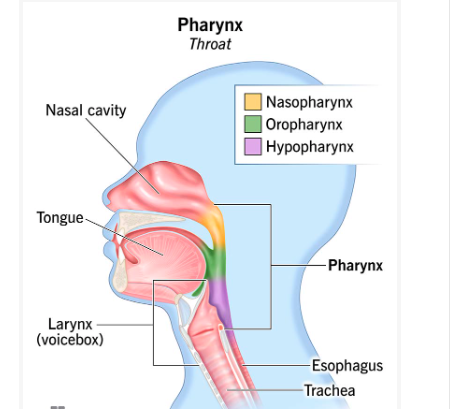
Composed of the pharynx (throat) and larynx (voice box) ;
responsible for warming air, filtering foreign particles , and humidifying air.

Cardiopulmonary Function
how well the heart and lungs work together to deliver oxygen to the body and remove carbon dioxide.
pharynx subdivisions
-nasopharynx
-oropharynx
-laryngopharynx or hypopharynx
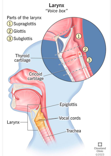
Larynx (voice box)
Structure located at the top of the trachea that houses the vocal cords.
Lower airway structures
Includes the trachea, bronchi, bronchioles, and alveoli of the lungs
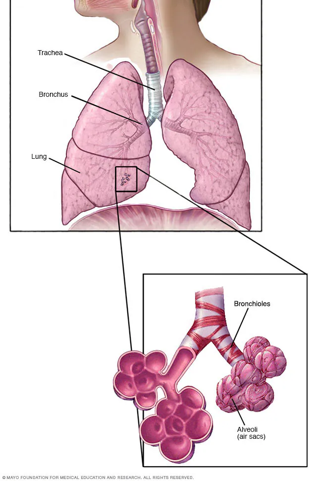
Alveoli
Structures at the end of the bronchioles where actual gas exchange occurs.

Lobar differences between right and left lungs
Right = three lobes
Left lung = two lobes.
(think same as av valves. tricuspid 3 right, bicuspid 2 left)
Neural control of bronchial diameter
The parasympathetic and sympathetic nervous systems regulate bronchoconstriction and bronchodilation.
Pleural cavity and fluid function
Located within the visceral and parietal pleural membrane layers; produces fluid that allows the lungs to expand and contract smoothly.
bronchi vs bronchioles
Bronchi are larger main airway passages extending from the trachea, while bronchioles are smaller branches leading directly to the alveoli

Mechanics of inhalation
Diaphragm and intercostal muscles contract, expanding the thorax and creating negative pressure inside the lungs to draw air in.
Mechanics of exhalation
Diaphragm and intercostal muscles relax, decreasing thoracic size and expelling gas from the lungs.
Diaphragm
Muscle separating the chest (thoracic) cavity from the abdominal cavity, controlled by the autonomic nervous system, that contracts during inhalation.
Surfactant
Lubricant produced in the lungs that keeps the alveoli from collapsing during exhalation.
Atelectasis
Collapse of alveoli or areas of lung tissue, resulting in reduced lung volume and impaired ventilation. It commonly occurs after surgery due to general anesthesia, opioids, or shallow breathing.
Primary structures involved in gas exchange
Bronchioles and alveoli, which deliver gas to the pulmonary capillaries via diffusion.
Ventilation
The flow of air into and out of the alveoli, transporting oxygen into the lungs and carbon dioxide out.
Perfusion
The flow of blood driven by the cardiopulmonary system into alveolar capillaries to exchange deoxygenated blood for oxygenated blood.
Pulmonary capillaries
Blood vessels located in the walls of the alveoli where diffusion and gas exchange occur.
Role of the brainstem in respiratory regulation
Monitors the body's oxygen demand and carbon dioxide levels, signaling the respiratory system to respond to excesses or undersupplies.
Effect of fever or exercise on respiration
Increases the rate and depth of respirations, causing more carbon dioxide to be exhaled and more oxygen to be inhaled.
Respirations
The number of breaths taken per minute. (12-20 on a normal adult)
Peripheral chemoreceptors location
Located in the aortic arch and carotid arteries; they sense changes in blood CO2 and O2 levels.
Central chemoreceptors location
Located in the medulla oblongata within the brainstem.
Role of chemoreceptors in respiration control
Peripheral chemoreceptors detect changes in blood CO2 and O2, signaling central chemoreceptors to adjust the rate and depth of respirations.
Emphysema
A type of COPD where the alveoli are damaged and lose their elasticity, causing air to become trapped in the lungs and making it difficult to exhale.
The alveoli walls are damaged/destroyed
The alveoli become larger, floppy, and less elastic (less recoil)
Air still enters, but it has trouble getting back out
This causes air trapping
Lung compliance
The ability of the lungs to expand (stretch) in response to pressure inside the alveoli.
Think:
Higher compliance = lungs expand more easily
Lower compliance = lungs are harder to expand
For example:
Emphysema → ↑ compliance (lungs are overly stretchy → easy to expand, hard to empty)
Pulmonary fibrosis → ↓ compliance (lungs are stiff)
Airway resistance
The pressure or opposition of the tissues in the airway to the flow of air.
Inspiratory reserve volume (IRV)
The additional amount of air that can breathed in after a normal inspiration.
Think of it as:
Take a normal breath in → this is tidal volume (TV).
Keep inhaling beyond that normal breath → that extra air is inspiratory reserve volume (IRV).
One full forced deep breath is TV + IRV
Expiratory reserve volume (ERV)
The additional amount of air expelled after a typical normal breath.
Think of it as:
Take a normal breath out → this is tidal volume (TV).
Keep exhaling beyond that normal breath → that extra air is expiratory reserve volume (ERV).
One forced deep exhale is TV + ERV
Tidal volume
The amount/volume of air inspired or expired with each normal unforced breath.
Residual volume
The amount of air remaining in the alveoli after expiration.
Think of the sequence:
Normal breath in → Tidal Volume (TV)
Normal breath out → TV
Force yourself to exhale MORE → Expiratory Reserve Volume (ERV)
After you have exhaled as much as physically possible, air STILL remains → Residual Volume (RV)
So:
ERV = air you CAN get out after normal exhale
RV = air you CANNOT get out even after forceful exhale
Forced vital capacity (FVC)
(WHAT ATI SAYS)
The amount of air that can be expelled from the lungs in 1s during forced expiration.
What it should say:
Forced Vital Capacity (FVC) → how much air comes out during the entire forced exhalation.
Forced Expiratory Volume in 1 Second (FEV₁) → how much air comes out during the first 1 second.
Tidal volume (TV) + Expiratory Reserve Volume (ERV) = Forced Vital Capacity (FVC).
Vital capacity (VC)
The maximum amount of air that is expelled after maximal inspiration.
Maximal inspiration → take the biggest breath possible.
Then exhale as much as possible.
The total amount you exhale = Vital Capacity (VC).
Tidal Volume (TV) + Inspiratory Reserve Volume (IRV) + Expiratory Reserve Volume (ERV) = Vital Capacity (VC)
Total lung capacity
The amount of air remaining in the lungs after maximal (forced) inspiration.
Think of it as:
Take the biggest breath possible → your lungs are filled with the maximum amount of air.
That maximum amount of air in your lungs = Total Lung Capacity (TLC).
This includes all the air in your lungs, even the air that cannot be exhaled (Residual Volume RV).
Formula:
Total Lung Capacity (TLC) = Vital Capacity (VC) + Residual Volume (RV)
Myocardial blood flow function
Supplies oxygen and essential nutrients to the heart muscle, enabling proper functioning of both pulmonary and systemic circulation.
Valvular state during diastole
Mitral and tricuspid valves are open to allow ventricular filling from the atria, while aortic and pulmonic valves remain closed.
Valvular state during systole
Mitral and tricuspid valves close, and the aortic and pulmonic valves open to permit blood ejection from the ventricles.
First heart sound (S1)
A sound produced at the onset of systole by the closure of the mitral and tricuspid valves.
Second heart sound (S2)
A sound produced at the end of systole when ventricular pressure drops, causing closure of the aortic and pulmonic valves.
Cardiac murmur cause
A blowing or whooshing sound caused by turbulent backflow of blood through incompetent heart valves.
Cardiac output (CO)
The volume of blood ejected by the left ventricle in one minute, typically ranging from 3 to 6L/min in a healthy resting adult.
CO=HR×SV ,
cardiac output x heart rate = stroke volume.
Stroke volume (SV)
The volume of blood ejected from the left ventricle with each individual contraction or cardiac cycle.
Factors influencing stroke volume
Preload, afterload, and contractility.
Preload (End-Diastolic Volume)
The volume of blood in the left ventricle at the end of diastole, immediately prior to the next contraction.
Frank-Starling mechanism
The physiological principle stating that increased ventricular stretching from higher end-diastolic volume produces a stronger contraction and greater stroke volume.
Afterload
The amount of resistance or force the left ventricle must overcome to eject blood into circulation during contraction.
Front: What is the effect of athletic training on cardiac output?
Athletes develop an increased resting Stroke Volume (SV). This allows them to maintain a normal resting Cardiac Output (CO) with a lower resting Heart Rate (HR).
During exercise, athletes can achieve a much higher maximal Cardiac Output (CO).
Stroke volume seen in athletes
xtreme athletes can have a cardiac output of up to 35 L/min during exercise.
Cardiac Conduction Electrical Pathway
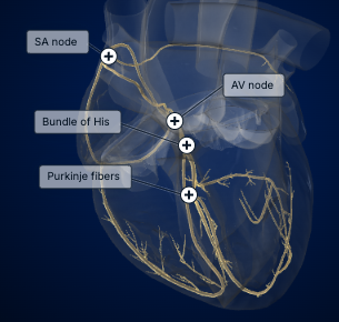
the electrical signal traveling through the heart responsible for the normal sinus rhythm—the expected heart rhythm for adults
SA node (pacemaker) → starts the electrical impulse
AV node → briefly delays the impulse
Bundle of His → carries the impulse into the ventricles
Right & left bundle branches → carry it down the septum
Purkinje fibers → spread the impulse through the ventricles → ventricles contract
Ambient room air oxygen fraction
Contains 21% oxygen.
Target oxygen saturation (SpO2) ranges
95% to 100% for healthy individuals, and 88% to 92% for clients with specific respiratory illnesses.
Complications of unhumidified nasal cannula therapy
Drying of nasal mucous membranes and skin breakdown from friction and mucosal dryness.
Clinical benefits of humidified oxygen
Prevents mucosal desiccation and keeps respiratory secretions moist, making them easier for the client to expectorate.
WHO SpO2 threshold for pediatric oxygen delivery
Oxygen delivery is recommended when SpO2<90% in children displaying signs of respiratory distress.
Target SpO2 range per American Heart Association AHA pediatric guidelines
Oxygen should be administered and titrated to achieve an SpO2 between 94% and 99%.
Preferred oxygen delivery method for infants with moderate to severe bronchiolitis
High-flow nasal cannula (HFNC) therapy, which is safer and more effective than low-flow nasal cannula therapy.
Recommendation on low-flow oxygen humidification in pediatric care
The use of heated or unheated humidification with low-flow oxygen delivery is not supported
Gastric distension in oxygen delivery
The swelling of the stomach with air or gas when supplemental oxygen or artificial ventilation is forced into the body, which can compress the diaphragm, impair lung expansion, and decrease ventilation and oxygenation.
Standard nasal cannula advantages and disadvantages
Advantages: Safe, simple, inexpensive, comfortable, decreases claustrophobia risk, allows eating and drinking, avoids rebreathing CO2, and delivers 24% to 44% oxygen at lower flow rates (1 to 6L/min).
Disadvantages: Causes dermatitis and nasal irritation, causes headaches and dry mucous membranes at flows above 4L/min, unsuitable for nasal obstructions (polyps, mucosal edema), and delivers less accurate oxygen percentage dependent on respiratory pattern.
High-flow nasal cannula (HFNC) advantages and disadvantages
Advantages: Simple to use, provides good humidification, carries minimal risk of gastric distention, and may help avoid intubation.
Disadvantages: More expensive than low-flow nasal cannula and requires a specialized flowmeter.
Simple face mask advantages and disadvantages
Advantages: Inexpensive and usable on mouth breathers.
Disadvantages: May cause claustrophobia, interferes with eating/drinking (critical in neonates, infants, and children), requires monitoring for nausea or vomiting, and retains CO2 (not recommended for clients at risk for CO2 retention).
and requires regular skin care during long-term use due to risk of skin breakdown.
Simple face mask flow rate and oxygen concentration
Delivers a medium concentration of 35%35% to 60%60% oxygen at a flow rate of 55 to 10 L/min10L/min.
Partial and nonrebreather masks advantages and disadvantages
Advantages: Recommended for short-term use in acute illness and trauma, delivering high oxygen concentrations (60% to 90%) at flow rates of 10 to 15L/min for clients with hypoxia.
Disadvantages: Cannot be used with humidification, requires a good facial seal, requires high flow rates to keep reservoir bag inflated, and carries risks of atelectasis and oxygen toxicity.
Venturi mask advantages and disadvantages
Advantages: Provides precise oxygen concentration independent of client breathing factors or flow rate, uses exchangeable Venturi barrels to deliver various concentrations, and reduces rebreathing of exhaled air.
Disadvantages: Can be noisy, can feel claustrophobic, and interferes with eating and drinking.