Portage Learning A&P Module 4, Articulations
1/208
There's no tags or description
Looks like no tags are added yet.
Name | Mastery | Learn | Test | Matching | Spaced | Call with Kai |
|---|
No analytics yet
Send a link to your students to track their progress
209 Terms
Joint (articulation)
Any place where adjacent bones (or bone and cartilage) meet to form a connection.
Classification of joints
Two ways joints are classified: structurally (by tissue/space) and functionally (by mobility).
Structural classes of joints
Fibrous, cartilaginous, synovial.
Functional classes of joints
Synarthrosis (immovable), amphiarthrosis (slightly movable), diarthrosis (freely movable).
Synarthrosis
Immovable; e.g., skull sutures, manubriosternal joint (often limited/none).
Amphiarthrosis
Slightly movable; e.g., intervertebral discs, pubic symphysis.
Diarthrosis
Freely movable; all synovial joints; mostly in the appendicular skeleton.
Basic joint motions
Linear (gliding), angular (incl. circumduction), rotation.
Uniaxial joint
Moves in one plane/axis; e.g., elbow (hinge).
Biaxial joint
Moves in two planes; e.g., metacarpophalangeal (knuckle).
Multiaxial (triaxial) joint
Moves in three planes incl. rotation; e.g., shoulder, hip.
Fibrous joint
Unites bones in a fibrous joint with dense fibrous connective tissue; no joint cavity.
Types of fibrous joints
Suture, syndesmosis, gomphosis.
Suture
Location: between skull bones; function: strong protection; mobility class: synarthrosis.
Fontanelle
Broad CT regions in infant skull; allow birth molding and rapid brain growth.
Synostosis
Fusion 'joined by bone'; frontal/maxillary halves fuse in childhood; late-life fusion of major sutures.
Syndesmosis
Two parallel bones linked by ligament or interosseous membrane; e.g., radius-ulna, tibia-fibula.
Syndesmosis functional class
Amphiarthrosis; forearm membrane flexible (allows rotation), leg membrane stabilizing (little motion).
Interosseous membrane
Unites bones + broad surface for muscle attachment.
Gomphosis
Tooth-to-socket joint; periodontal ligament anchors root; synarthrosis.
Cartilaginous joints
Unites bones in cartilaginous joints with hyaline cartilage or fibrocartilage; no joint cavity.
Types of cartilaginous joints
Synchondrosis and symphysis.
Synchondrosis
Bone joined to hyaline cartilage (temporary or permanent); synarthrosis.
Temporary synchondrosis
Classic example: Epiphyseal plate (growth plate): hyaline cartilage between diaphysis and epiphysis.
Synostosis
Fusion of growth plate to bone (plate replaced by bone at maturity).
Childhood pelvic synchondroses
Ilium-ischium-pubis fuse into single coxal bone (synostosis).
Permanent synchondrosis
Key thoracic example: First sternocostal joint (1st rib to manubrium).
Symphysis
Bones joined by fibrocartilage; strong yet slightly movable; amphiarthrosis.
Narrow symphyses
Examples: Pubic symphysis; manubriosternal joint.
Wide symphyses
Major example: Intervertebral discs; unite vertebrae, allow small motion, provide cushioning.
Synovial joint
Uniquely defined by a fluid-filled joint cavity between articulating bone surfaces.
Functional class of all synovial joints
Diarthrosis (freely movable).
Articular (joint) capsule
Two layers: outer fibrous layer; inner synovial membrane.
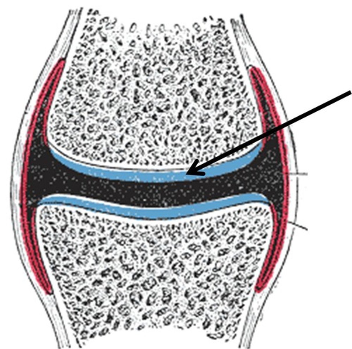
Articular cartilage
Hyaline cartilage covering articulating surfaces; reduces friction, protects bone.
Synovial membrane
Produces synovial fluid: lubricates, nourishes avascular cartilage, absorbs shock.
Diffusion from synovial fluid
Aided by compression/relaxation 'pumping.'
Ligaments
Three positional types: Extrinsic (outside capsule), intrinsic (part of capsule), intracapsular (inside capsule).
Tendons
Role at joints; muscle to bone; dynamic stabilizers that resist forces across joints.
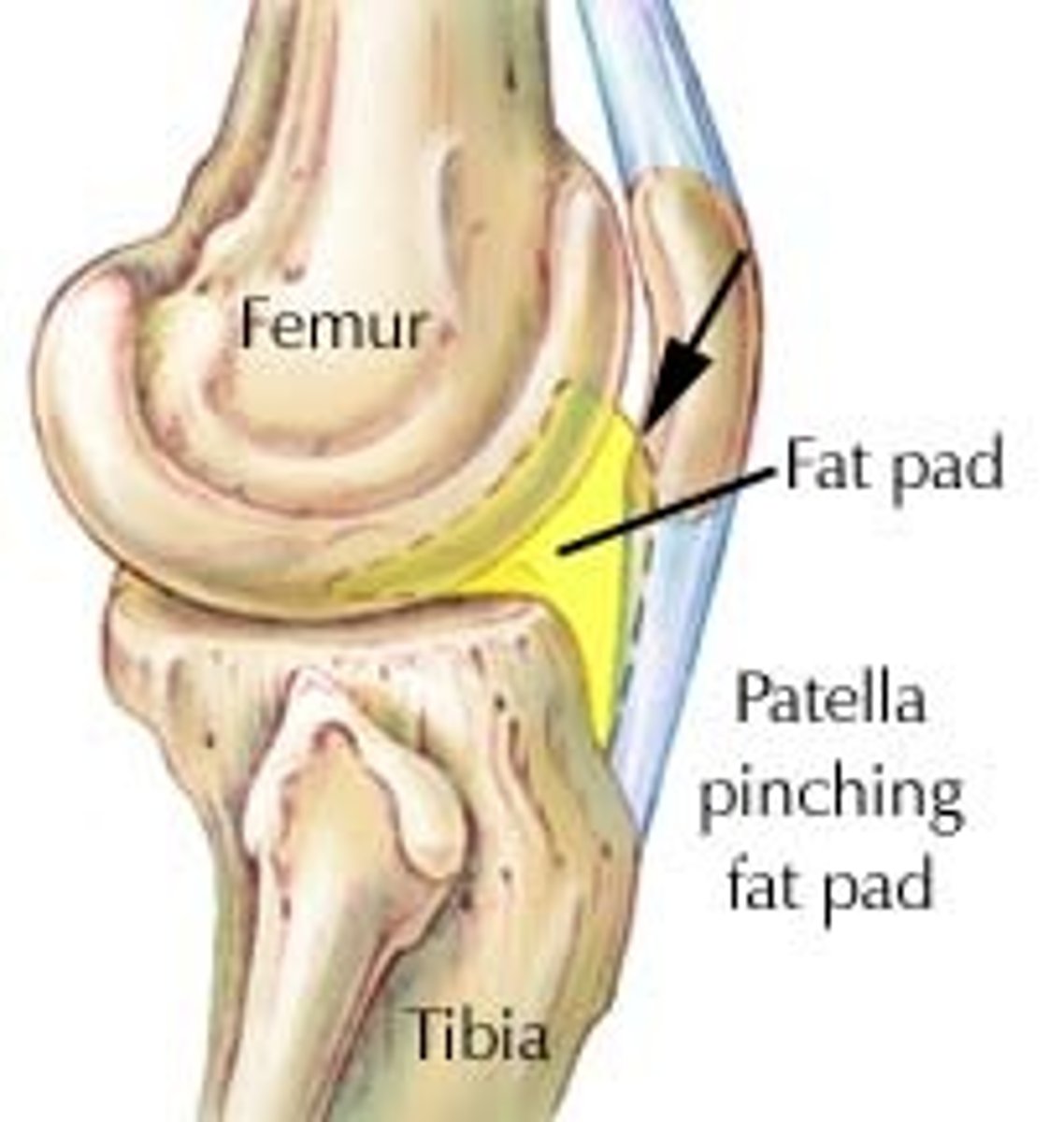
Fat pads
Fill spaces and cushion.
Articular disc vs meniscus
Disc: small, oval (e.g., TMJ, sternoclavicular). Meniscus: larger, C-shaped (knee) for stability + shock absorption.
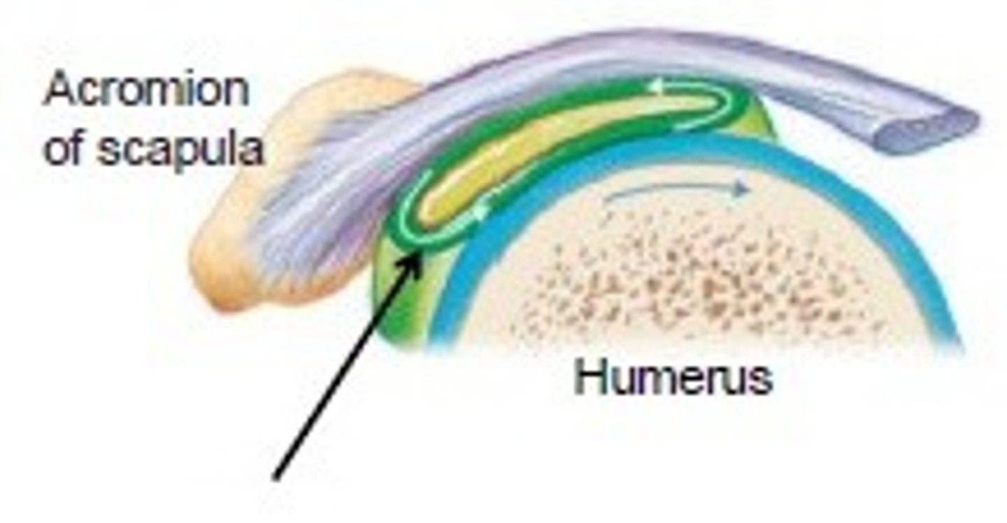
Bursa
Small fluid-filled sac reducing friction where tissues rub.
Tendon sheath
Elongated bursa around a tendon; reduces friction during motion.

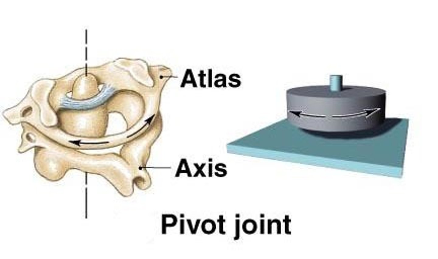
Pivot joint
Movement: rotation around one axis; example: atlantoaxial (C1-C2), proximal radioulnar.
Hinge joint
Movement: flexion/extension in one plane; examples: elbow, interphalangeal, knee (primarily hinge).

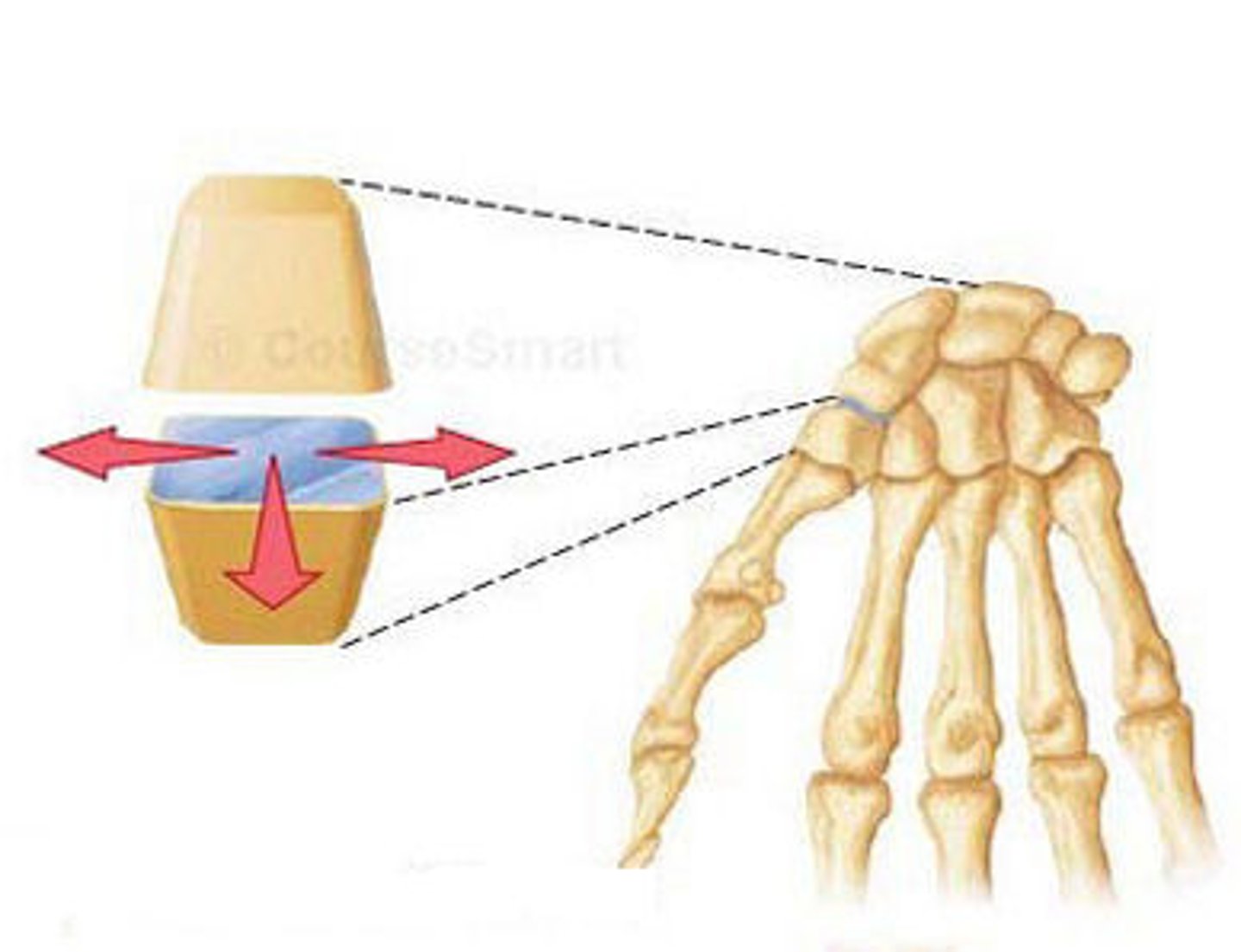
Condyloid (ellipsoid) joint
Movement: biaxial: flex/extend + abduct/adduct; examples: MCP (knuckles), radiocarpal (wrist).
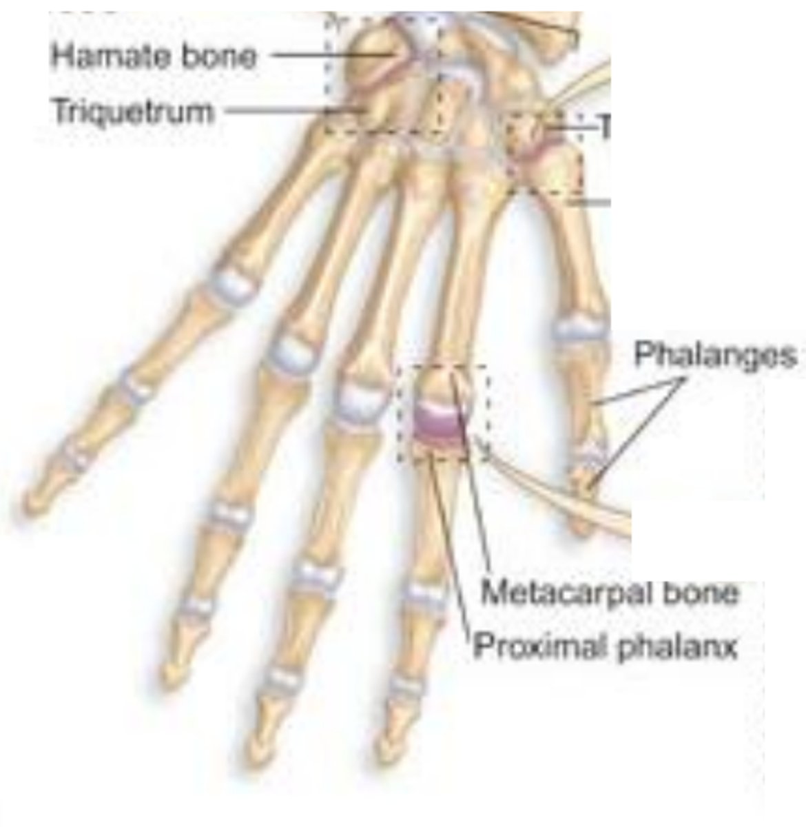
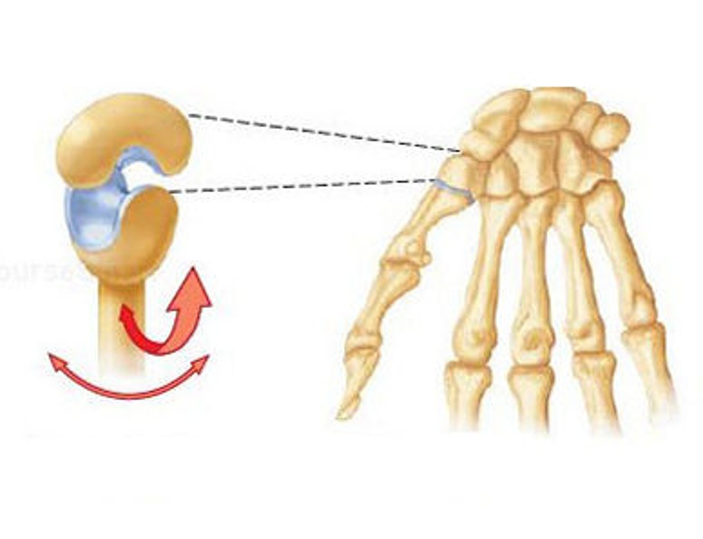
Saddle joint
Movement: biaxial with added range; example: 1st carpometacarpal (thumb).
Plane (gliding) joint
Movement: sliding/translation; examples: intercarpal, intertarsal, acromioclavicular.
Ball-and-socket joint
Movement: multiaxial including rotation; examples: shoulder, hip.
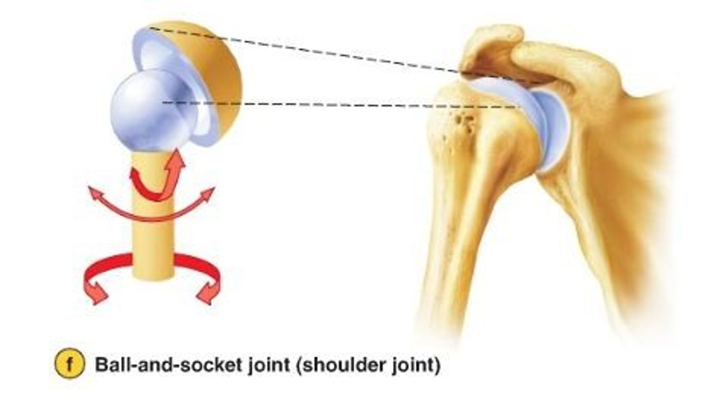
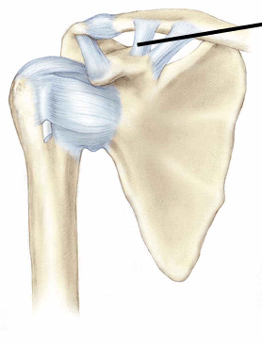
Shoulder joint
Bones involved: clavicle, scapula, humerus (glenohumeral joint).
Shoulder mobility vs stability
Highly mobile but less stable due to large humeral head vs shallow glenoid cavity; relies on ligaments + muscles.
Glenohumeral ligaments
Reinforce capsule, connect glenoid to humeral head.
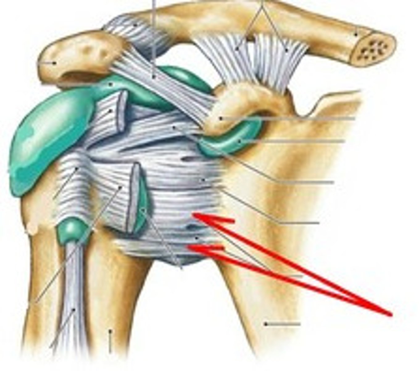
Coracoacromial ligament
Connects coracoid process to acromion of scapula.

Coracoclavicular ligament
Connects coracoid process of scapula to clavicle.
Glenoid labrum
Fibrocartilage rim that deepens glenoid socket.
Rotator cuff
Muscle/tendon group providing primary dynamic stability to shoulder.
Hip joint
Bones: head of femur + acetabulum; type: multiaxial ball-and-socket.
Hip vs shoulder stability
Hip = deeper socket, stronger ligaments, less motion.
Acetabular labrum
Fibrocartilage lip deepening socket for femoral head.
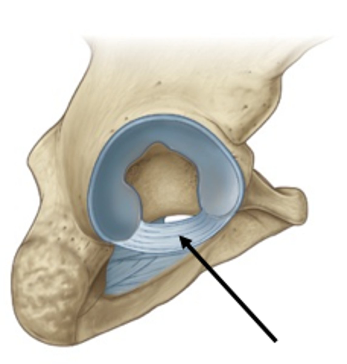
Iliofemoral, pubofemoral, ischiofemoral
Major hip ligaments; tighten in extension to stabilize upright posture.
Knee joint
Type: modified hinge joint; motion: flexion and extension.

Patella
Bone type: sesamoid bone in quadriceps tendon; role: protects tendon, increases leverage.
Menisci
Names: medial & lateral; role: fibrocartilage for shock absorption + stability.
Medial meniscus injury
More common due to strong attachment to tibial collateral ligament → less mobility.
Collateral ligaments
Names: fibular (lateral) and tibial (medial).
Cruciate ligaments
Names: ACL & PCL; role: prevent anterior/posterior displacement of femur on tibia.
shoulder joint
Most dislocated major joint; reason: shallow socket and high mobility.
'Terrible triad' of the knee
Tibial (medial) collateral ligament, medial meniscus, and ACL.
Rotator cuff tear risk factors
Age >60, repetitive lifting or abduction sports, trauma.
Bursitis
Definition: inflammation of bursa due to overuse, trauma, pressure, RA, gout, infection.
Osteoarthritis
Main cause: wear-and-tear degeneration of articular cartilage, common after 60.
Hands, spine, knees, hips, feet.
Osteoarthritis most affected joints
Rheumatoid arthritis
Mechanism: autoimmune inflammation destroying synovial joints + cartilage.
Gout
Cause: uric acid crystal buildup; typical location: often big toe.
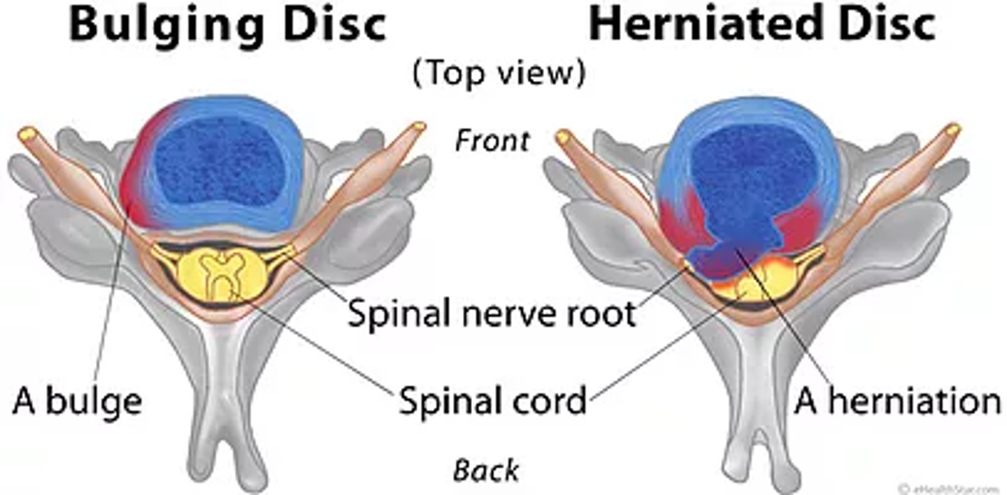
Aging + spine disc changes
Loss of water in nucleus pulposus → decreased height and elasticity.
Bulging vs herniated disc
Bulging = annulus protrudes; Herniated = nucleus pulposus breaks through annulus.
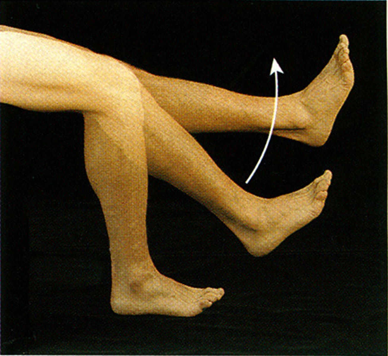
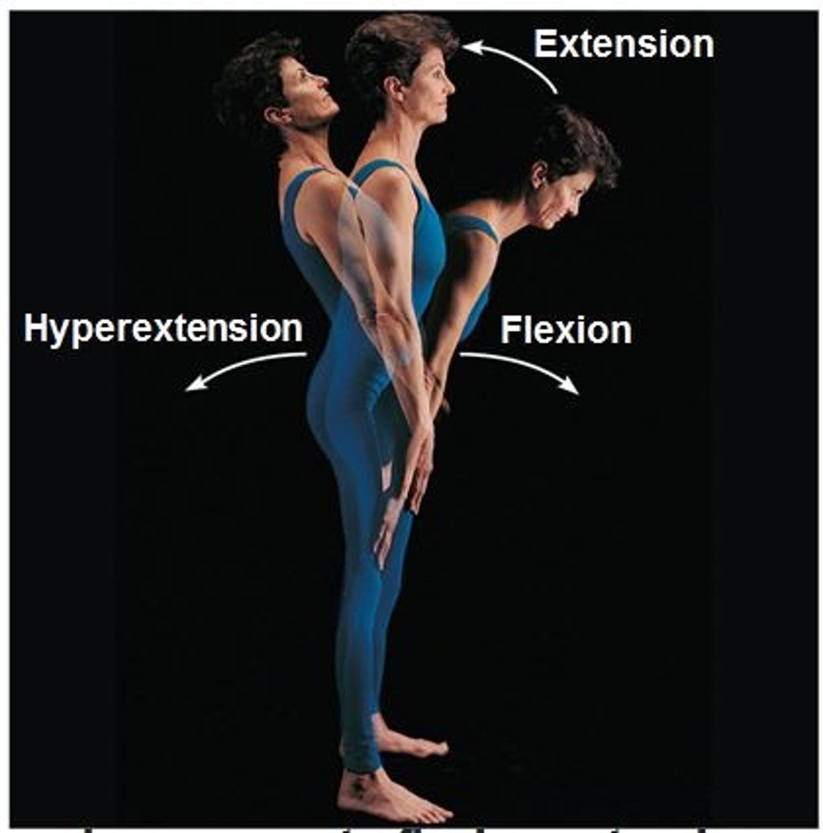
Flexion
To bend. Decreases the angle between the bones of a joint. Examples: Elbow, knee, neck, ankle. Joint Type: Hinge, Condyloid, Saddle, Ball-and-Socket.
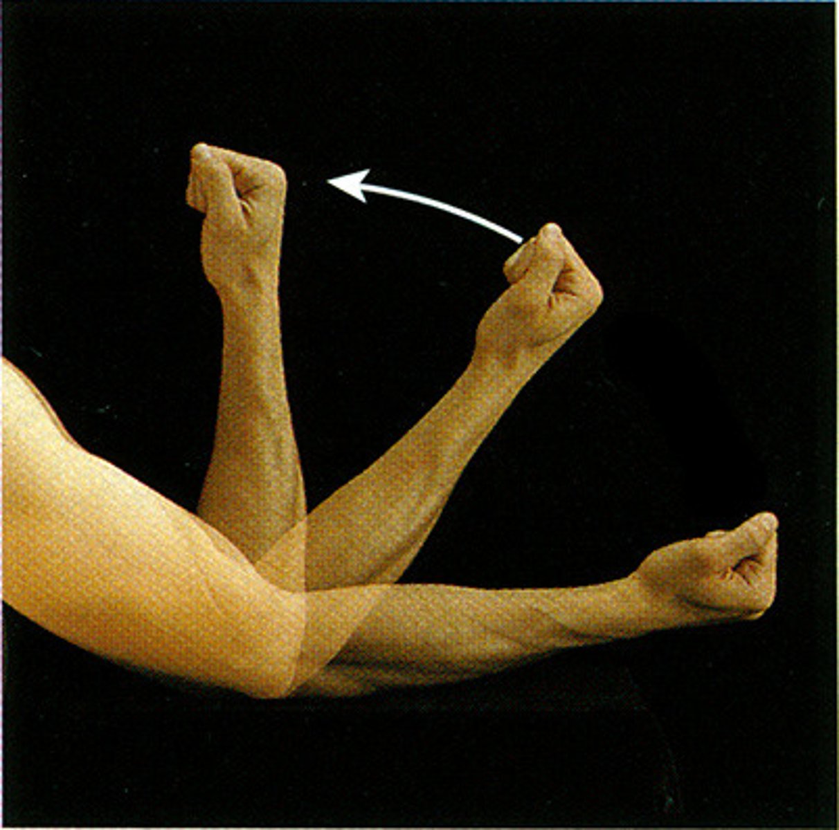
Extension
To stretch out. Increases the angle between the bones of a joint. Examples: Elbow, knee, neck, ankle. Joint Type: Hinge, Condyloid, Saddle, Ball-and-Socket.
Hyperextension
Beyond or excessive. Extension beyond anatomical position (angle >180°). Examples: Wrist, neck. Joint Type: Hinge, Condyloid, Saddle, Ball-and-Socket.
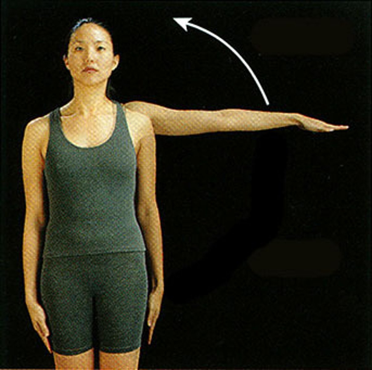
Abduction
To lead away. Movement away from the midline. Examples: Arms, legs, digits. Joint Type: Condyloid, Saddle, Ball-and-Socket.
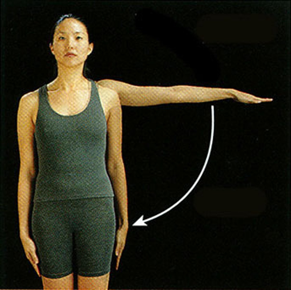
Adduction
To lead toward. Movement toward the midline. Examples: Arms, legs, digits. Joint Type: Condyloid, Saddle, Ball-and-Socket.
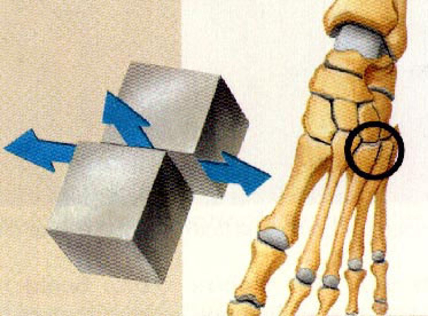
Gliding
Nearly flat bone surfaces slide or glide over each other. Example: Carpal bones. Joint Type: Plane Joint.
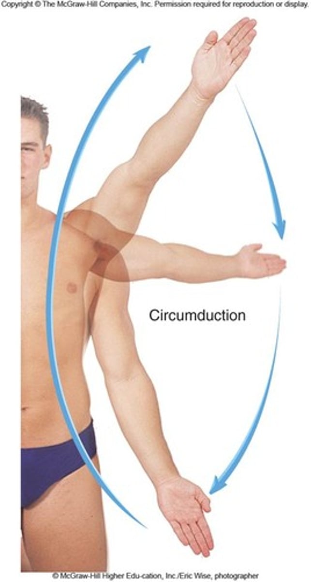
Circumduction
"Circ" = circle. Move distal part of an appendage in a circle while the other end remains stationary (no rotation). Examples: Shoulder, hip, knuckle. Joint Type: Condyloid, Ball-and-Socket.
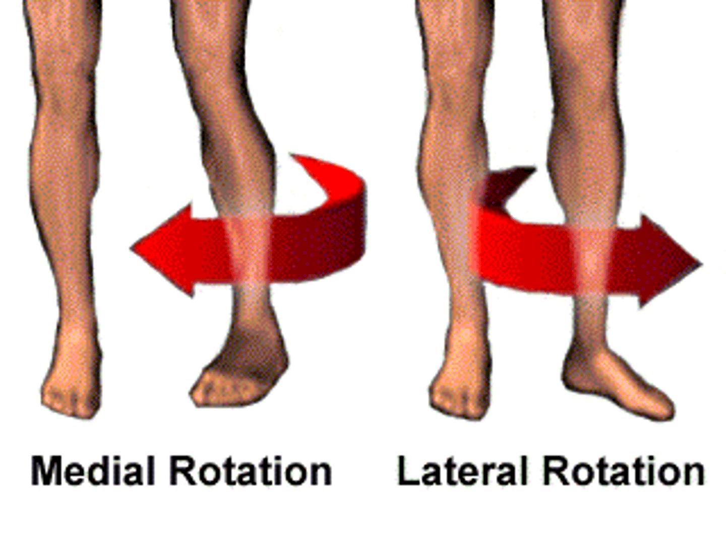
Medial Rotation
"Rota" = revolve. Rotation toward the midline (internal rotation). Examples: Neck, arm, leg. Joint Type: Pivot, Ball-and-Socket.

Lateral Rotation
Rotation away from the midline (external rotation). Examples: Neck, arm, leg. Joint Type: Pivot, Ball-and-Socket.
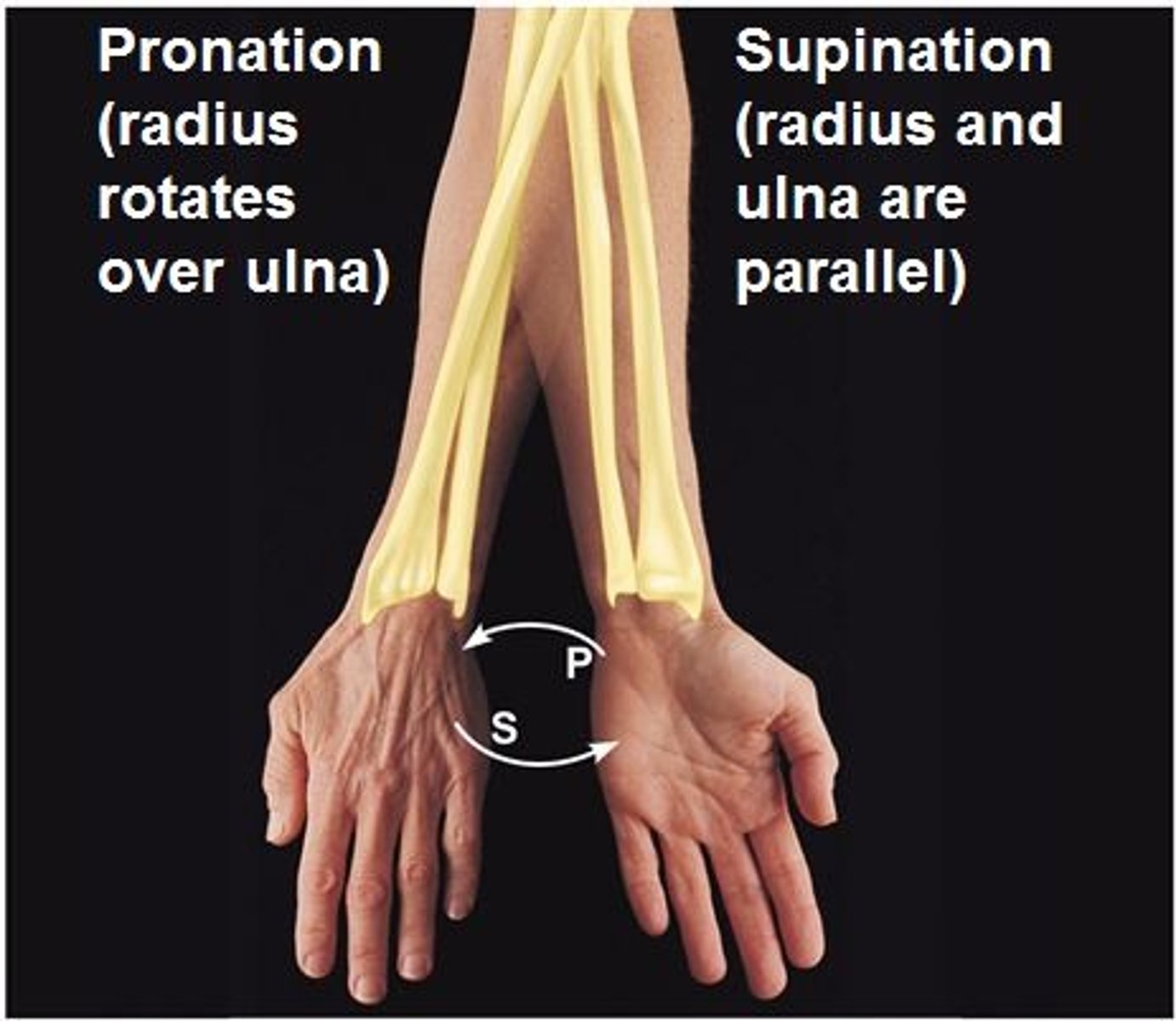
Pronation
"Pronate" = lying face down. Rotation of forearm so the palm faces posteriorly; radius crosses over ulna. Example: Radioulnar joint. Joint Type: Pivot.

Supination
"Supine" = lying on back. Rotation of forearm so palm faces anteriorly; radius and ulna are parallel. Example: Radioulnar joint. Joint Type: Pivot.
Inversion
Turns sole of foot medially. Example: Foot.
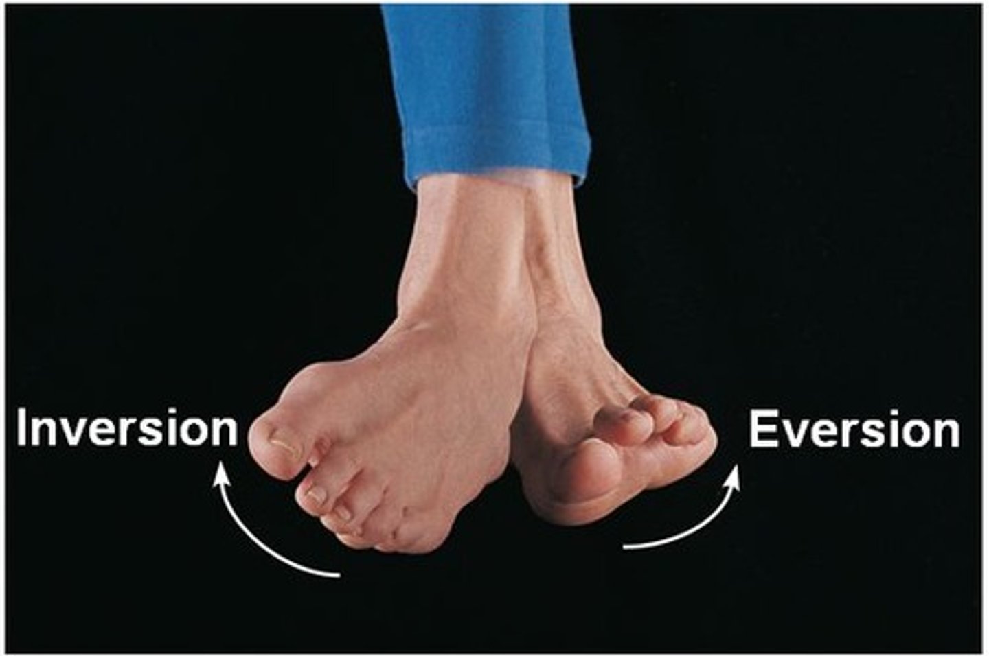
Eversion
Turns sole of foot laterally. Example: Foot.
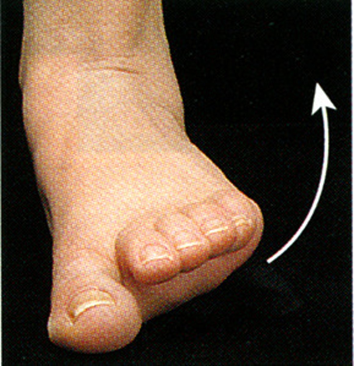
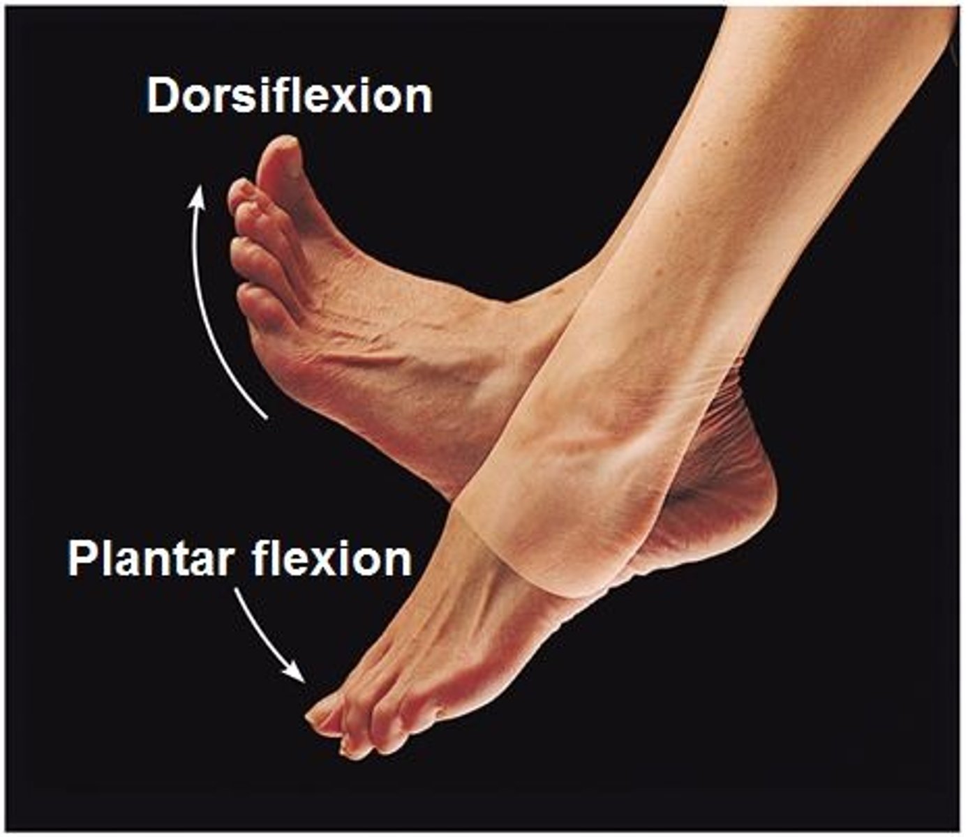
Dorsiflexion
Flexion at ankle lifting toes upward. Example: Foot.
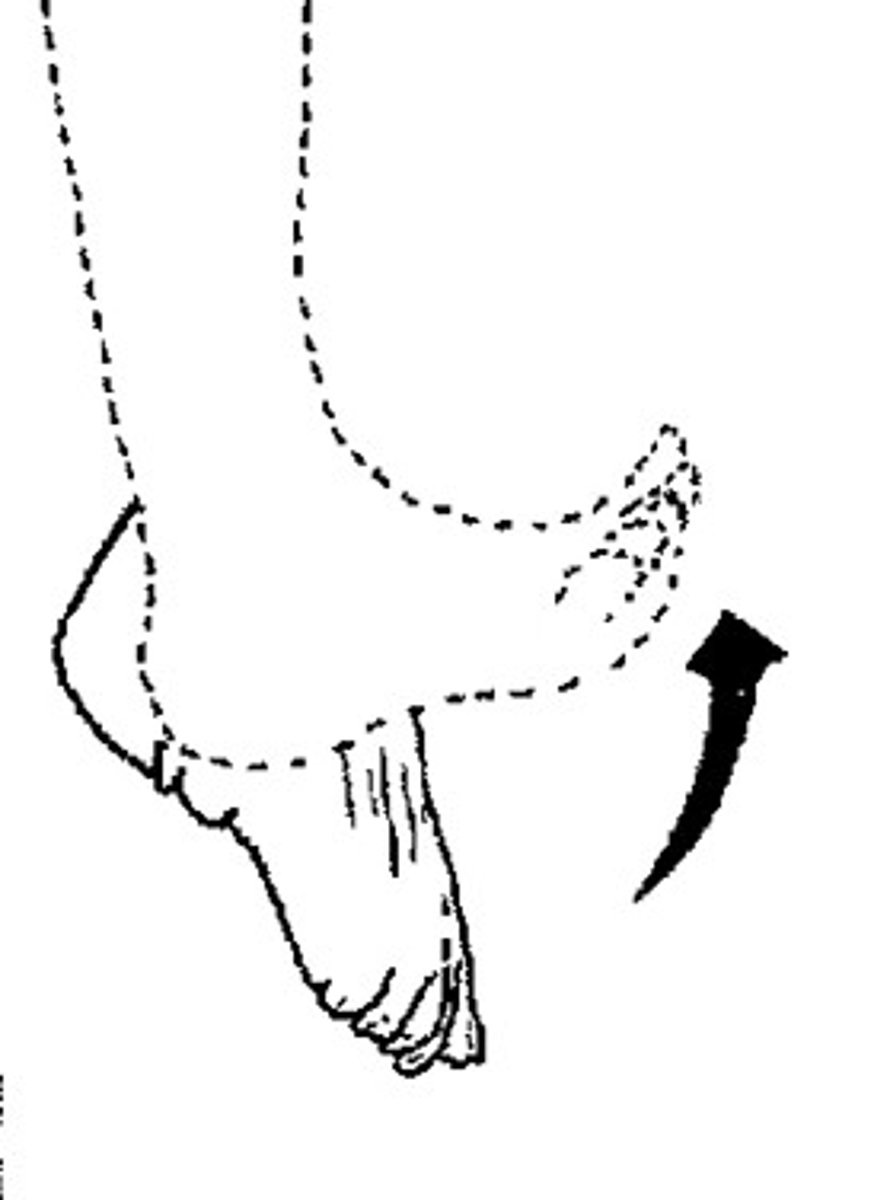
Plantar Flexion
Extension at ankle pointing toes downward. Example: Foot.
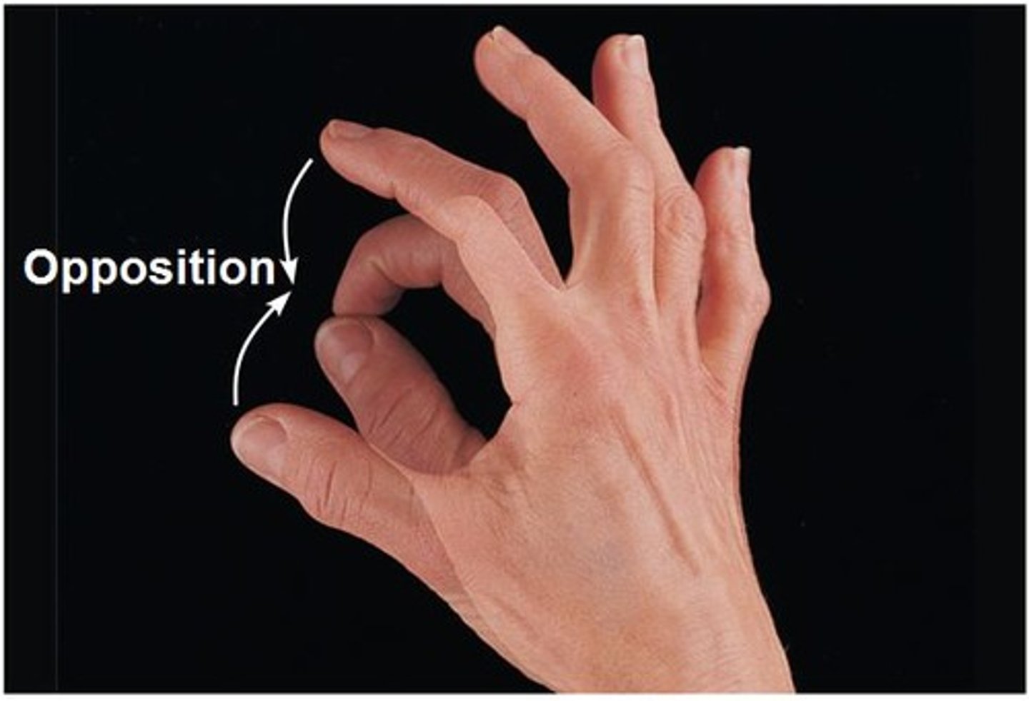
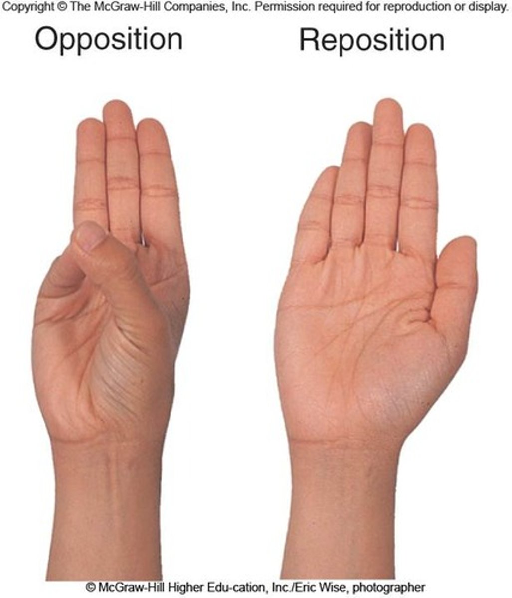
Opposition
Thumb moves toward palm or other fingers. Example: Thumb.
Reposition
Movement returning thumb to anatomical position. Example: Thumb.
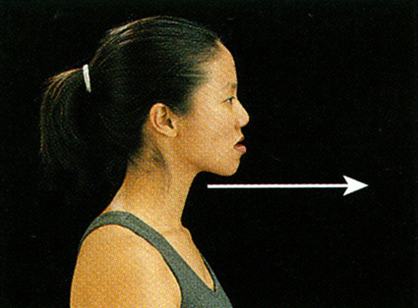
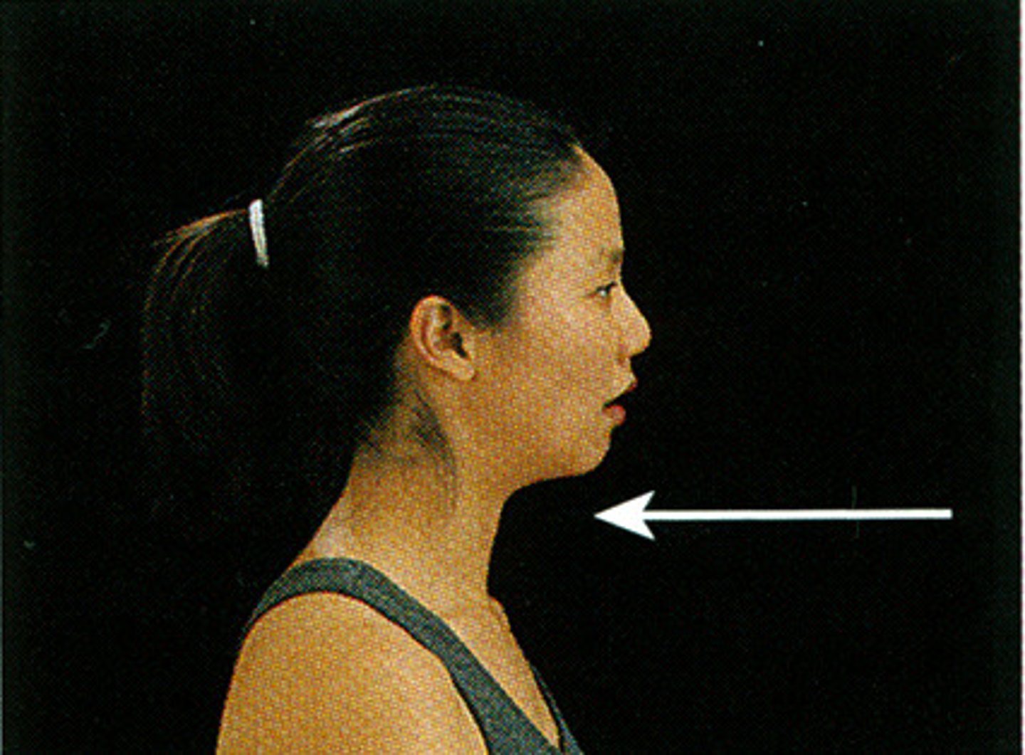
Protraction
Anterior movement in the horizontal plane (forward). Example: Scapulothoracic joint.
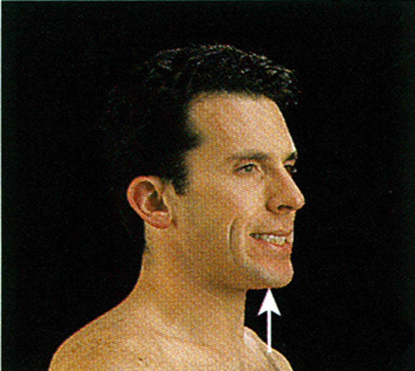
Retraction
Posterior movement in the horizontal plane (backward). Example: Scapulothoracic joint.
Elevation
Movement superiorly (upward). Examples: Scapula, mandible (chewing).
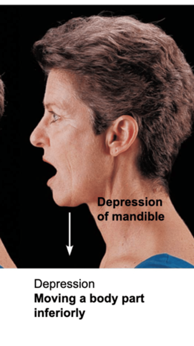
Depression
Movement inferiorly (downward). Examples: Scapula, mandible.
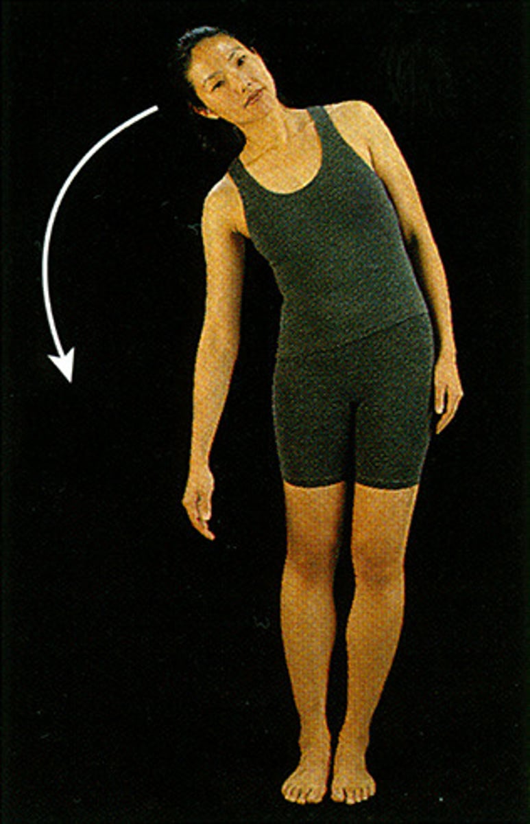
Lateral Flexion
Bending of the vertebral column to the side. Example: Neck or trunk.
Sutures, Syndesmosis, Gomphosis
3 types of fibrous joints
Synchondrosis, Symphysis
2 types of cartilaginous joints
Amphiarthrosis
Term for slightly mobile joint