Bone development and Healing
1/16
There's no tags or description
Looks like no tags are added yet.
Name | Mastery | Learn | Test | Matching | Spaced | Call with Kai | Chat |
|---|
No analytics yet
Send a link to your students to track their progress
17 Terms
Osteoblast
Development and repair
Osteocyte
Maintain bone structure
Osteoclast
Breakdown bone matrix, allowing remodelling
Bone marrow
Tissue where haematopoiesis occurs
Primarily in long bones
Long bone epiphysis
Proximal (top) end and distal (bottom) of bone
Intramembranous Ossification
Bone develops within two layers of fibrous connective tissue, may result in flattened bone
Flat bones i.e. skull, ribs, scapula and pelvis
Endochondrial Ossification
Bone develops within cartilage framework, replacing cartilage with bone as animal skeletally matures
Bones e.g. long bones femur, humerus, short bones - carpal bones
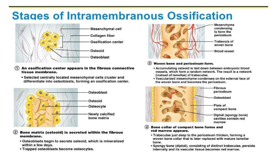
Intramembranous Ossification stages
Skeletal anatomy of foetus begins as soft fibrous tissue, allow growth development.
Once bone has been deposited, structure fixed in size, small adjustments due to remodelling
As bone fills space between 2 parallel membranes, shape characteristically flat appearance
Endochondrial Ossification
Dynamic, continuing months after animal born, affected by lifestyle, environment and nutrition and genetics
Endochondrial Ossification stages 1-3
Cartilage framework - foetus initially produces hyaline cartliage skeleton. Temporary skeleton complete prior birth
Primary Ossification - (long bones) primary centre of ossification forms with diaphysis. Osteoblasts replace cartilage with bone. Blood vessels infiltrate outer layers of cartilage framework, periosteum develops
Bone modelling - Amount of bone increases within main shaft, separating long bone into diaphysis (ossification site) and epiphyses at ends of long bone (initially no ossification). Structure of bone develops, more bone deposited within outer edges (cortical bone), blood vessels infiltrate into centre of bone (medullary cavity)
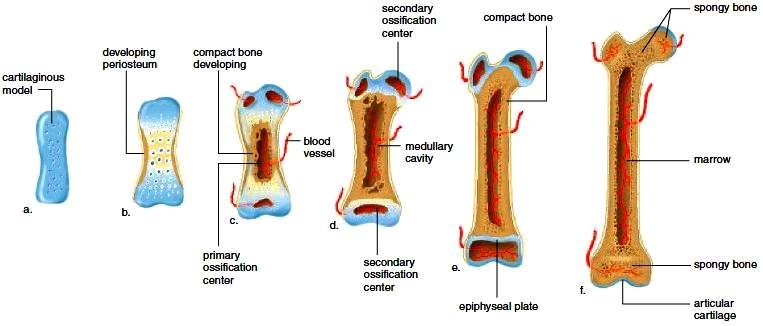
Endochondrial Ossification stages 4-6
Secondary Ossification - Occur within proximal and distal ends of bone - epiphyses. As with diaphysis, bone cells replace cartilage. Dense bone created at the edges (cortical bone), no medullary cavity formation. Cancellous (spongy) bone develops instead, becoming important site for bone marrow
Epiphyseal (Growth) Plates - As primary secondary site of ossification expan, eventually remains only a band of cartilage between diaphysis and each of epiphyses - epiphyseal plate. Bone continues to be deposited within cartliage, allowing animal to grow
Skeletal maturity - Growth ends when epiphyseal plates fused, becoming completely bone. Distal growth plate fuses first, then proximal plate. Age at which close depends on factors e.g. nutrition, although mainly influenced by genetics
Breed
Affects development of bone
Great Dane 18-24 months to skeletally mature, epiphyseal plates remaining open for extended period
Chihuahua 6m
Chondrodysplatic e.g. dachshund have growth plates calcifying earlier than other breeds - result shortened bones, curved appearance
Bone
Bone is a dynamic tissue, blood supply and exchange of nutrients occuring as with other tissues. Natural cell death requires continual replacement of bone within skeletal system
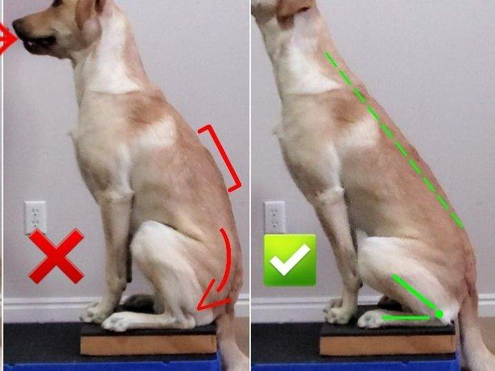
Bone density
Can naturally alter to postural changes
Place more weight on one hip siting/standing, developing posture that places more strain on some joints
Often seen after injury, e.g. stifle ligament rupture (cruciate), dog uses on hindlimb to previous injured
Cause muscle strain, discomfort, physical changes can occur in dependent limb (bearing weight), increased bone deposition in joints in response to increased pressure
Bone Healing
Endochondrial Ossification adapted to ensure bone repairs to function as load-bearing structure, capable of support animal weight
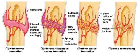
Bone healing stages
Haematoma - After impact, bone fragments cause damage to surrounding tissue - haematoma formation - collection of blood outside blood vessels
Inflammation - Haematoma replaced by granulation tissue, osteoblasts and stem cells. Migrate into fracture gap (esp. around periosteum), new blood vessels migrate to area and medullary cavity
Callus - Tissue formed. Consists fibrous tissue, cartilage, immature bone, increasing density of tissue in area and stabilising to fracture site
Bone Infiltration - As bone heals, amount of fibrous tissue decreases, bone and cartilage increase to provide greater stability. Chondroclasts resorb cartilage within callus, osteoblasts line surfaces with new bone to create mineralised matrix
Clinical union - Bone fragements become rigidly united by callus. Typically 12-16 weeks in adult cat/dogs with fractures in young animals healing more quickly
Remodelling - When bone stabilises, callus remodels, replaced by new, more organised bone. Can take years
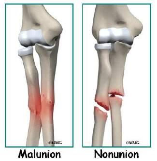
Incomplete healing
Incomplete healing
Imperfect fracture heal - non-unions.
May be associated with limb length discrepancy, infection and stiff joints
Fractures can heal in incorrect position with shortening, angular deviation or rotation. Malunion
If bone fragment does not ‘glue’ back, small fragment can turn into sequestrum, acts as foreign body