3007 wk 2 lec
 Call Kai
 Learn
Learn Practice Test
 Spaced Repetition
 Match
Match Flashcards
 Knowt Play
1/52

There's no tags or description
Looks like no tags are added yet.
Name | Mastery | Learn | Test | Matching | Spaced | Call with Kai | Chat |
|---|
No analytics yet
Send a link to your students to track their progress
53 Terms
Key points wk 1 lab
Hand Hygiene
Before and after many different tasks, interactions etc.
Glove use
When there is a risk of blood contact
Avoid routine use where possible, unless risk is present
ANTT
Aseptic Non-Touch Technique
Avoid touching KEY PARTS
even if sterile gloves are used
Maintenance of Aseptic Field
Scrub key part for at least 15secs!
General use of sterile equipment and sterile technique
How to check patency of CVC line
1) Scrub the hub - 15secs
2) Let air dry
3) Follow ANTT
4) Aspirate 2 - 5mls to check that line is patent, and to ensure no drugs are left in the line
5) Remove syringe and discard
6) Flush using NS with pulsatile “start stop” motion
When you’re done, always remember to clamp at the end!
Considerations for pushing meds through CVC
The line has a certain amount of mL → it is generally much longer than the avg. PIVC
Therefore, the flush that you do after pushing a drug through HAS to go at the same rate (usually a line may be around 0.5mLs - so slow pushing around 2mLs will ensure the drug in the line is pushed into vascular system safely)
What happens if a CVC bung falls of or is removed?
#1 = check pt if they are stable
Risk of air embolism
Risk of infection
Risk of blood loss through open lumen
→ Therefore, ensure clamping bung when not in use to protect against potential incidents like this!
Tunnel vs Non-tunnelled CVC
Tunnelled = Goes straight into vasculature
Non-tunnelled = Goes into and UNDERNEATH subcut tissue before entering the vascular space (often,
WEEK 2 RESPIRATORY FAILURE
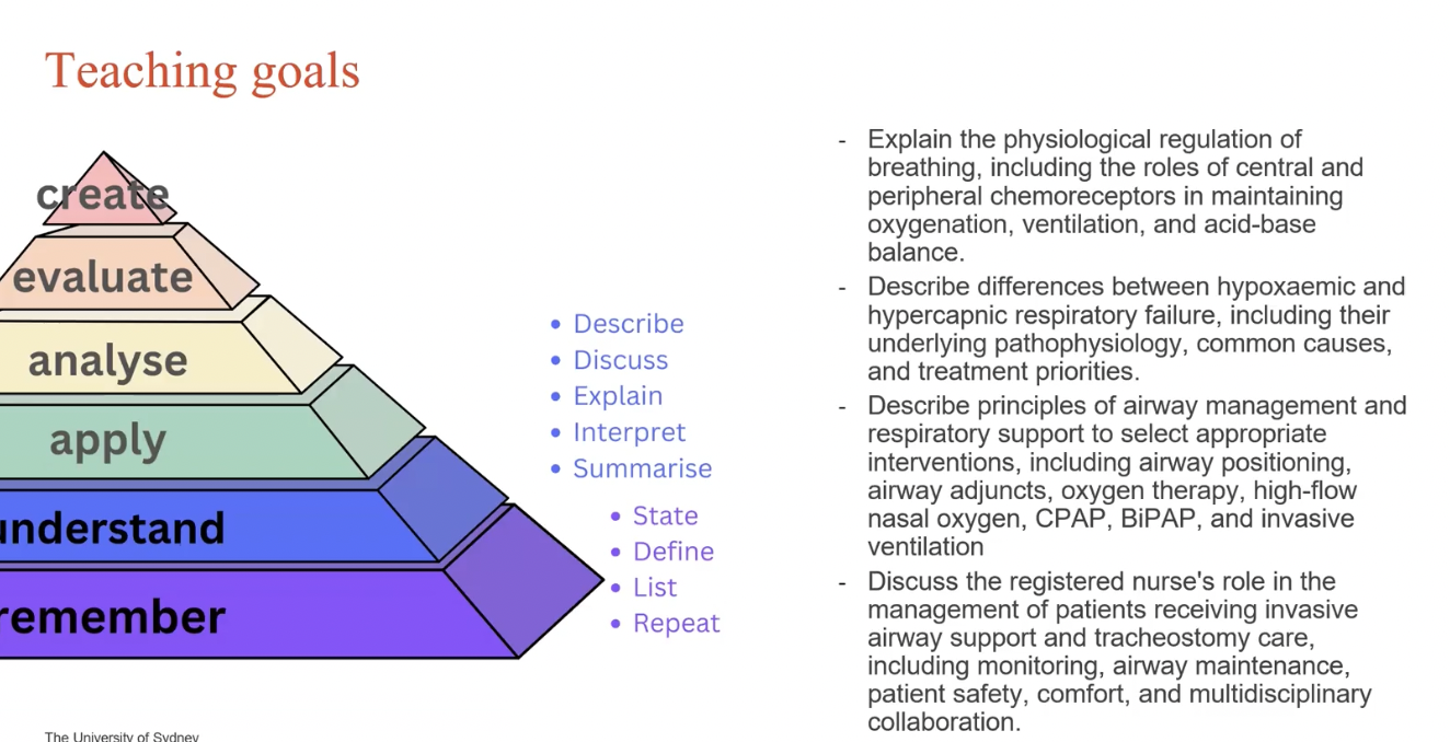
Diffusion
Random movement of free molecules in solution
→ Often driven by concentration gradient (high conc to low conc to balance out)
No energy req
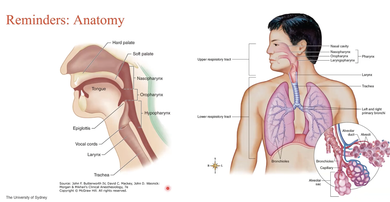
Anatomy reminder
Normal gas exchange
Ventilation
Diaphragm contracts
Increasing total space inside chest cavity
Pressure decreases within to be less than outside pressure
Air passively enters the lungs
Air slowly exhales as diaphragm slowly relaxes
Diffusion
Blood diffuses in alveoli
CO2 enters alveoli
O2 is released from alveoli in exchange for CO2
Perfusion
CO2 is released from organs and passed through blood stream to alveoli
O2 is released from alveoli into blood stream for perfusion
Tidal Volume
Volume of each breath in mLs
How do we breathe? What is our respiratory control?
Central chemoreceptors
In brain medulla
Responds to alterations in CO2
Responding to pH changes
Increase H = acidic → higher RR and TV
Decrease H = basic → lower RR and TV
Peripheral chemoreceptors
In Carotid and aortic bodies (neck / heart)
Responds to low O2
2 Main breathing aspects
Oxygenation
Delivery of oxygen to lungs → circulation → tissues
SpO2
Haemoglobin
Does not account for CO2 levels
Ventilation
Process of inspiration and expiration
Air moving in and out of lungs
Removal of CO2 from body
Measures as minute ventilation = RR x TV
Visual of perfusion and gas exchange
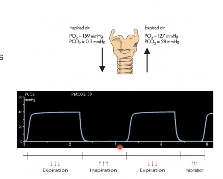
PCO2 is much lower in the environment
PCO2 is higher when it is being expired as the result of energy use from within our body (since CO2 is byproduct of energy production)
Consider diffusion in this context!!
The body is seeking O2 initially, the CO2 is exchanged and released
Hence the highest O2 uptake at the beginning, then highest CO2 output on expiration

Conditions that influence Respiratory Failure
General lung function
O2 delivery
Cardiac Output
Baseline metabolic rate (e.g. fever? Sepsis?)
Acute resp failure
Sudden onset
→ Impaired gas exchange becoming life-threatening
Type 1 Respiratory Failure
Due to OXYGENATiON
Failure to oxygenate
PaO2 < 60 - 70mmHg (less than SIX SEVENNN ty)
OR Decreased SaO2 / SpO2
Causes of T1RF
Pneumonia
Left HF
Pulmonary oedema
Acute Respiratory Distress Syndrome (ARDS)
Treatment of T1RF
1) Increase FiO2 (dial up O2 therapy)
2) Apply pressure to airway to adjust diffusion gradient for improved diffusion
Type 2 Respiratory Failure
Failure to remove CO2 from the body
Hypercapnic
Failure to ventilate = insufficient removal
PaCO2 > 50 mmHg within the body (greater than FIFTY)
The issue here is that air is not moving!!
Causes of T2RF
COPD
Asthma
CNS depression
Trauma impacting ventilation
Treatment of T2RF
1) Increase minute ventilation ( = TV x RR)
2) Increase RR / Increase TV
Hypoxaemic RF
T1RF
1) Mismatch between ventilation and perfusion
V / Q mismatch
→ Blood flow may be perfused, but is not ventilating
e.g. Sputum in alveoli preventing airflow from diffusing into bloodstream OR PE / blood clot preventing blood from perfusing well due to stagnation
2) Shunt
Blood completely bypasses the lungs, and flows from the R) side heart → L) side heart
No diffusion with alveoli, leaving blood poorly oxygenated
3) Diffusion limitation
Oxygen has difficulty crossing into blood stream
Poor diffusion from alveoli into blood stream
4) Alveolar hypoventilation
Inadequate ventilation
Alveoli not achieving enough air flow → decreased oxygen and increased CO2 levels (because poor inhalation and poor exhalation)
Hypercapnic RF
Imbalance between:
1) Ventilation supply
Generally affected by airflow obstruction or trapping
e.g. Asthma, COPD (generally conditions that cause air to stop moving)
2) Ventilation demand
Amount of ventilation required to keep PaCO2 within normal limits
Assessing clinical deterioration
RR is a key indicator of a compensatory mechanism for malfunctioning physiological processes!!!
e.g.
RR > 27 is a key predictor for cardiac arrest in adults in hospital
Inspection
Auscultation
Palpation ?
Percussion (for signs of fluid overload or density)
CO2 monitoring
Measuring amount of CO2 exhaled
→ Want to ensure that CO2 levels are maintained within the body
PCO2 is what we use to measure this!!! (view image)
- Exhalation when there is a reading, inhalation when reading is minimal (because we don’t breathe in CO2 lol)
EVIDENT S&S of Hypoxaemia
1) SPo2 < 80%
2) Late cyanosis
Dyspnoea
Tachypnoea
Drowsiness increasing
S&S of Hypercapnia
1) Dyspnoea
2) Tachypnoea with shallow breathing
Decrease TV
Decrease Minute Ventilation
Common O2 goals in critical care
PaO2 70 - 100mmHg
PaCO2 35 - 45mmHg
SPo2 92% - 96%
Range can be 88% - 92% for someone with Hx of chronic respiratory diseasse
NOTE: The more O2 we give (we can’t just automatically give 100% O2 → the higher risk of death). Therefore, we often lower the sats threshold in order to provide oxygen that is adequate enough to sustain living capacity
Main things to monitor in critically ill respiratory patients
Mental status
Ventilation rate
SpO2
ABGs (if available → this is pretty much a blood test but taken from an artery to measure O2, CO2, pH etc. from an oxygenated blood line)
Generally keep using this until PaO2 & PaCO2 returns to normal baseline
Non-invasive Positive Pressure ventilation
HF nasal prongs
Pushing large amounts of air to keep lungs open
leads to lower levels of Positive End-Expiratory Pressure (PEEP)
→ CONSIDER PEEP as pressure that exists in the air sacs after expiration! You don’t want too much because too much pressure is bad. Lower PEEP can be preferred in conditions where air trapping or over-pressure is the issue
Allows for improved oxygenation as more SA is present when air sacs are kept open
Prevents lung collapse
Reduces breathing effort
Mask
Delivers large amounts of air when patient is breathing spontaneously (on their won)
Positive Pressure Ventilation because there is a positive pressure going into the lungs to maintain breathing
When should we not use PPV Mask?
1) NOT for patients with excessive secretions
2) Decreased LOC
3) High O2 requirements
4) Facial traumas (becuase how is the PPV going to work effectively)
What can Masks deliver?
CPAP
Constant Positive Airway Pressure
PEEP
Supports oxygenation
BiPAP
Bilevel Positive Airway Pressure
Different pressure levels for inspiration and expiration (IPAP & EPAP)
Supports oxygenation and ventilation
When to use non-invasive PPV
Increased dyspnoea
Severe tachypnoea (RR > 24)
Increased WOB, use of accessory muscles
Muscle / breathing fatigue
OR
PaCO2 > 45mmHg
PaO2 < 60 mmHg
Contraindications for Non-invasive PPV
LOC → unable to breathe independently and on their own
would consider invasive in this instance
Mask doesn’t fit appropriately (e.g. facial trauma)
Hypotensive shock
Because increasing and forcing pressure into the lungs can cause great increases in thoracic pressure which can cause DECREASED BP because of the pressure
Upper GI bleeding
Because it causes high risk for vomiting and increased GI distress
Inability to protect or maintain airway
Poor toleration or ineffectivity of non-invasive PPV
What are things that can compromise airways?
FOOD!
Inflammation (e.g. tonsilitis)
Secretions
Mucus
Consider tongoue positioning with LOC (because decreased LOC can cause the tongue to drop back and block the airway)
Nursing interventions for Airway compromise?
Effective / assisted coughing
Adequate hydration
Airway suctioning
Pt positioning (sitting them up)
Use of airway adjuncts in maintaining airway
1) Oropharyngeal Airway (OPA)
CONTRAINDICATIONS:
Consciousness
Can cause gag reflex
Oral trauma
Intact gag reflex (can consider NPA if this is the case)
2) Nasopharyngeal Airway (NPA)
CONTRAINDICATIONS:
Nasal trauma
Basal skull #
3) Supraglottic airway
CONTRAINDICATIONS:
Limited mouth opening
→ Often used for promptly secure airway TEMPORARILY
4) Endotracheal tube
CONTRAINDICATIONS:
Facial trauma / bleeding
Total airway obstruction (Consider Trachy in this instance)
5) Tracheostomy
Bag Valve Mask
Used a rapid intervention for increased conc of oxygen or to provide breathing (when a patient is failing to breathe on their own)
It is often a short-term intervention used before intubation to
Airway adjuncts may be used to keep airway patent and allow O2 to flow in effectively
Provided at 12 - 15L/min
- Should see visible rise and fall of chest (5 - 7mL / kg in adults)
What to be aware of in BVM!
Hyperventilation!
Because BVM when used aggressively causes too high PEEP → leading to circulatory complications because the lungs are used to using negative pressure to breathe
Forced Positive Pressure Ventilation can cause disruptions to the body’s normal mechanisms of breathing
Therefore, don’t just bag them TF up → bag them according to regular breathing as opposed to just constantly squeezing and releasing
Indications for INVASIVE Airway Support
Pretty much when non-invasive is contraindicated
e.g.
No LOC
Cardiac arrest
Evident medical instability
Endotracheal Intubation
Rapid Sequence Intubation (RSI)
Plan
Prepare (drugs equipment, monitoring nec.)
Protect cervical spine
Positioning appropriate for intubation
Pre-oxygenation
Pre-treatment (e.g. sedation medications)
Paralysis to ensure effective intubation placement
Placement with proof!
Post-intubation management (often to ICU)
Plan for Intubation
Observing the airway to measure the degree of difficulty to get tube through
Class 1 - 4
Grade 1 - 4
Higher grade = more difficult to intubate, smaller passageway

Endotracheal Tube Securement
Take note of the number it is at when inserted (how deep it is in to track whether the tube is staying in place)
Record the number of teeth a patient has
Confirmation of ETT placement
Confirmed with x ray
Position should be 3 - 5cm above Carina (the upside down Y region just before it splits into the lungs)
Too high = risk of damaging vocal cords
Too low = risk of ONLY RIGHT LUNG intubation (leaving the left without oxygen)
Note that ETT can still slightly move internally
Risks of ETT
Thirsty
Risk of dental injury
Difficulty with neck / head movement
Impaired swallowing
Nursing management of ETT
Ensure correct tube placement
Secure ETT
Maintain its placement and position
Ensure cuff inflation (near the end of the tube to prevent air leaks and ensure that oxygen pushed through is going into the lungs, rather than shifting the ETT from its place)
Imagine pushing air through it → it can risk causing the ETT to go upwards because of air pressure
Ongoing and cautious monitoring of oxygenation and ventilation
Understanding weaning plan and goals for care
Comfort and active communication with patient - understanding this difficult and often frustrating experience
Pain management
1:1 nursing care
Regular oral hygiene care

Things to note in ETT care
GCS for verbal would be 1T = Tube
Is a pt is paralysed as a means for intubation, motor is measured as NT (not testable)
When will we use a tracheostomy?
1) Example: Imagine major trauma to face and jaw → meaning intubating would be inappropriate
A hole would then need to be cut to bypass this issue (it also goes by the vocal cords)
2) For improved secretion removal in the mouth
3) If prolonged ventilation is required
You simply wouldn’t have an ETT in place for extended durations because it risks damaging the vocal cords
Benefits of Trachy
Decreased WOB and airway resistance
Easier, safer access to mouth
Improved oral hygiene
More comfortable for patient
Nursing tracheostomy care
Be very careful with new trachys (< 7 days)
Check security ties, placement, positioning
2 person procedure
Maintain cuff pressure
Maintain patency
Clean stoma (the hole in their windpipe)
Emotional support and comfort