AEB - Anatomy

0.0(0)
Studied by 0 people
call kaiCall Kai
Locked
learnLearn
examPractice Test
spaced repetitionSpaced Repetition
heart puzzleMatch
flashcardsFlashcards
GameKnowt Play
Card Sorting

1/283

encourage image

There's no tags or description

Looks like no tags are added yet.

Last updated 1:19 PM on 9/23/26
Name
Mastery
Learn
Test
Matching
Spaced
Call with Kai
Chat

No analytics yet

Send a link to your students to track their progress

284 Terms

1
New cards
Cranial bones
bones of the skull that enclose and protect the brain and meninges, meeting at sutures (immovable fibrous joints).
2
New cards
Calvaria (skull cap)
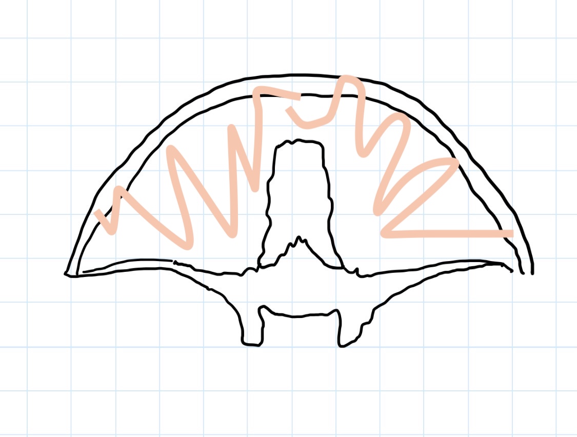
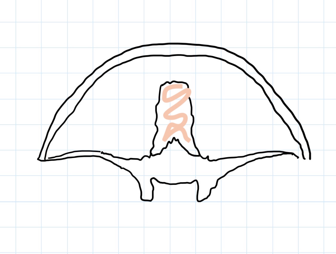
upper dome of the skull forming the roof of the cranial cavity.
3
New cards
Cranial cavity
protected space inside the skull that houses and supports the brain.
4
New cards
Pterion
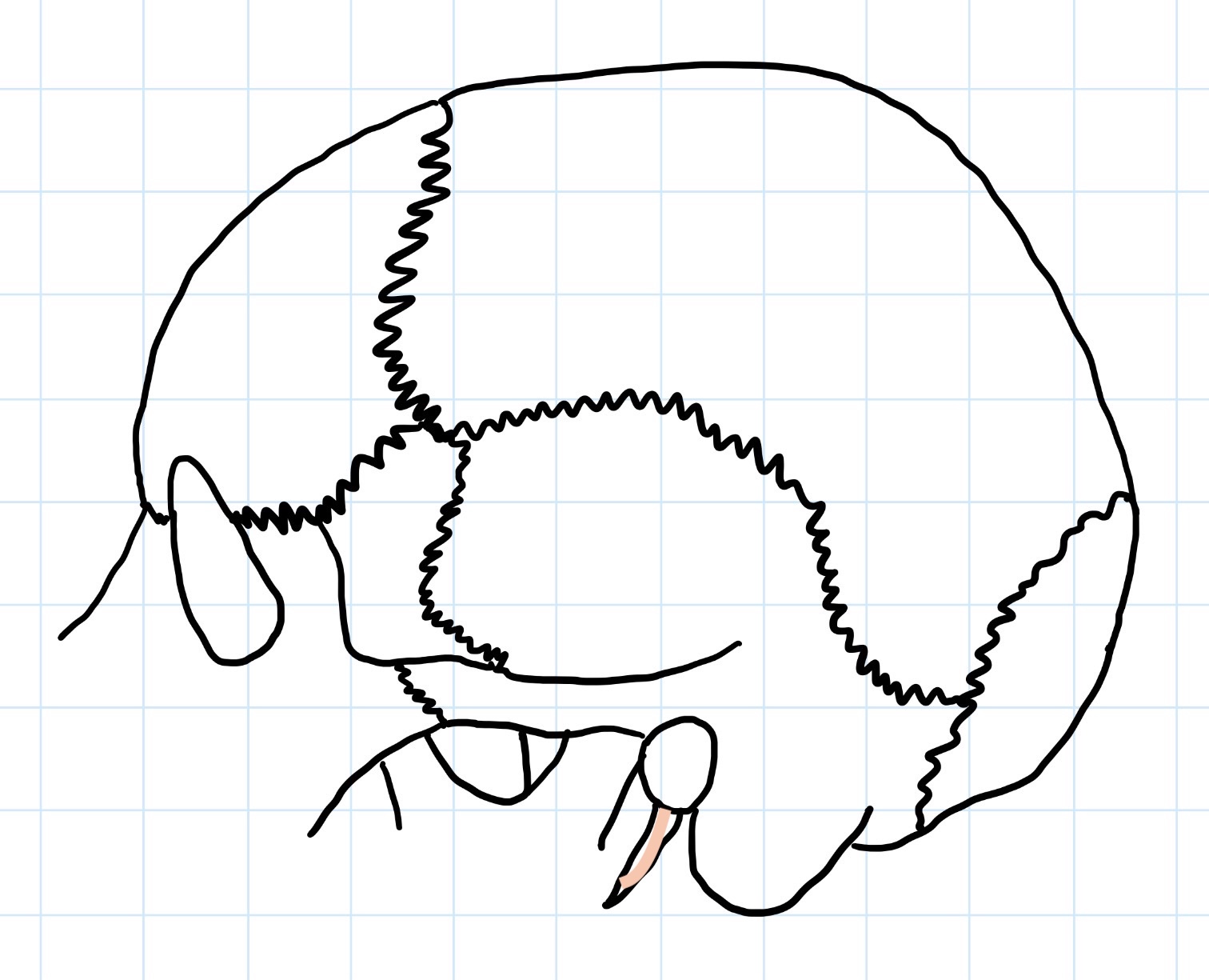
formation of sutures at the lateral aspect of the skull joining frontal, parietal, sphenoid, and temporal bones overlying a branch of the middle meningeal artery.
5
New cards
Middle meningeal artery
a branch of what artery is below the pterion, meaning fracture of thin temporal bone can cause life threatening extradural haemorrhage?
6
New cards
Fontanelles
soft, membranous regions of the foetus/neonate where developing bones have not fully ossified allowing flexibility of skull in childbirth and as brain grows.
7
New cards
Anterior fontanelle
closes at 18-24 months, forming the bregma landmark.
8
New cards
Posterior fontanelle
closes at 3 months, forming the lambda landmark.
9
New cards

Frontal bone


10
New cards

Greater wing of sphenoid bone


11
New cards

Temporal bone


12
New cards

Styloid process


13
New cards

Mastoid process


14
New cards

Occipital bone


15
New cards

Parietal bone


16
New cards

Pterion


17
New cards

Lambda


18
New cards

Lambdoid suture


19
New cards

Sagittal suture


20
New cards

External occipital protuberance


21
New cards

Coronal suture


22
New cards

Bregma


23
New cards
Anterior cranial fossa
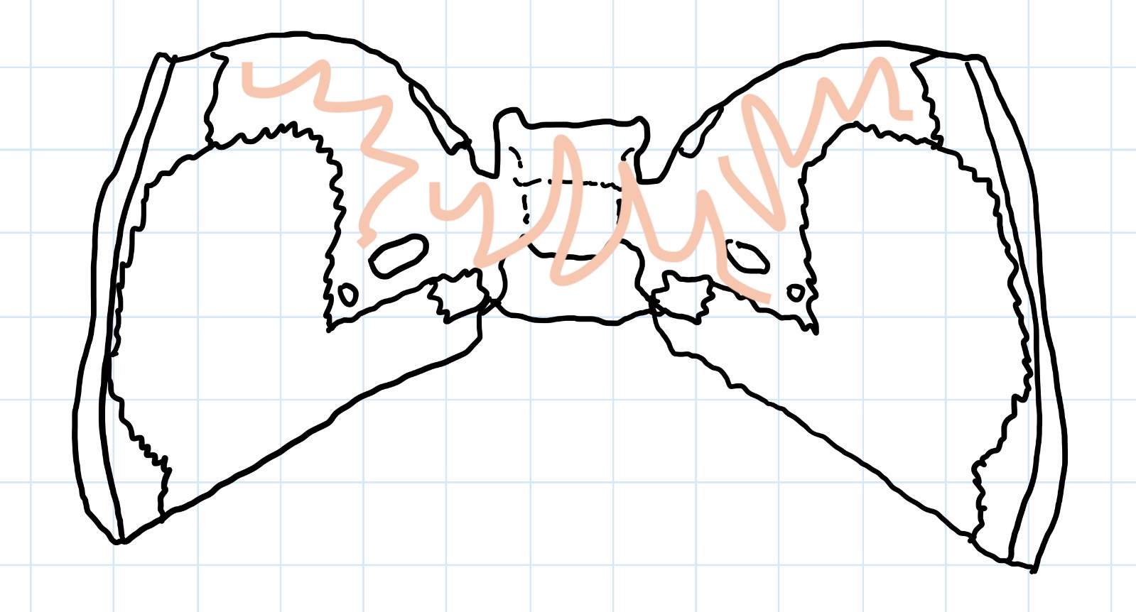
depression of the internal skull base formed from the frontal, ethmoid, and sphenoid bones, containing the frontal lobes.
24
New cards
Middle cranial fossa
depression of the internal skull base formed from the sphenoid and temporal bones, containing the temporal lobes.
25
New cards
Posterior cranial fossa
depression of the internal skull base formed from the temporal and occipital bones, containing the cerebellum.
26
New cards

Frontal bone


27
New cards

Cribiform plate of ethmoid bone


28
New cards

Lesser wing of sphenoid bone


29
New cards

Pituitary (hypophysial) fossa


30
New cards

Sphenoid bone


31
New cards

Temporal bone


32
New cards

Clivus


33
New cards

Occipital bone


34
New cards

Internal occipital protuberance


35
New cards

Telencephalon

embryological term for cerebrum, arising from the forebrain.

36
New cards

Diencephalon

embryological term for thalamus and accompanying structures, arising from the forebrain.

37
New cards

Mesencephalon

embryological term for the midbrain.

38
New cards

Rhombencephalon

embryological term for hindbrain, forming the pons, cerebellum, and medulla.

39
New cards
Cerebral cortex
outermost layer of cerebrum composed of packed nerve cells (grey matter) covering in gyri and sulci responsible for motor, sensor, and higher order functions.
40
New cards
Insula cortex
hidden structure of the cerebral cortex located deep to the lateral fissure, responsible for audition, pain, and homeostasis.
41
New cards
Nuclei
clusters of nerve cell bodies located deep inside the central nervous system.
42
New cards
Projection fibres (up/down)
nerve axon bundles connecting cerebral cortex with lower parts of the central nervous system.
43
New cards

Association fibres (forward/back)

nerve axon bundles connecting different cortical areas (gyri) in the same cerebral hemisphere.

44
New cards
Commissural fibres (right/left)
nerve axon bundles connecting both hemispheres, allowing interhemisphere communication.
45
New cards
Corpus callosum
largest commissural bundle.
46
New cards
Central sulcus
groove separating frontal from parietal lobe, mainly present on the lateral aspect of the cerebrum.
47
New cards

Lateral fissure

deep groove separating temporal from frontal and parietal lobes, mainly present on the lateral aspect of the cerebrum.

48
New cards

Parieto-occipital sulcus

groove separating parietal from occipital lobes, mainly present on the medial aspect of the cerebral hemispheres.

49
New cards
Calcarine sulcus
groove dividing occipital lobe holding the primary visual cortex, mainly present on the medial aspect of the cerebral hemispheres.
50
New cards
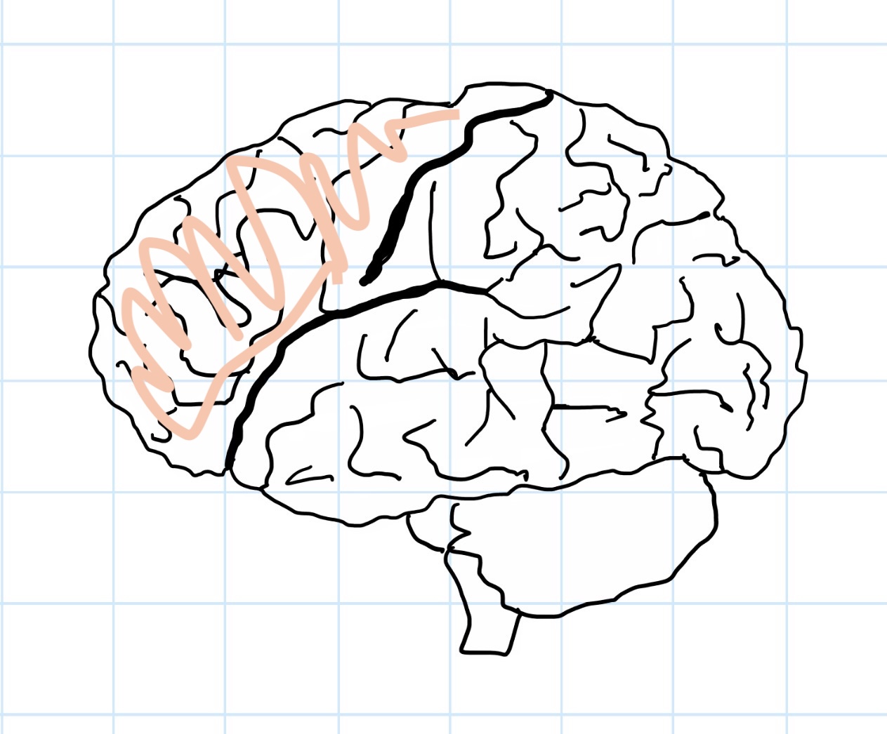
Frontal lobe
cerebral lobe responsible for executive function, behaviour, memory, and motor control.
51
New cards
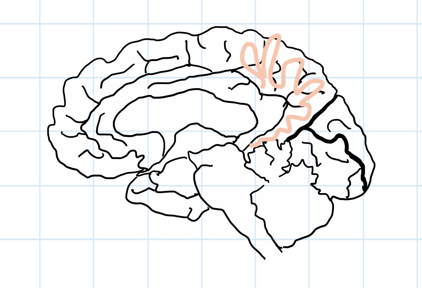
Parietal lobe
cerebral lobe responsible for somatosensory function and sensory integration.
52
New cards
Occipital lobe
cerebral lobe primarily responsible for vision.
53
New cards
Temporal lobe
cerebral lobe responsible for audition, memory, and learning.
54
New cards
Limbic lobe
cerebral lobe responsible for emotion, behaviour, memory, and olfaction.
55
New cards
Brain stem
stalk composed of the **midbrain, pons, and medulla oblongata** containing motor and sensory white matter tracts and nuclei supplying head face with other cardiovascular and respiratory regulatory functions
56
New cards
Cerebellum
hind brain structure composed of central portion (vermis) and 2 hemispheres for maintenance of balance, posture, and unconscious motor movements
57
New cards
Peduncles
group of 3 stalks connecting motor regions of the brain and spinal cord to the cerebellum.
58
New cards

Frontal lobe


59
New cards

Central sulcus


60
New cards

Parietal lobe


61
New cards

Occipital lobe


62
New cards

Temporal lobe


63
New cards

Lateral fissure


64
New cards

Frontal lobe


65
New cards

Limbic lobe


66
New cards

Parietal lobe


67
New cards

Occipital lobe


68
New cards

Parieto-occipital sulcus


69
New cards

Calcarine sulcus


70
New cards

Diencephalon


71
New cards

Temporal lobe


72
New cards

Thalamus


73
New cards

Hypothalamus


74
New cards

Midbrain


75
New cards

Pons


76
New cards

Medulla oblongata


77
New cards

Brain stem


78
New cards

Cerebellum


79
New cards
Meninges
layers of connective tissue which cover and protect the brain and spinal cord, formed of the dura, arachnoid, and pia mater from superficial to deep.
80
New cards
Dura, arachnoid, pia
Recall the meningeal layers from superficial to deep.
81
New cards
Dura mater
tough, thick outermost membrane layer of the meninges composed of the outer periosteal layer adhered to the skull, and the inner meningeal layer.
82
New cards
Periosteal layer
outer layer of dura mater, adhered to the skull.
83
New cards
Meningeal layer
inner layer of dura mater, lining arachnoid mater.
84
New cards
Dural folds
meningeal layer separates from the periosteal layer, projecting downward to divide the cranium into different compartments.
85
New cards
Falx cerebri
large, sickle-shaped dural fold separating the hemispheres of the brain.
86
New cards
Tentorium cerebelli
large, crescent-shaped dural fold separating the cerebrum from the cerebellum.
87
New cards
Falx cerebelli
small, sickle-shaped dural fold separating the lobes of the cerebellum.
88
New cards
Diaphragma sellae
small, circular dural fold forming a roof over the pituitary fossa.
89
New cards
Dural venous sinuses
blood channels between dural layers at the dural folds.
90
New cards
Subarachnoid space
space between arachnoid mater and pia mater bridged by thin filaments and containing cerebrospinal fluid and blood vessels.
91
New cards
Thin filaments
strands of connective tissue that span across the subarachnoid space.
92
New cards
Arachnoid granulations
small, fluid-filled pouches of the arachnoid mater that protrude through the dura mater into the dural venous sinuses allowing CDF to enter blood.
93
New cards
Ventricles
four connected, fluid-filled spaces deep within the brain that produce and circulate cerebrospinal fluid.
94
New cards
Cerebrospinal fluid
cushioning fluid produced by the choroid plexus, also for provision of nutrients, removal of wastes, immunity, and lightening head.
95
New cards
Choroid plexus
specialised tissue in ventricles producing cerebrospinal fluid.
96
New cards
Lateral, third, cerebral aqueduct, forth
Recall the path cerebrospinal fluid flow through the ventricles.
97
New cards
Apertures into subarachnoid space, central canal
Describe the 2 exit points for cerebrospinal fluid from the forth ventricle.
98
New cards
Lateral ventricles
ventricles associated with the cerebrum.
99
New cards
Third ventricle
ventricle associated with the thalamus.
100
New cards
Cerebral aqueduct
ventricle associated with the midbrain.