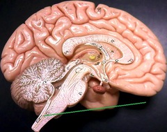
Normandale Human Anatomy Lab Practical 1 Diagram
 Call Kai
 Learn
Learn Practice Test
 Spaced Repetition
 Match
Match Flashcards
 Knowt Play
1/107

There's no tags or description
Looks like no tags are added yet.
Name | Mastery | Learn | Test | Matching | Spaced | Call with Kai | Chat |
|---|
No analytics yet
Send a link to your students to track their progress
108 Terms
ocular lens
revolving nosepiece
objective lens
mechanical stage
diaphragm adjustment lever
base
light switch
mechanical stage adjustment knobs
variable light control
stage aperture
condenser
condenser height adjustment knob
light source
coarse focus knob
arm
fine focus knob
nucleus
control center of the cell

cytoplasm
A jellylike fluid inside the cell in which the organelles are suspended

plasma membrane
A selectively-permeable phospholipid bilayer forming the boundary of the cells

lumen
cavity inside a tube, blood vessel, or hollow organ

apical surface
A: upper free surface exposed to the body exterior or the cavity of an internal organ
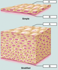
basal surface
B: bottom layer of epithelial tissue that attaches to the basement membrane
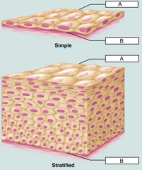
basement membrane
layer between epithelium and underlying connective tissue
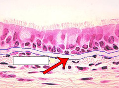
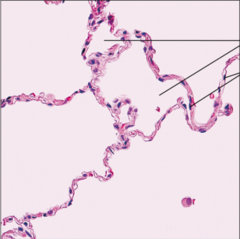
simple squamous epithelium
name the tissue: single layer of flat cells with disc-shaped nuclei
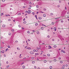
simple cuboidal epithelium
name the tissue: single layer of cube-like cells with large spherical nuclei
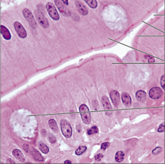
simple columnar epithelium
name the tissue: singe layer of tall cells with round to oval nuclei; many cells bear microvilli; some bear cilia; contains goblet cells
microvilli
fingerlike extensions of the plasma membrane of apical epithelial cells
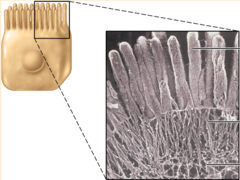
brush border
B: surface of a cell covered with microvilli. increases surface area of a cell for absorption
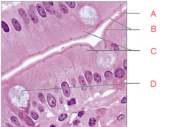
goblet cells
D: a column-shaped cell; produces mucin; secrets mucus

cilia
whiplike, highly motile extensions of the apical surface membranes of certain epithelial cells
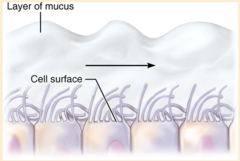
pseudostratified ciliated columnar epithelium
name the tissue: single layer of cells of differing heights, some not reaching the free surface; nuclei seen at different levels; may contain mucus-secreting cells and bear cilia

stratified squamous epithelium
name the tissue: thick epithelium composed of several cell layers; basal layers are cuboidal or columnar; surface layer are squamous
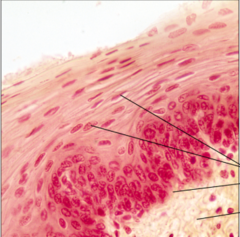
transitional epithelium
name the tissue: resembles both stratified squamous and stratified cuboidal; basal cells cuboidal or columnar; surface cells dome shaped or squamous-like
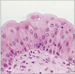
areolar connective tissue
name the tissue: gel-like matrix with all three fiber types; cells: fibroblasts, macrophages, mast cells, and some white blood cells
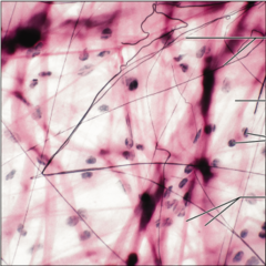
fibroblast
C: a cell in connective tissue that produces the extracellular matrix

extracellular matrix
A,B,C&D: composed of some type of ground substance embedded with protein fibers.
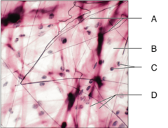
ground substance
B: unstructured material that fills the space between the cells and contains the fibers; found be soft, gel-like substance that holds tissue fluid in tissues or hard, calcified by inorganic calcium salts in bones

collagen fibers
D: thick, strongest and most abundant type of fiber; resist tension and gives strength
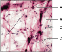
elastic fibers
A: rubberlike protein; ability to recoil back to its original form after being stretched.

adipose
name the tissue: gel-like matrix with all three fibers types but very sparse; closely packed fat cells; nucleus pushed to the side by large fat droplet
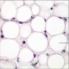
adipocyte
A: fat cell
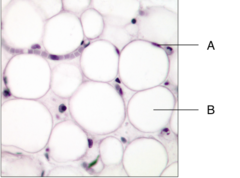
fat vacuole
B: a droplet of fat

dense regular connective tissue
name the tissue: primarily parallel collagen fibers; a few elastic fibers; major cell type is the fibroblast

dense irregular connective tissue
name the tissue: primarily irregularly arranged collagen fibers; some elastic fibers; major cell type is the fibroblast

hyaline cartilage
name the tissue: amorphous but firm matrix; collagen fibers form an imperceptible network; chondroblasts produce the matrix and when mature lie in lacunae
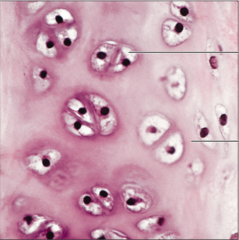
chondrocyte
A: "cartilage cell"

lacuna
B: "little-lake" a cavity occupied by a cell

perichondrium
C: membrane of fibrous connective tissue that surrounds the external surface of cartilage

elastic cartilage
name the tissue: amorphous but firm matrix; extensive elastic fibers in matrix

fibrocartilage
name the tissue: amorphous but less firm matrix; thick collagen fibers predominate

bone
name the tissue: hard, calcified matrix containing many collagen fibers; osteocytes lie in lacunae. Very well vascularized.
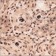
osteocyte
B: "bone-cell" seen here within a lacuna

canaliculus
D: very small tube or channel, found connecting lamella to central canal in compact bone.
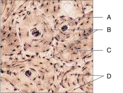
central canal
A: Haversian canal, runs through the core of each osteon, contains small blood vessels and nerve fibers
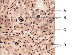
lamella
C: "little-plate" a layer of bone tissue that encircles the perimeter of the bone

osteon
tube-shaped unit in mature, compact bone; consists of concentric layers of bone lamellae surrounding a central canal; also called Haversian system.

blood
name this tissue: red and white blood cells in a fluid matrix
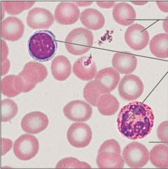
erythrocyte
A: red blood cells
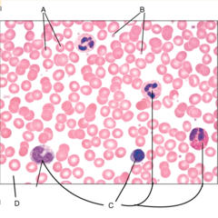
leukocyte
C: white blood cells

platelets
B: thrombocytes
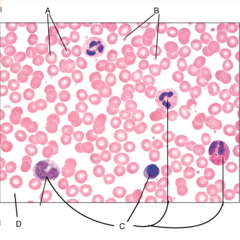
plasma
D: the nonliving, fluid component of blood, within which the blood cells are suspended.

nervous tissue
name the tissue: neurons are branching cells; processes may be long, extending from the cell body

neuron
nerve cell
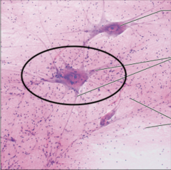
dendrites
B: a neuron's bushy, branching extensions that receive messages and conduct impulses toward the cell body
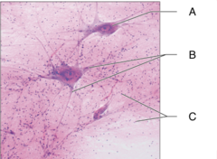
cell body
A: largest part of a typical neuron; contains the nucleus and much of the cytoplasm

neuroglia
C: supporting cells in nervous tissue
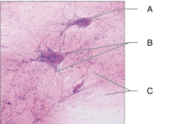
skeletal muscle
name the tissue: long, cylindrical, multinucleate cells; obvious striations

sarcolemma
plasma membrane of a muscle fiber
sarcoplasm
cytoplasm of a muscle fiber
striations
alternate dark and light bands found on skeletal and cardiac muscle

cardiac muscle
name the tissue: branching, striated, generally uninucleate cells that connect at specialized junctions

intercalated discs
complex junctions that interconnect cardiac muscles cells in the wall of the heart

smooth muscle
name the tissue: spindle-shaped cells with central nuclei; no striations; cells arranged closely to form sheets

epidermis
a keratinized stratified squamous epithelium that contains four distinct types of cells: keratinocytes, melanocytes, tactile epithelial cells, and dendritic cells
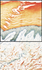
stratum corneum
A: "horny layer" most external part of the epidermis

stratum lucidum
B: occurs in thick skin but not thin skin
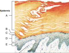
stratum granulosum
C: flattened keratinocytes; abundant pre-keratin intermediate filaments; waterproofing glycolipid that is secreted into the extracellular space
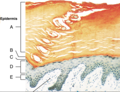
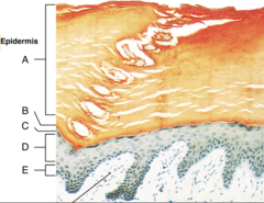
stratum spinosum
D: several cell layers thick; name from the many spinelike extensions of its keratinocytes; contain thick bundles of intermediate filaments

stratum basale
E: deepest epidermal layer; one row of actively mitotic stem cells; some newly formed cells become part of the more superficial layers
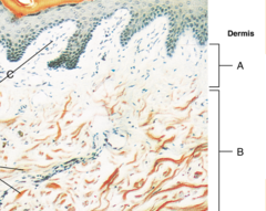
dermis
second major region of the skin, is a strong, flexible connective tissue; typically consist of connective tissue proper: fibroblasts, macrophages, mast cells, and scattered white blood cells

papillary layer
A: superficial 20% of the dermis, is areolar connective tissue containing very thin collagen and elastic fibers

dermal papillae
C: fingerlike projections that extend into the overlying epidermis

reticular layer
B: 80% of the thickness of the dermis, is dense irregular connective tissue
hypodermis
"below the skin" subcutaneous tissue; consists of both areolar and adipose connective tissue; stores fat; anchors the skin to the underlying structures

trachea
the windpipe; a passage through which air moves in the respiratory system.

lungs
main organs of the respiratory system

diaphragm
a dome-shaped, muscular partition separating the thorax from the abdomen in mammals

heart
a hollow, muscular organ that pumps blood throughout the body.
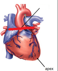
aorta
largest artery in the body

kidney
filters waste from the blood like urea, water, salt and proteins.

ureter
a duct leading from the kidney to the urinary bladder.

urinary bladder
saclike organ in which urine is stored before being excreted

esophagus
a muscular tube that connects the mouth to the stomach.

stomach
large muscular sac that continues the mechanical and chemical digestion of food

small intestine
digestive organ where most chemical digestion and absorption of food takes place

large intestine
the last section of the digestive system, where water is absorbed from food and the remaining material is eliminated from the body
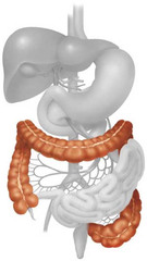
liver
largest gland in the body, produces bile

spleen
organ near the stomach that produces, stores, and eliminates blood cells
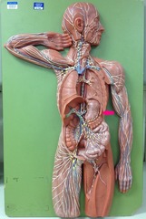
brain
mass of nerve tissue that is the main control center of the nervous system

spinal cord
nervous tissue within the spinal cavity