Neuro
1/11
There's no tags or description
Looks like no tags are added yet.
Name | Mastery | Learn | Test | Matching | Spaced |
|---|
No study sessions yet.
12 Terms
Neurolocalizations for intracranial Dz:
Forebrain
Cerebellum
Brainstem
Neurolocalizations for spinal cord Dz:
C1-C5
C6-T2
T3-L3
L4-S3
Neurolocalizations for neuromuscular Dz:
Myopathies
Junctionopathies
Neuropathies
List the sections of the canine spinal cord and how many vertebrae there are in each section:
8 cervical
13 thoracic
7 lumbar
3 sacral
5 caudal (variable)
List the sections of the feline spinal cord and how many vertebrae there are in each section:
8 cervical
13 thoracic
7 lumbar
3 sacral
variable caudal
What cranial nerves are involved in the menace response?
Menace response: entails closing the eyelids or retracting the globe in response to a threatening gesture aimed at each eye
CN II (optic nerve):
CN VII (facial nerve): innervates the muscles of facial expression; blink response
What cranial nerves are involved in the PLR (pupillary light reflex) response?
Before assessing PLR, first assess the pupils for symmetry (unequal size = anisocoria). Then, shine a light in one eye and look for pupillary constriction; direct response. The other pupil should simultaneously constrict to a lesser degree; indirect/consensual response. Repeat for the other eye.
CN II (optic nerve): sensory (afferent) pathway for vision and the PLR
CN III (oculomotor nerve): the efferent arm of this reflex is mediated by CN III (parasympathetic). CN III has both parasympathetic and somatic motor functions. Damage to this nerve or associated nucleus causes ipsilateral pupillary dilation; the pupil will not constrict when the ipsilateral or contralateral eye is stimulated.
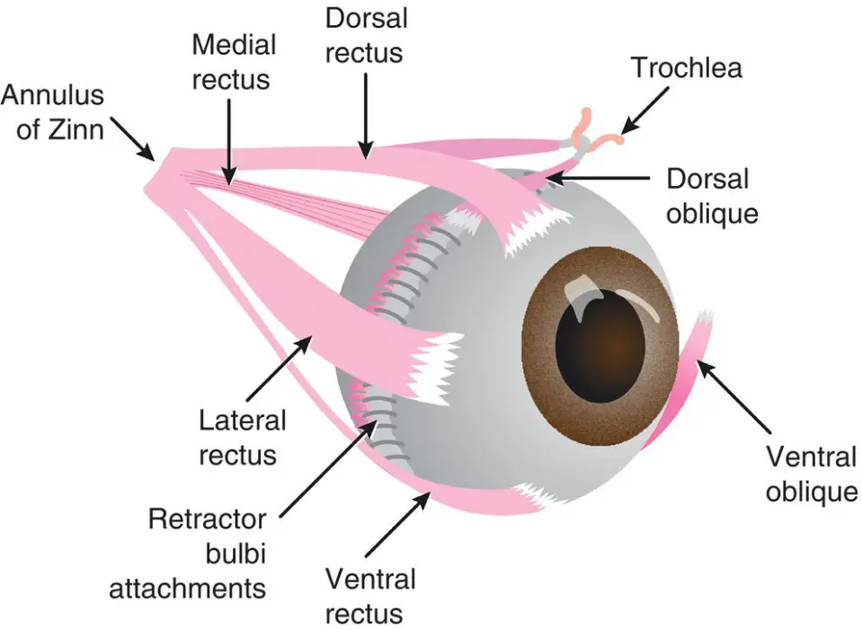
(The somatic portions of CN III innervate the ventral oblique mm and the eye’s dorsal, ventral, and medial rectus mm. A lesion affecting this part of the nerve or associated nucleus leads to paralysis of the innervated extraocular mm and ventrolateral deviation of the eye; “strabismus”.)
What cranial nerves are involved in the palpebral reflex response?
CN V (trigeminal nerve): has both sensory and motor fxn. Ophthalmic, maxillary, and mandibular branches provide sensory innervation to the face. The ophthalmic branch is stimulated by touching the medial canthus of the eye and assessing for a blink (palpebral reflex). The maxillary branch is best tested by connecting the nasal mucosa with a blunt instrument.
(The motor branch of the trigeminal nerve innervates the muscles of mastication, of which the masseter and temporal mm are most prominent.)
CN VII: innervates the mm of facial expression. Facial nerve lesions usually cause loss of the palpebral reflex and drooping of the lip and ear on the affected side. An animal with a CN VII lesion will also be unable to blink in response to a menace gesture.
(The nerve also supplies parasympathetic innervation to the lacrimal gland and the sublingual and mandibular salivary glands. Decreased tear production may occur and can be demonstrated using a Schirmer tear test. It also provides sensory fibers for the taste to the cranial two-thirds of the tongue.)
What cranial nerves are involved with the muscles of mastication and facial sensation?
CN V: The trigeminal nerve has both sensory and motor function. Ophthalmic, maxillary, and mandibular branches provide sensory innervation to the face. The ophthalmic branch is stimulated by touching the medial canthus of the eye and assessing for a blink (palpebral reflex). The maxillary branch is best tested by connecting the nasal mucosa with a blunt instrument. The motor branch of the trigeminal nerve innervates the muscles of mastication, of which the masseter and temporal muscles are most prominent.
What cranial nerves are involved in the oculovestibular reflex?
The oculovestibular reflex, also known as the "doll's eye reflex" in dogs, is a neurological test that assesses the function of the brainstem, vestibular system, and extraocular muscles. It involves observing eye movements when the dog's head is moved, with a normal response being the eyes moving in the opposite direction of the head movement to maintain gaze on a stationary point.
CN III (oculomotor nerve): The somatic portions of the oculomotor nerve innervate the ventral oblique muscle and the eye's dorsal, ventral, and medial rectus muscles. A lesion affecting this part of the nerve or associated nucleus leads to paralysis of the innervated extraocular muscles and ventrolateral deviation of the eye, known as strabismus.
CN IV (trochlear nerve): motor nerve that controls the movement of the dorsal oblique muscle of the eye. It is responsible for the depression and medial rotation of the eye.
CN VI (abducens nerve): Provides motor function to the lateral rectus extraocular muscle and retractor bulbi. Examined by touching the globe and observing for retraction (also tests V for sensory). Responsible for physiologic nystagmus when turning head (also involves III, IV, and VIII)
CN VIII (vestibulocochlear nerve): vestibular dysfxn is suggested by the presence of a head tilt, leaning, falling or rolling, ataxia, and nystagmus. Pathologic nystagmus is characterized by the direction of the oscillations (horizontal, rotary, vertical) and typically has distinct fast and slow phases. Nystagmus is named according to the direction of the fast phase. When moving the head from side to side, nystagmus develops in normal animals with the fast phase in the direction of head movement. This normal physiologic nystagmus or oculovestibular reflex may be depressed in animals with vestibular disease. Strabismus can also be seen with vestibular disease, and it is often elicited by changing the position of the head. Cranial nerves innervating the extraocular muscles of the eye (CN III, IV, and VI) are closely connected to the vestibular system. These pathways function to maintain the position of the eyes concerning the head and body.
What cranial nerves are involved in the gag reflex?
CN IX (glossopharyngeal nerve): innervates the pharynx for swallowing (with X). Also innervates some salivary glands and provides taste innervation from caudal tongue. Examine by eliciting a gag reflex and observing for dysphagia (difficulty swallowing).
CN X (vagus nerve): innervates the larynx, esophagus, and pharynx. Also provides parasympathetic innervation to the heart and viscera. Tested with a gag reflex (along with CN IX).
CN XI (spinal accessory nerve): innervates cranial cervical (neck) muscles
What cranial nerves are involved in the motor of the tongue?
CN XII (hypoglossal nerve): innervates the muscles of the tongue. Dysfunction of the hypoglossal nerve may lead to difficulty eating, prehending food, and muscle atrophy.