lab test 3
1/125
There's no tags or description
Looks like no tags are added yet.
Name | Mastery | Learn | Test | Matching | Spaced | Call with Kai |
|---|
No analytics yet
Send a link to your students to track their progress
126 Terms
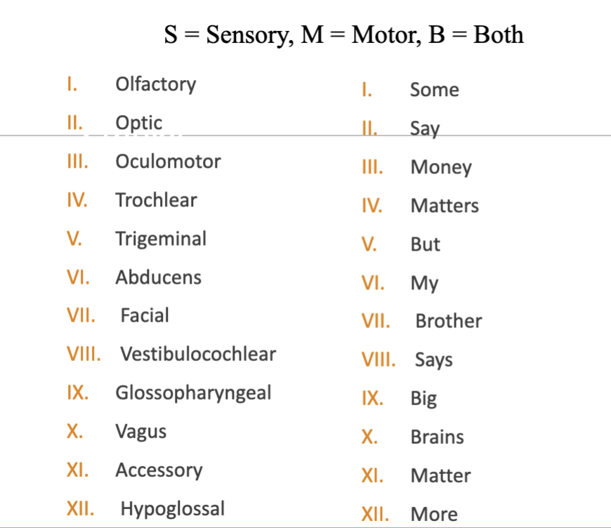
cranial nerves numbered
frontal lobes
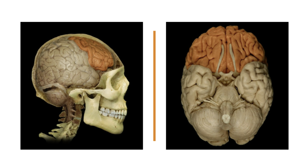
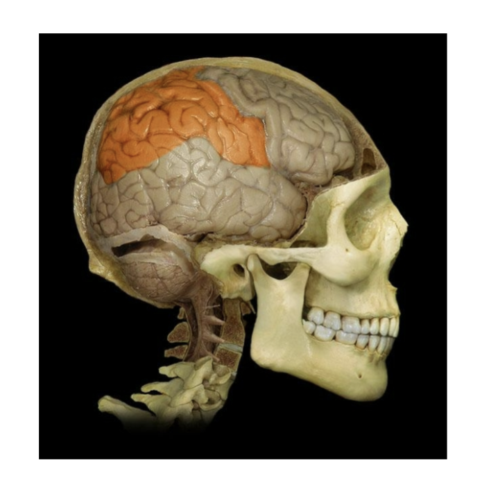
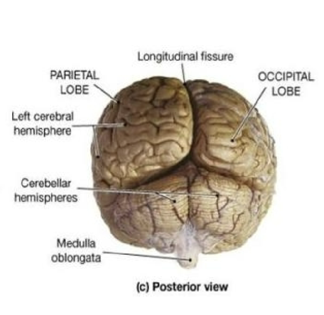
parietal lobe
temporal lobe
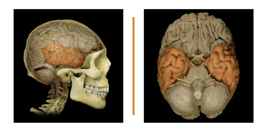
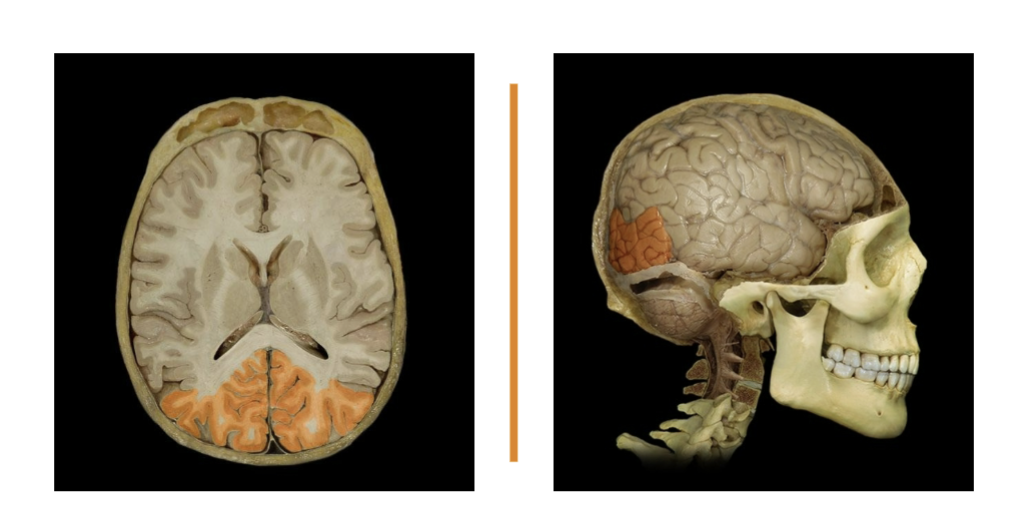
occipital lobe
longitudinal fissure
separates left and right cerebral hemispheres
extends along the midsagittal plane
falx cerebri is found here
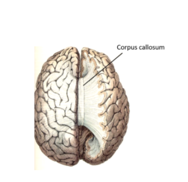
corpus callosum
connects L/R cerebral hemispheres and is their main mode of communication
last resort tx for seizures is to sever corp[us callosum
cerebellum and arbor vitae
Little brain
Arbor vitae: tree of life
Cerebellum is responsible for muscle memory, equilibrium, posture, proprioception
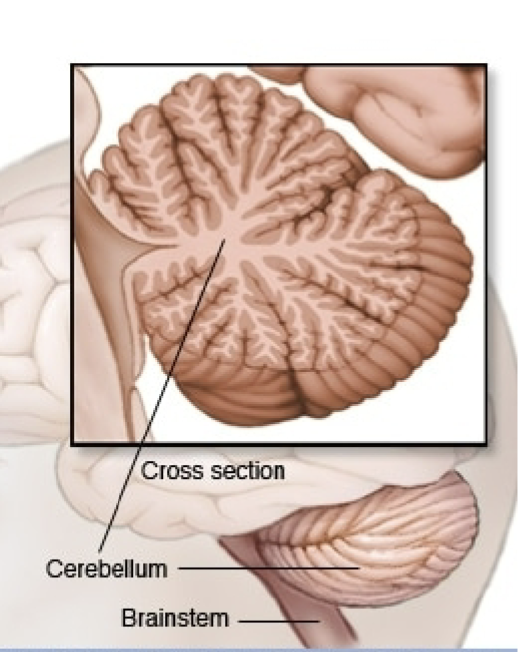
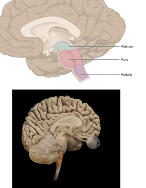
Pons
Brainstem includes: midbrain, pons, medulla oblongata
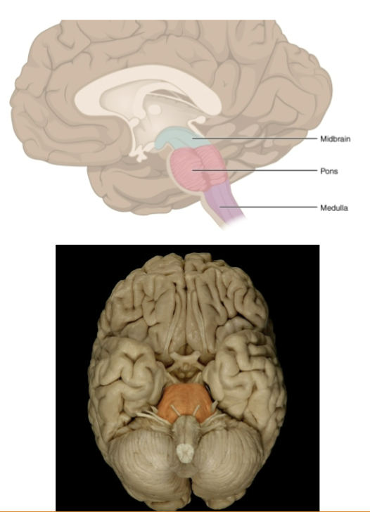
Medulla Oblongata
Continuous with spinal cord, motor tracts cross to the opposite side of the brain
police snipers aim for tip of the nose or base of ear lobe
functions include sensory relay for cranial nerves, relay for thalamus, cardiac center, vasomotor center, respiratory center
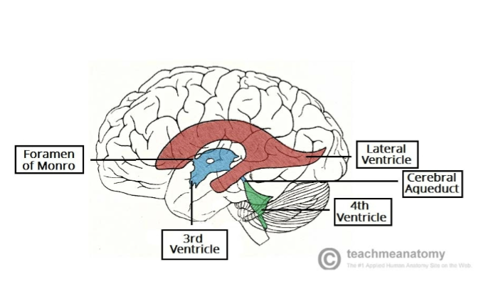
Ventricles
Little cavity
Ventricles are continuous with each other and with the central canal of the spinal cord
4 ventricles: lateral (x2), 3rd, 4th
Lateral: largest
3rd: located within diencephalon (w/ thalamus, hypothalamus, epithalamus
4th: between the pons and the cerebellum. Narrows at the caudal end before merging with the central canal in the spinal cord
choroid Plexi
in all ventricles
formed by ependymal cells that line the ventricles and nearby blood capillaries
Meninges
connective tissue that surround and separate portions of the brain
dura mater, arachonoid mater, pia mater
dura mater
tough mother
most superficial layer
extends as flat partitions in the cranial cavity at several locations providing additional support
Dura Mater - falx xerebri
in midsagittal plane
anterior attach- crista galli
posterior attach- superior portion of tentorium cerebelli
Dura Mater - tentorium cerebelli
tent, little brain
separates occipital lobe and temporal lobe from cerebellum
Arachnoid mater
spider mother
delicate web of collage and elastic fiber
Pia Mater
delicate mother
deepest layer of cranial meninges
delicate connective tissue adheres to brain tightly, difficult to distinguish from brain
Optic nervies, optic chiasm, optic tract
optic= to see, chiasm= x-shaped, optic nerves- cn II
Optic nerves enter the optic foramen and then converge to form the optic chiams
Optic Chiasm is superior to sella turcica and pituitary gland
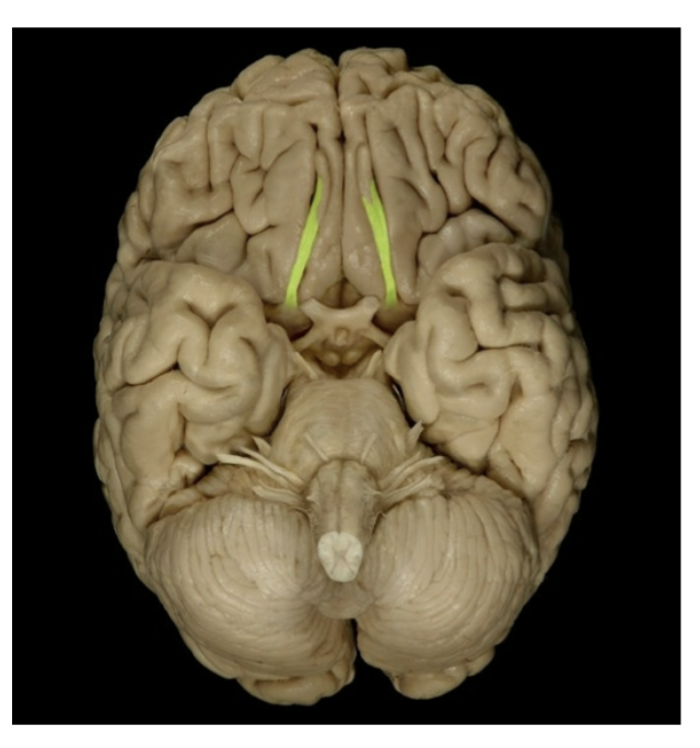
Olfactory bulb
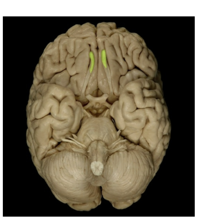
Olfactory Tract
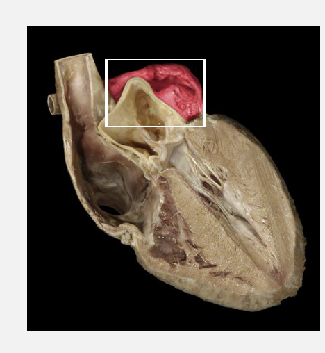
Left Atrium
Right Atrium
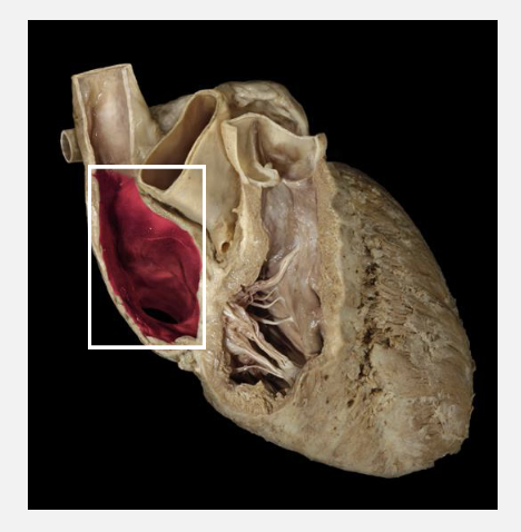
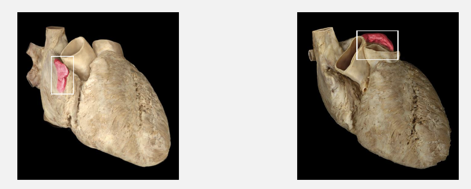
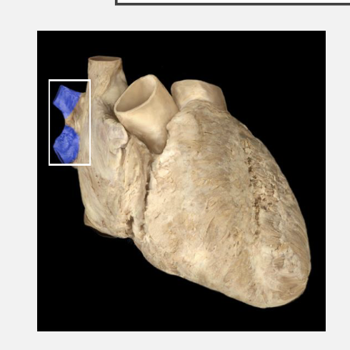
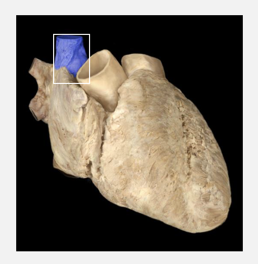
R/L Auricles
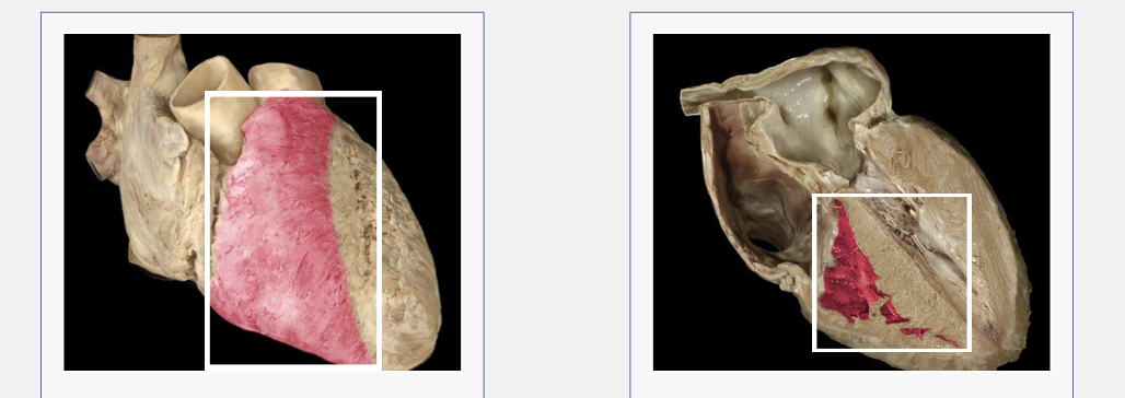
Left Ventricle (closed/open)
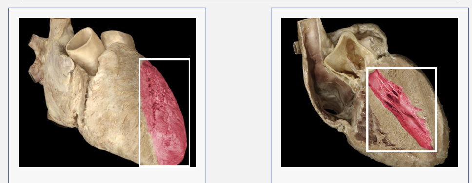
Right Ventricle (closed/open)
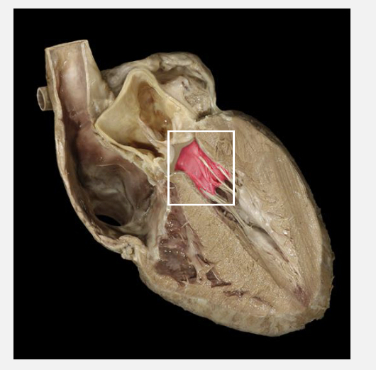
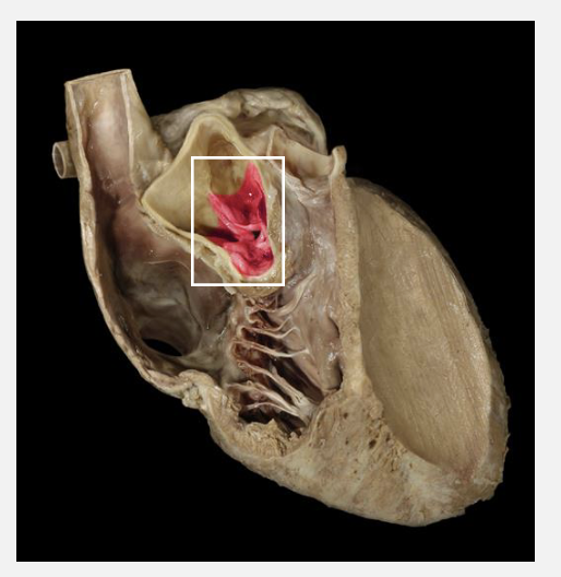
Tricuspid
Bicuspid
Pulmonary Semilunary Valve
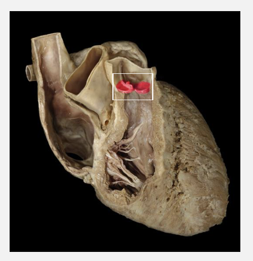
Aortic Semilunar Valve
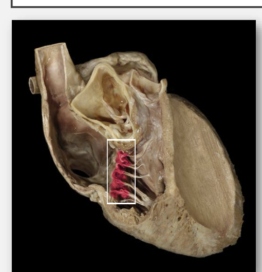
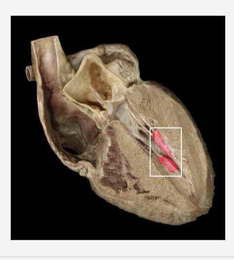
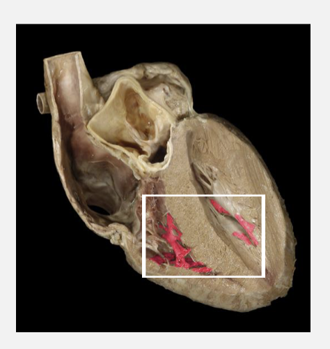
Chordae Tendineae
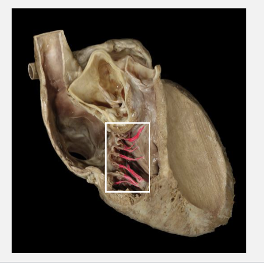
Papillary Muslces
Trabeculae Carneae
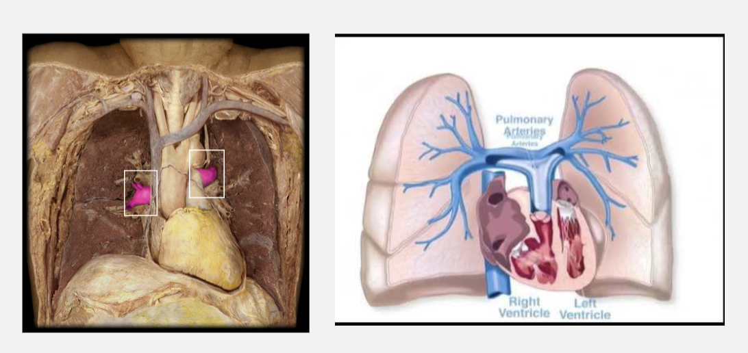
Pulmonary Arteries
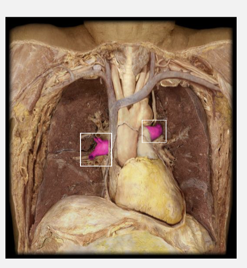
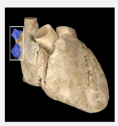
Pulmonary Veins
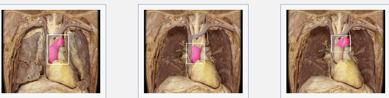
Pulmonary Trunk and Pulmonary Artery
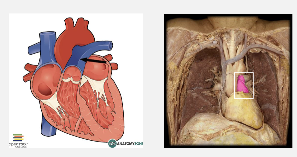
Pulmonary Arteries
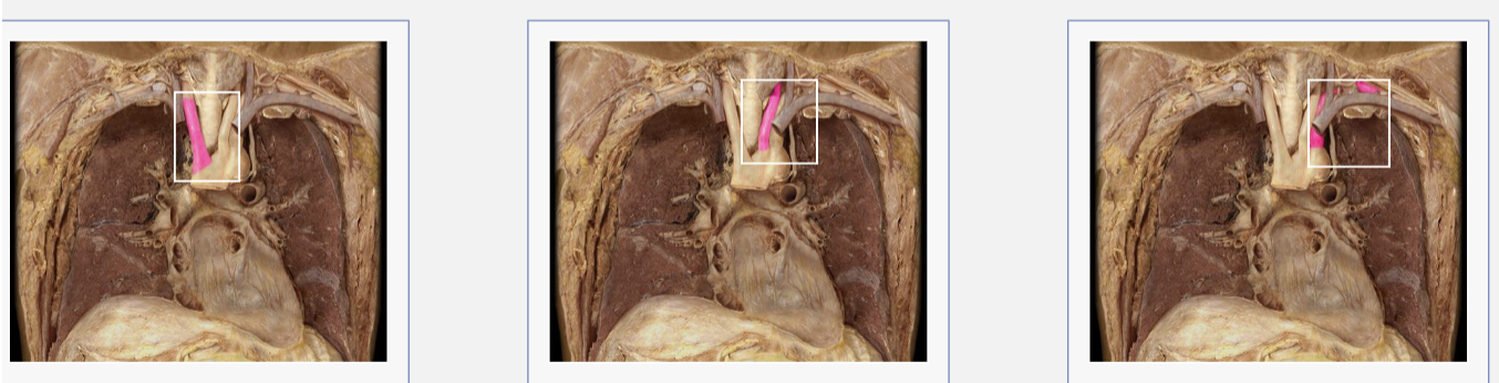
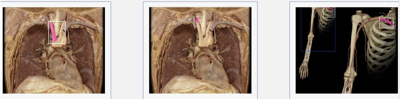
Aorta (asceneding/arch)
aortic arch: 3 arteries that arise from the aortic arch
brachiocephalic trunk, left common carotid artery, left subclavian
Brachiocephalic trunk: 2 arteries that branch off the trunk
Brachiocephalic trunk, right common carotid artery, right subclavian artery
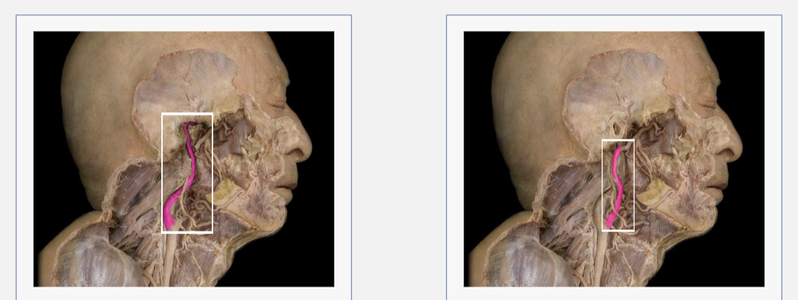
internal/external carotid artery
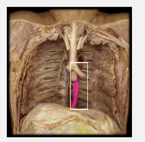
descending thoracic aorta
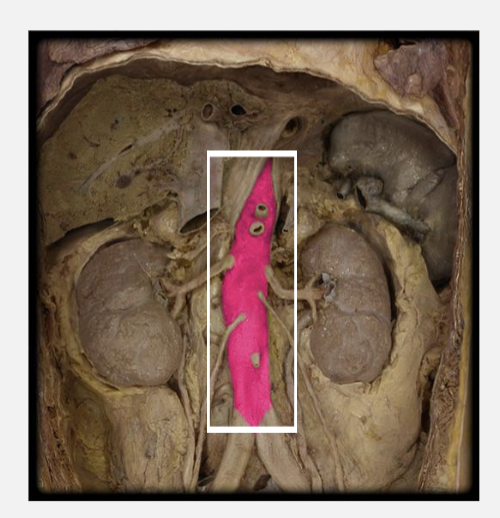
Descending abdominal aorta
L/R common iliac artery
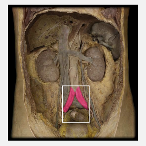
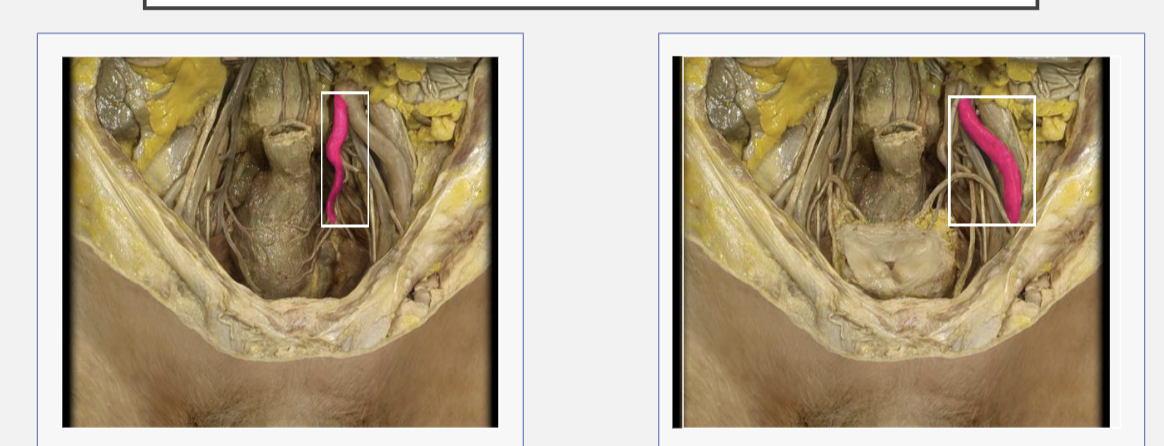
internal/external iliac arteries
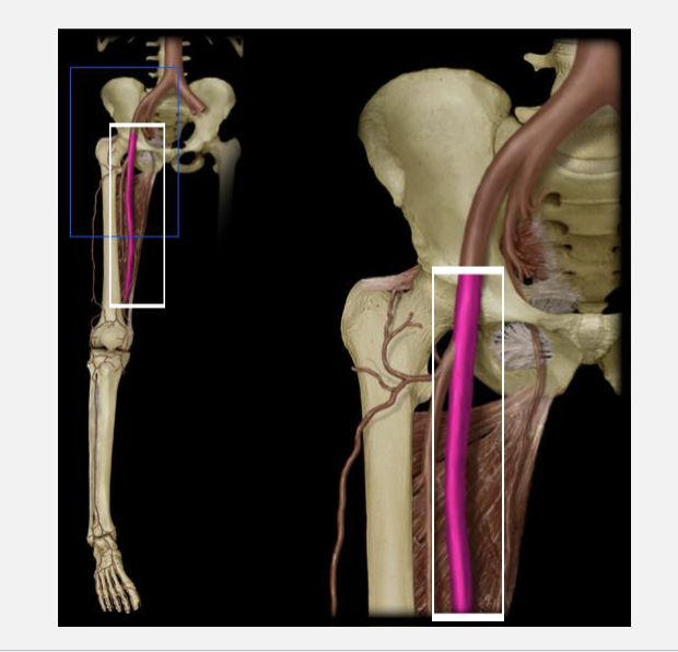
femoral artery
L/R pulmonary veins
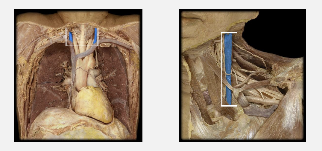
Superior vena cava
internal jugular vein
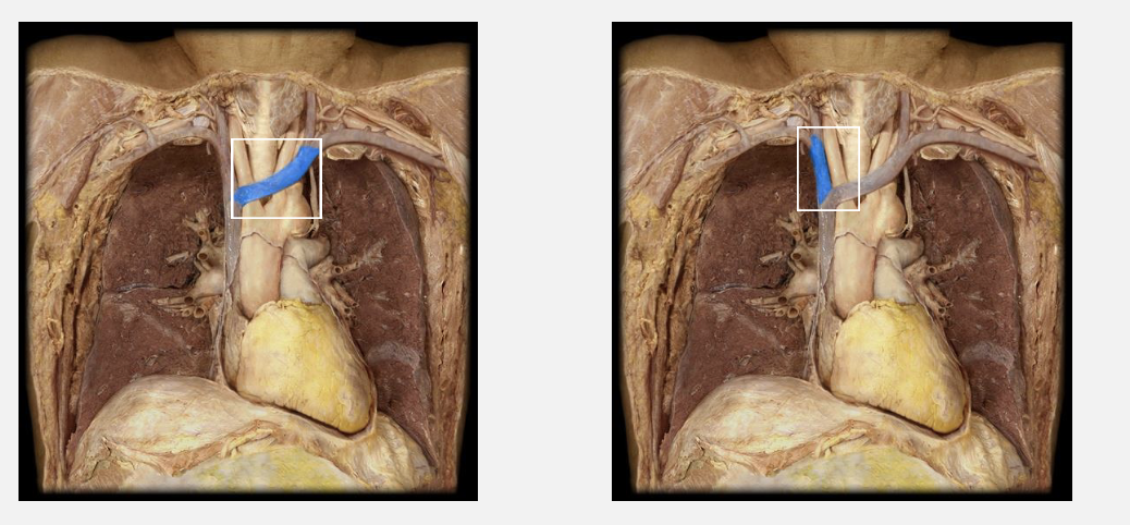
L/R Brachiocephalic vein
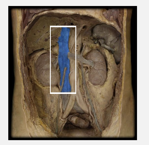
inferior vena cava
R/L common iliac veins
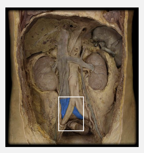
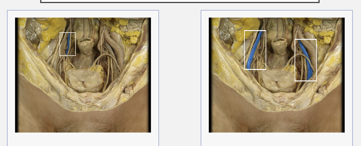
internal/external iliac veins
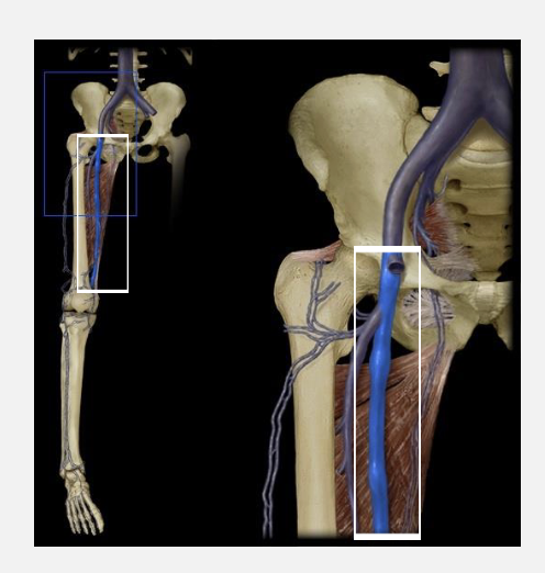
Femoral Vein
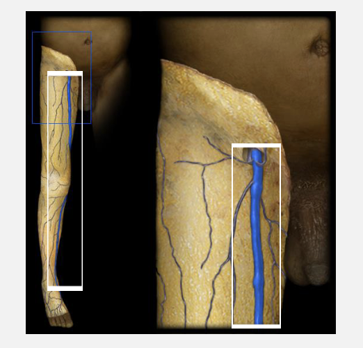
Great Saphenous vein
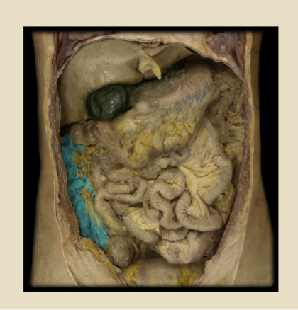
Spleen
largest organ in the lymphatic system. it is an important organ for keeping bodily fluids balanced, but it is possible to live without it. the spleen is located under the ribcage and above the stomach in the left upper quad of the abdomen. A spleen is soft and generally looks purple
Nares
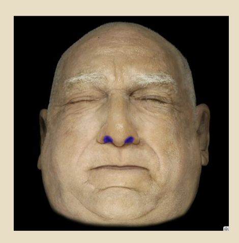
vestibule
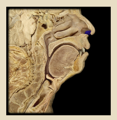
hair in nose
vibrissae
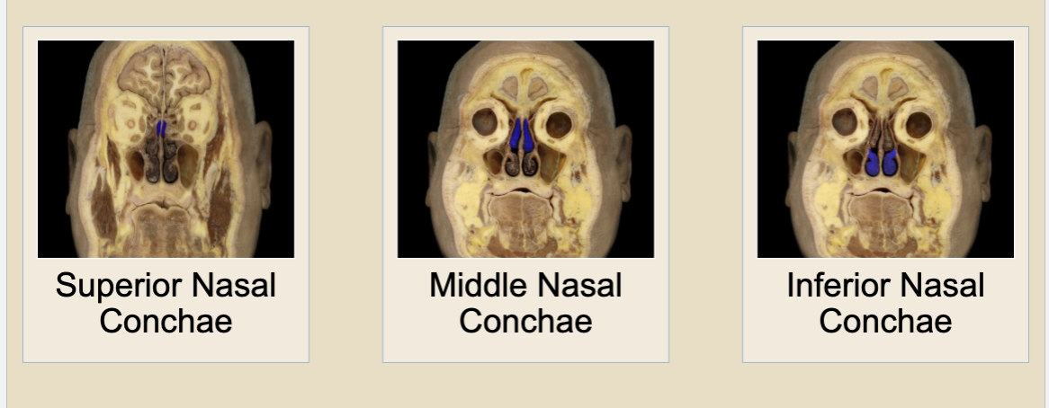
nasal conchae
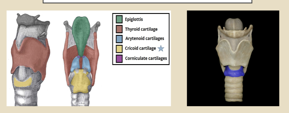
epiglottis
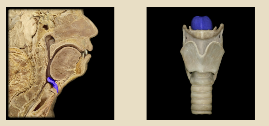
thyroid cartilage
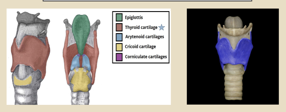
cricoid cartilage
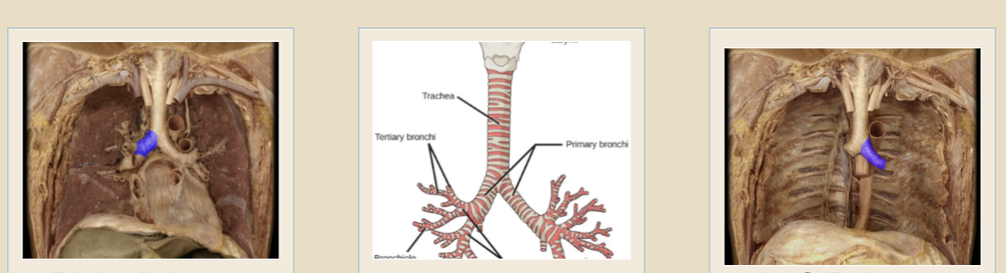
trachea
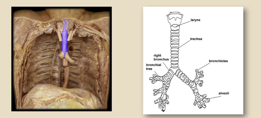
bronchi
R/L
Right Lung
Left Lung
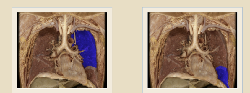
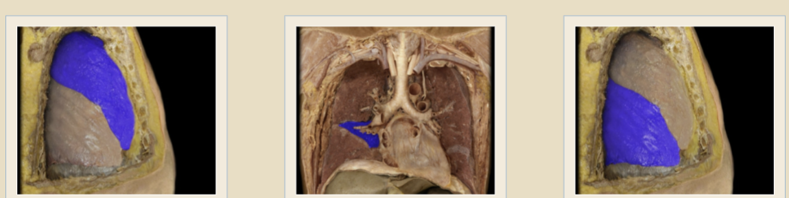
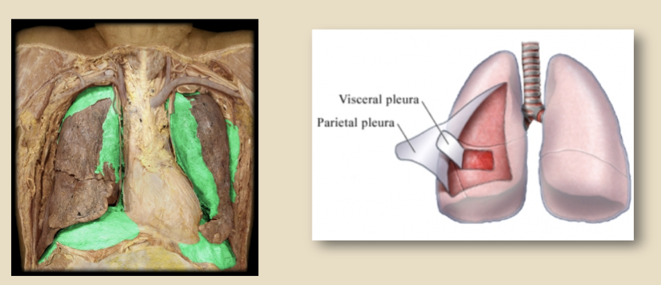
Visceral Pleura
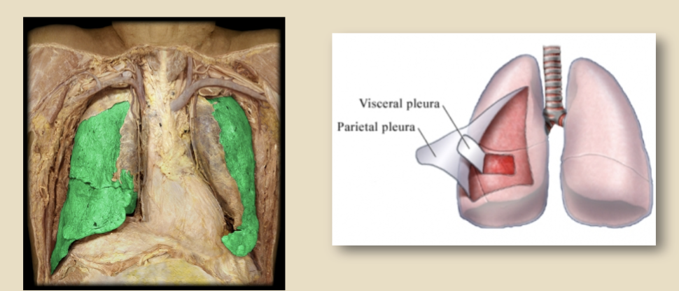
Parietal Pleura
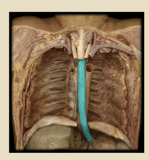
esophagus
muscular tube connecting pharynx w/ stomach
runs behind windpipe and heart, in front of spine
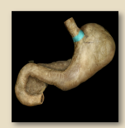
cardia of stomach
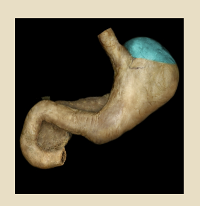
fundus of the stomach
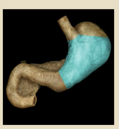
body of stomach
pylorus of the stomach
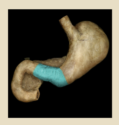
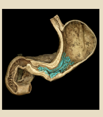
rugae of the stomach (folds)
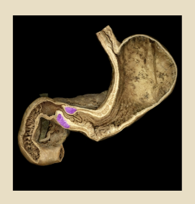
pyloric sphincter
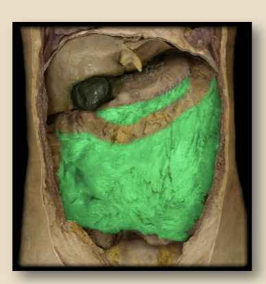
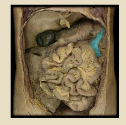
greater omentum
lesser omentum
mesentery
liver
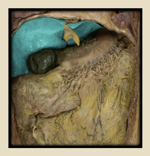
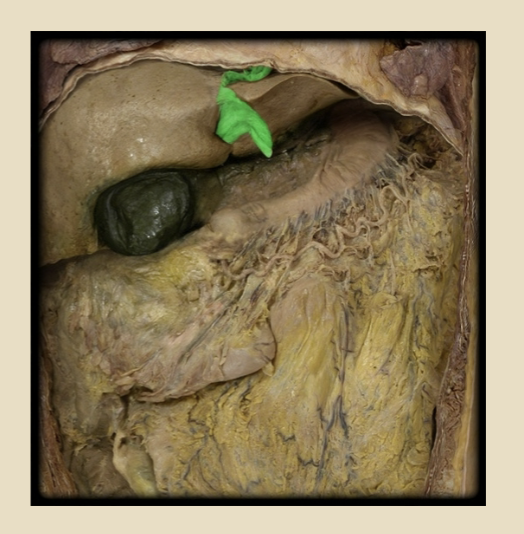
falciform ligament
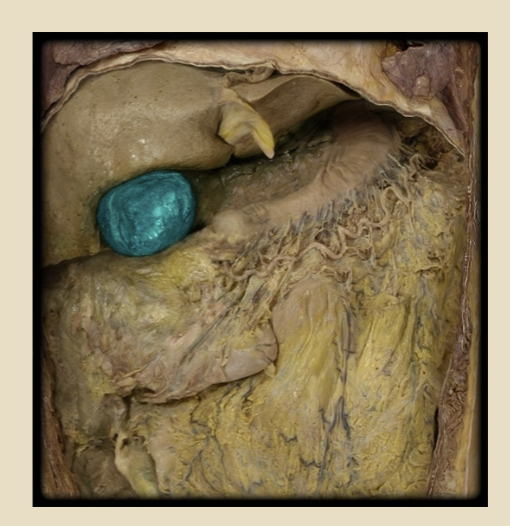
gallbladder
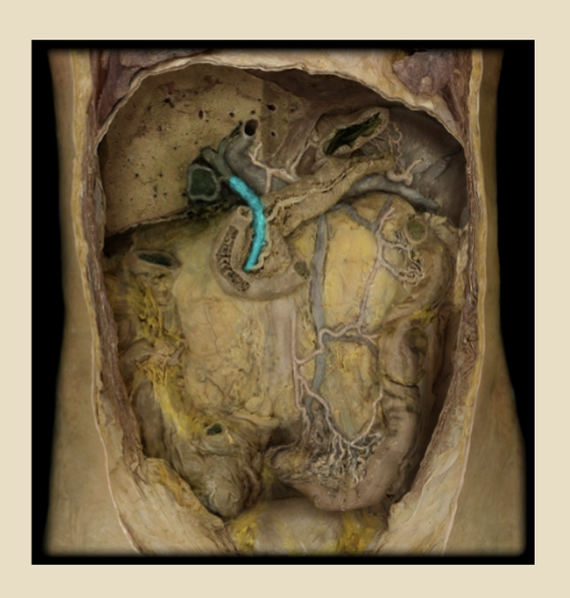
bile duct
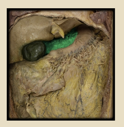
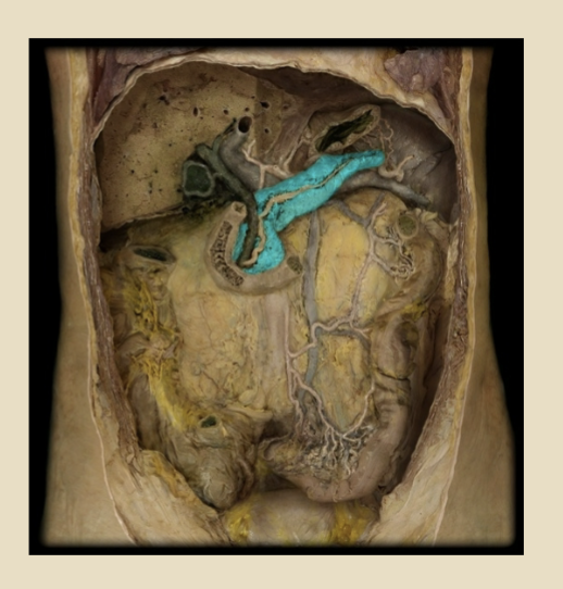
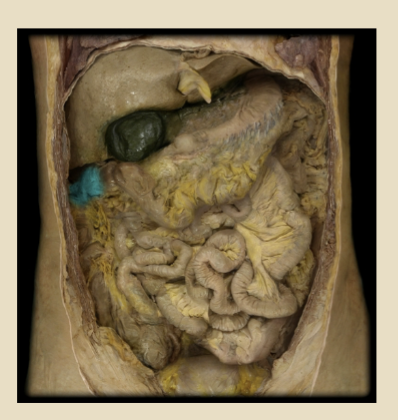
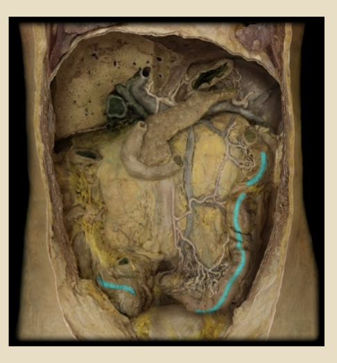
pancreas
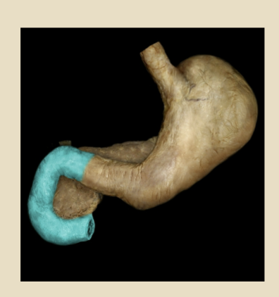
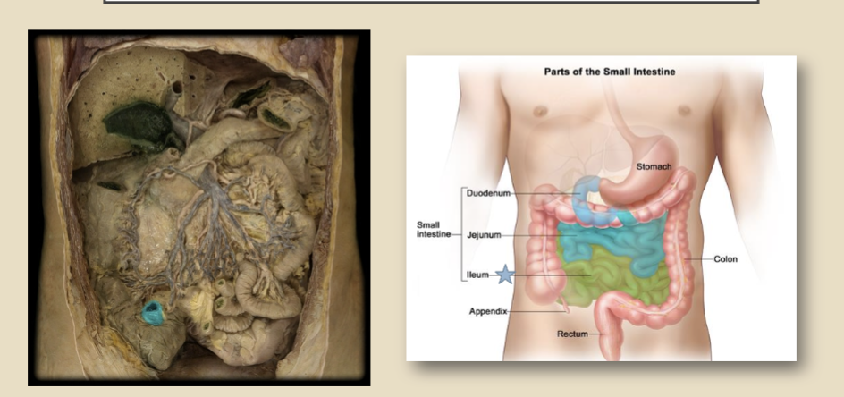
duodenum (small intestine)
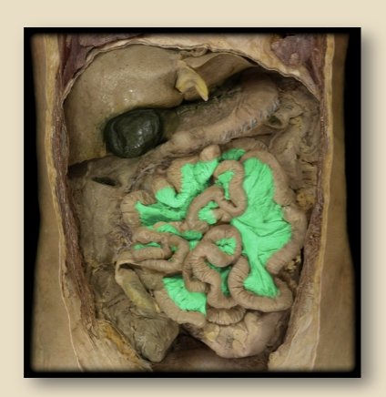
jejunum
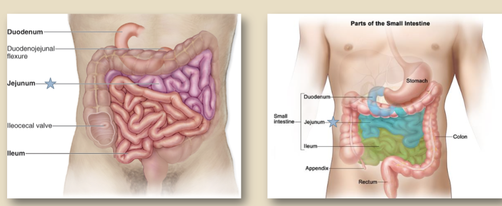
Ileum (small intestine)
cecum
vermiform appendix
ascending colon
hepatic flexure (right colon flexure)
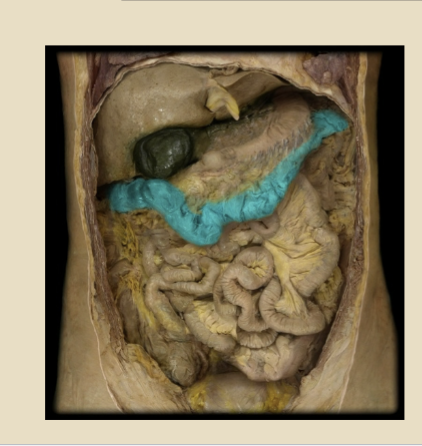
Transverse colon
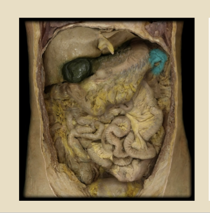
splenic flexure
descending colon
Sigmoid colon

Taeniae Coli
Haustra
small segmented pouches of the bowl separated by the haustral folds. formed by circumferential contraction of the inner muscular layer of the colon. the outer longitudinal muscular layer is organized into 3 bands (taeniae coli) which run from the cecum to the rectum.
rectum
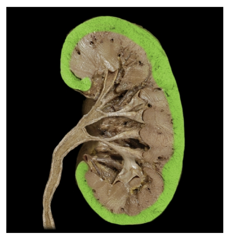
Kidneys
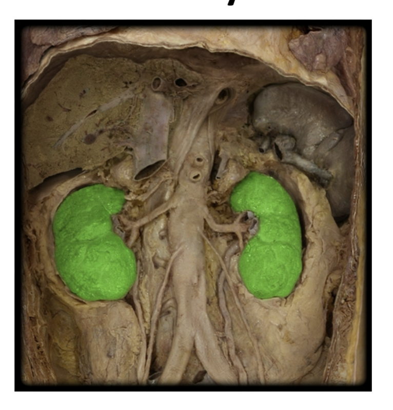
renal cortex