CNS Infections, protozoal, fungal
 Call Kai
 Learn
Learn Practice Test
 Spaced Repetition
 Match
Match Flashcards
 Knowt Play
1/38

There's no tags or description
Looks like no tags are added yet.
Name | Mastery | Learn | Test | Matching | Spaced | Call with Kai | Chat |
|---|
No analytics yet
Send a link to your students to track their progress
39 Terms
Describe Prions
AKA?
Differenty Types?
Transmission
Describe the healthy protein version
Genetics?
Expressed in?
Function?
Describe the changes that makes the protein pathologic
Prions
Transmissable Spongiform Encephalopathies (TSE)
Creutzfeld-Jakobs Disease (CJD)
Bovine Spongiform Encephalopathy (BSE)
Kuru (humans), scrapie (sheep)
Chronic wasting disease (CWD)
Transmission
ingestion of contaminated meat
PrPc (healthy protein)
PRNP gene on chromosome 20
Expressed in:
neuronal cells, Ganglia, Peripheral Nerve Cells
tonsils, kidneys, heart, lungs, & spleen
Function (suggested):
Maintenance of neuronal integrity in the brain
Possible role in Cu metabolism & cellular response to oxidative stress
Pathologic PrPc
PrPc -> PrPsc
α-helical > β-sheets
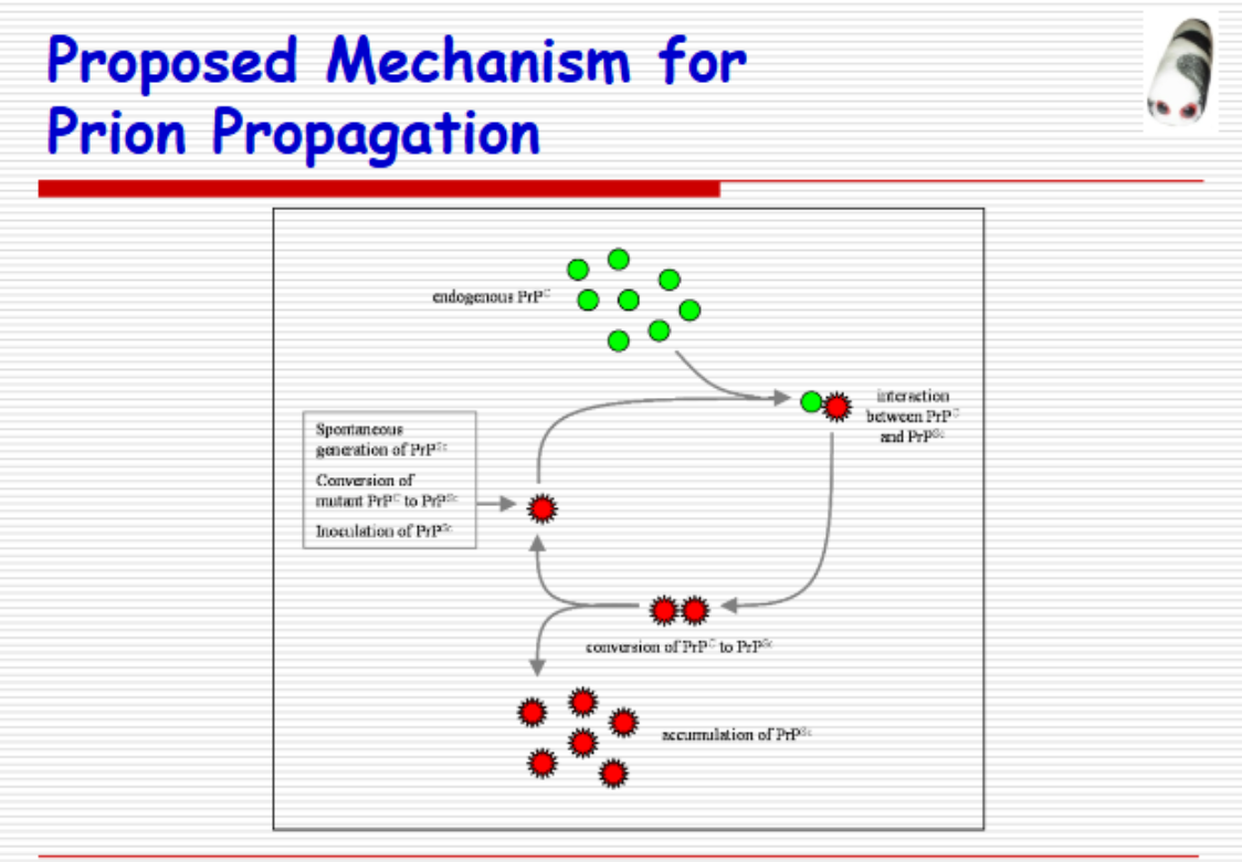

What happens in Prion Disease
Symptoms
Describe Kuro
Geography
Epidemology
What happens in Prion Disease
Vacuolization of neuronal cytoplasm
-> sponge-like appearance of brain parenchyma
Symptoms
Personality changes
Depression
Lack of coordination
Jerky movements
Insomnia, confusion, memory problems
Later stages
Severe mental impairment
Inability to move or speak
Kuru
Geography:
Restricted to the highlands of New Guinea
Fore (for-ay) Tribe
Epidemology:
epidemic levels in the 1950’s and 60’s
Practiced ritualistic cannibalism
Relatives prepared and ate the tissues (including the brain) of deceased family members
Brain tissue is highly infectious
Transmission was through eating or contact through open sores or wounds
Describe T. Gondii
Stats
3 infectious stages
Life Cycle
Normal vs immunocompromised response
STATs
Most common parasitic infection of the CNS
Worldwide zoonosis
In US, 20-40% of population seropositive for T. gondii
In Europe, as much as 90% are seropositive (esp. France)
Cultural habits of eating raw or undercooked meat
3 infectious stages
Tachyzoites
Rapidly multiply in any cell
Seen in acute stages of infection
Bradyzoites
tissue cysts
neural and muscular tissues, brain, eyes, skeletal and cardiac muscles
Multiply slowly – in chronic stages
Sporozoites
Found in oocysts
Shed in feces
Life Cycle
organisms ingested -> Tachyzoites released -> GI Cells -> Multiply, rupture, Infect contiguous cells -> lymphatics -> body dissemination -> Tachyzoites proliferate -> necrotic foci surrounded by cellular response
Normal vs Immunocompromised:
Normal:
Immune response neutralizes and removes tachyzoites
Compromised
-> necrotizing encephalitis
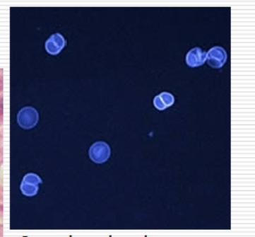
Sporulated and unsporulated oocysts of T Gondi
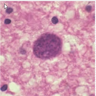
Cysts in brain tissue
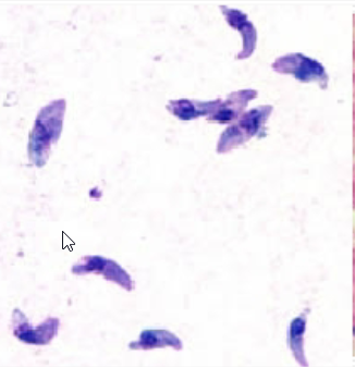
Tachyzoites
List the symptoms of T. Gondii
Immunocompetent
Compromised
Congenital
Symptoms:
Immuno Competent (asymptomatic 80-90%)
Fever, malaise, night sweats, sore throat
Retroperitoneal & mesenteric lymphadenopathy
Chorioretin
Immunocompromised (CNS disease in 50%)
Encephalitis,
meningoencephalitis,
mass lesions
Hemiparesis,
seizures,
mental status changes
Myocarditis,
pneumonitis
Congenital (Severe if maternal infection occurs early in preg )
CSF pleocytosis & elevated protein levels
Microcephaly
Survivors:
Mental retardation
Visual defects
Seizures
What causes Cerebral malaria and the consequences
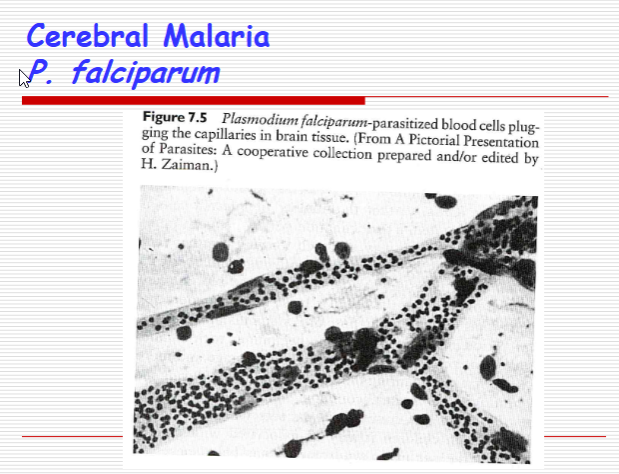
Cerebral malaria
Deadliest complication of P. falciparum
Consequences:
Encephalitic syndrome
Ataxia,
seizures,
hemiplegia,
coma,
Death
residual neurological damage after successful antiparasite
Long term cognitive deficits
Memory, learning, and language impairments
Visuospatial and motor deficits
Psychiatric disorders
Describe Plasmodium falciparum
Symptoms
Key Feature
Symptoms
Gradual onset
Disoriented (or violent), severe headaches, coma
Abrupt onset
Can then suddenly lapse into a coma after asymptomatic
Key feature:
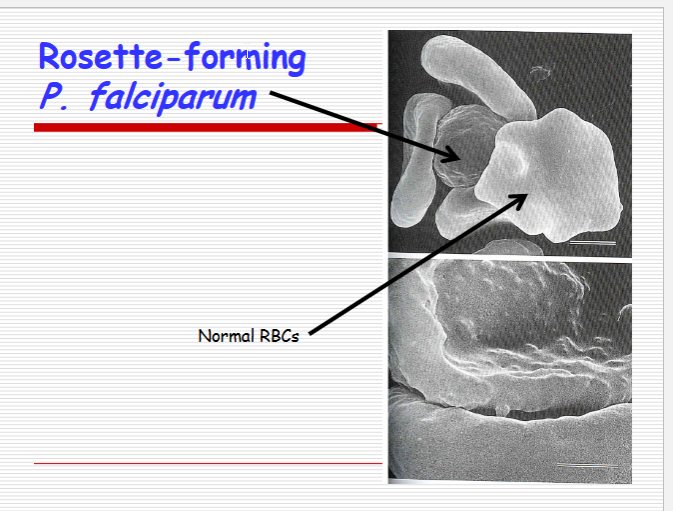
RBC rosette-forming P. falciparum
Parasitized RBC surrounded by 3 Normal RBCs
mediated by knobs seen on parasitized RBC

Describe the pathogenesis of cerebral malaria
Most serious Complication? Stats?
Pathogenesis
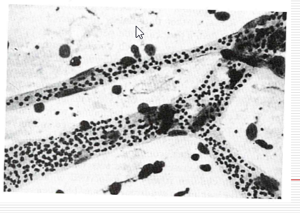
Sequestration of parasitized RBCs ->
increased brain volume
Compensatory vasodilation
In Cerebrum, cerebellum, and medulla oblongata
Most serious complication: Death
10% of all P. falciparum-infected patients
80% of all fatal malarial cases
Describe Naegleria fowleri
Disease Caused
Characteristics
Locations
Naegleria fowleri
Disease Caused:
Primary amebic meningoencephalitis (PAM)
mortality rate = 95%
N. fowleri Characteristics
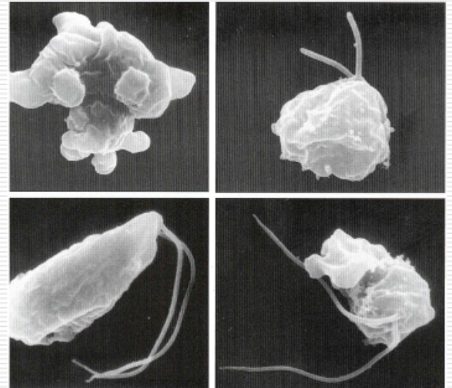
Thermophilic, free-living amoeba
N. fowleri Locations:
Waterways contaminated by thermal discharge of powerplants, heated swimming pools, hot springs (up to 45degC)
Inhabits fresh water ponds, lakes, rivers
Higher temperatures, low precipitation = decreased water depths -> proliferation of amoeba
Naegleria fowleri
Transmission/ Period between contact of Naegleria fowleri and onset of symptoms?
Symptoms
Early
Acute
Other?
Prognosis
PAM
Transmission:
amebic-contaminated water -> Nose (inhalation, aspiration, or aerosol) -> phagocytose olfactory epi. Cells -> olfactory mucosa -> Olf. Nerve
Period between contact of Naegleria fowleri and onset of symptoms
2-3 days to as long as 7-15 days
Early symptoms
Upper, respiratory distress
Headache, lethargy
Olfactory problems
Acute phase
Sore throat
Stuffy, blocked, or discharging nose
Severe headaches
Other symptoms
Pyrexia, vomiting, stiffness of neck
Mental confusion, coma
3-5 days before death
Cardiorespiratory arrest and pulmonary edema = causes of death
NOTE: Symptoms indistinguishable from bacterial meningitis
Prognosis
Only 5 survivors of PAM have been reported,
3% of reported cases
High mortality rate
Difficulty in diagnosis
Poor to marginal response of patients to therapy
Why N. Fowleri likes to be in CSF/Brain?
N. Fowleri Pathological Abilities?
Laboratory diagnosis
Why N. Fowleri likes to be in CSF/Brain?
Allows rapid growth
B/c it has mitochondria -> High O2 in CSF and Brain Tissue
N. Fowleri Pathological Abilities
highly phagocytic
RBCs/brain tissue
Severe hemorrhagic necrosis of brain
Produces amebostome (food cup)
Secretes lysosomal hydrolases and phospholipases into it
Produces/Secretes heat stabile hemolytic proteins, heat-labile cytolysin, phospholipase A, cysteine protease
Kill cells that come in contact
Laboratory diagnosis
Spinal Tap
Elevated protein
Normal or slightly reduced glucose levels
WBC high (400-26000/μL)
RBC high
CSF is hemorrhagic
Gram stain results in no bacteria
Wet mount positive for motile trophozoites
NOTE:
Early diagnosis is mandatory due to rapid course of infection following incubation of 1-2 weeks
Near immediate chemotherapy is essential for survival
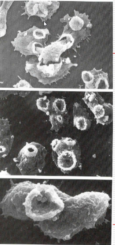
Naegleria fowleri
What are some possible treatments of N. Fowleri
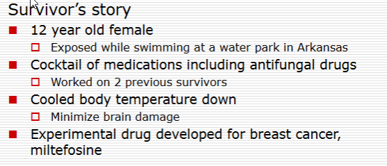
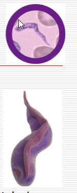
Human African Trypanosomiasis (HAT)
African Sleeping Sickness
Describe Trypanosomiasis
Causative Agents? What is the difference between the two?
What are kinetoplastids?
Trypanosomiasis
Causative Agent:
Trypanosoma brucei gambiense
West African trypanosomiasis
Exclusively human reservoir
Slow-progressing that can be self-limiting or develop into a chronic disease involving the CNS and lymphatic system
Trypanosoma brucei rhodesiense
East African (or Rhodesian) trypanosomiasis
Zoonotic disease (animal reservoir)
Rapidly progressing disease
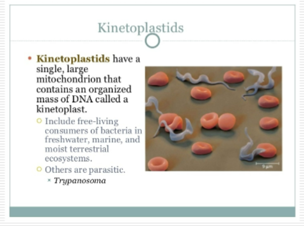
Kinetoplastids: Mitochondrial DNA
Describe (African Sleeping Sickness)
Disease Progression
NS impairment
Time line
MOA
Symptoms
Disease progression
1-2 week incubation period
Trypanosomal chancre (?)
Acute blood stage infection
Fever, headaches
Invasion of lymphatics
Enlarged lymph nodes
Weight loss, weakness, rash, itching
Continued, intermittent febrile attacks
Relapses occur
antigenic variation of trypanosomal surface
Life cycle exhibits different morphologies
Nervous system impairment
Timeline
Trypanosoma gambiense: 6-12 months after initial
Trypanosoma rhodesiense: within weeks with
MOA:
Crosses BBB -> meningoencephalitis
Symptoms:
Apathy, fatigue, confusion, motor changes
(tics, slurred speech)
Changes in sleep patterns
Extreme fatigue during day, extreme agitation during night
coma or death
What are Kinetoplastids?
Describe Taenia solium
Life cycle
Symptoms
how does it avoid immune system
Taenia solium
Life Cycle:
eggs ingested -> hatch in stomach -> brain via blood stream -> crosses BBB (unknown) -> attach to brain tissues of cavities of brain fluid -> dev. Cyst-like structures (Neurocysticercosis)
Symptoms:
Seizures
When larvae attach to brain tissues
Larval presence = brain activity uncontrolled
Altered mental states
Headaches, nausea, dizziness
When larvae blocks flow of brain fluid
Avoidance of the immune system
Enters BBB
Brain not easily accessible to immune cells
Cysts -> degrade Abs
Possible nutrient source
production of protein signals -> inhibit phagocytosis
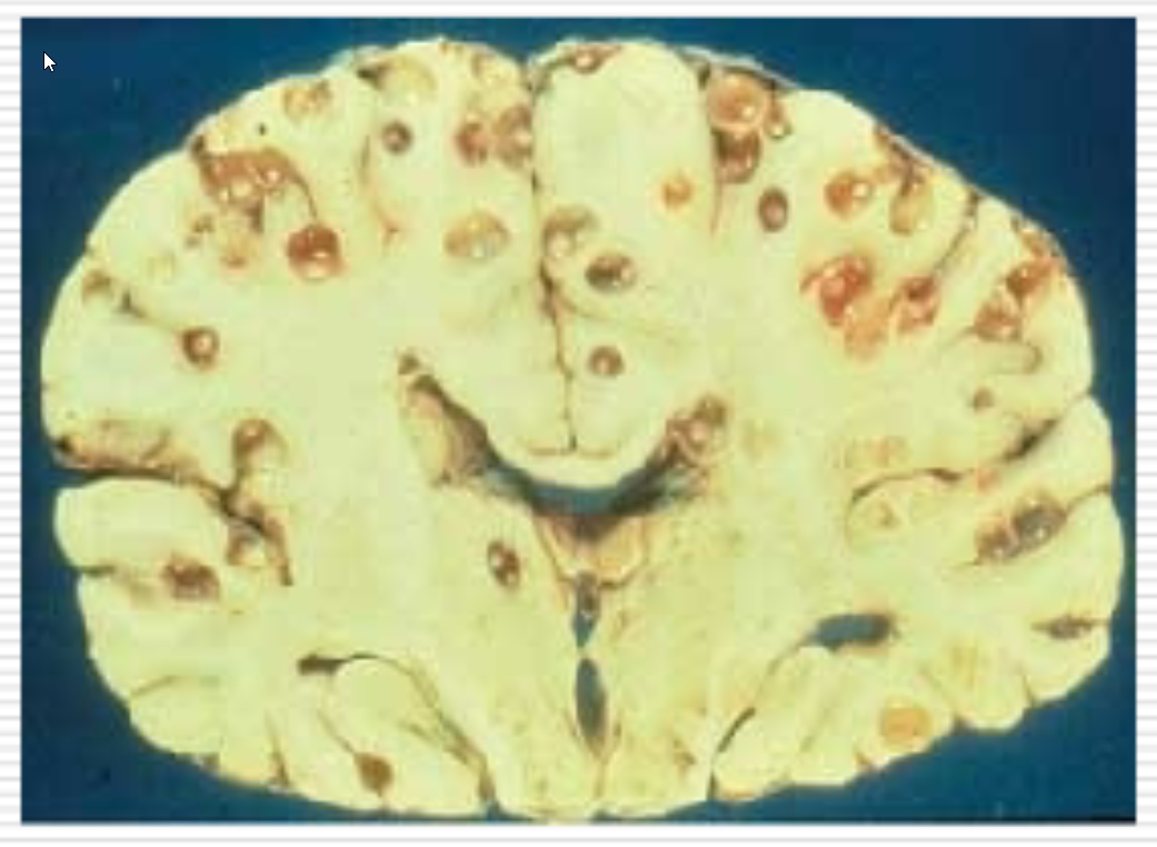
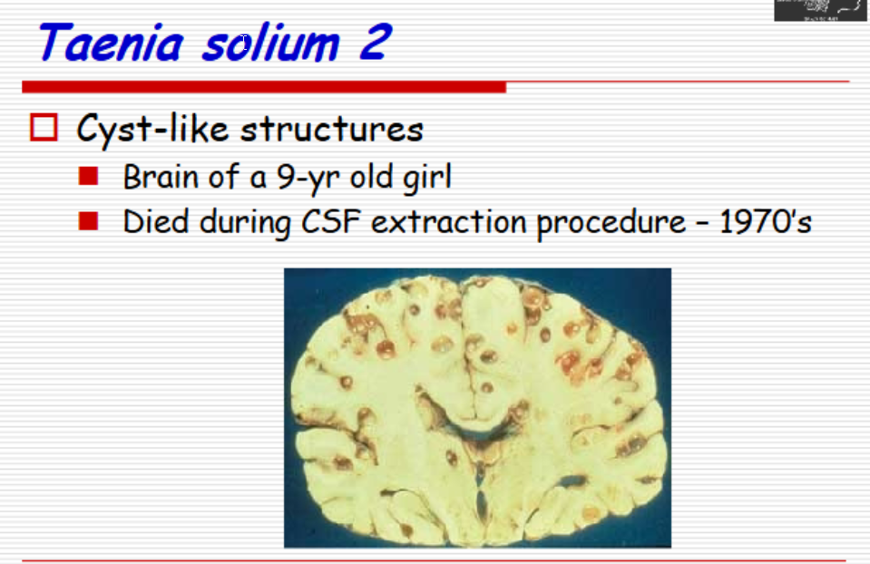
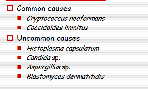

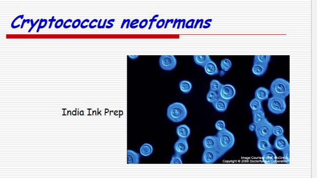
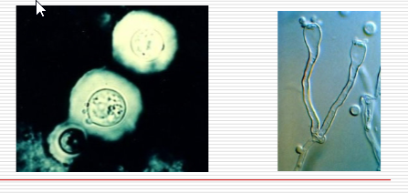
Cryptococcus neoformans
Describe Cryptococcus neoformans
Features
Major Env. Sources
Clinical Form
Virulence factora
Features:
Encapsulated yeast-like fungus w/ prominent capsule
capable of forming mycelia
Major environmental sources
Soil contaminated with pigeon droppings
Decaying wood in hollows of living trees
Clinical Form:
Meningoencephalitis
Neurotropic in nature
Virulence Factors
Polysaccharide capsule
Ability to grow at 37 degC
Production of phenol oxidase enzyme
Prevents formation of toxic hydroxy radicals
Protects organism against oxidative stress
describe cryptococcosis
Locations
Diagnostic
Complications
Pathogenesis
Cryptococcosis
Locations:
Wound or cutaneous
Pulmonary
Cryptococcal meningitis
dissemination from pulmonary infection
Diagnostic
Copious amounts of capsular material are produced
Detected in blood or CSF
Complications
Pts affected
Those w/ cell-mediated deficiencies
AIDS
Organ transplant recipients
Pathogenesis:
CNS infections involve both brain and meninges
Cryptococcal antigen in CSF -> changes osmolality -> changes flow/absorption -> Increase intracranial pressure
Headaches
Vision loss
Early death
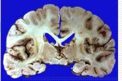
Cryptococcosis
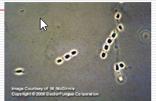
Coccidioides immitis
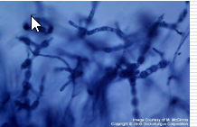
Coccidioides immitis
Describe Coccidioidomycosis
What is it?
Transmission
Describe the Coccidioides spp.
Two Types
STATs
Geography
Describe the pathology
Coccidioidomycosis
What is it?
True, systemic mycosis
Acquired by inhalation
Transmission:
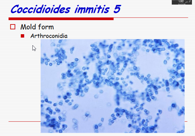
Dry arthroconidia carried by dust storms
Related to activities involving tillage of soil
Agriculture
Telephone post digging
Archaeology
Coccidioides spp.
Two Types:
Coccidoides immitus
Coccidoides posadasii
STATs
Most virulent of human mycotic agents
Geography:
Narrow region
San Joaquin Valley, California
Maricopa and Pima counties of Arizona
Southwestern Texas
regions that are characterized by low
rainfall, semiarid conditions
NOTE: Dimorphic fungus
Pathology
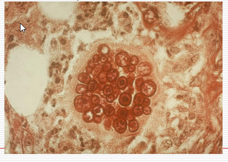
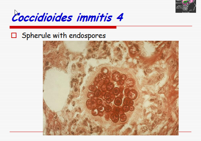
Mold form -> infectious athroconidia -> maturation -> airborne spores -> inhalation -> in lungs, forms multinucleated spherules -> ruptures, releases endospores -> new spherules

List the symptoms of Valley fever
Immunocompetent
Immuno compromised
Valley Fever
Symptoms:
Immunocompetent
Asymptomatic
Self-limiting; some antifungals may be needed
Confers specific immunity to reinfection
Immuno compromised
Pneumonia
Ruptured lung nodules
Disseminated disease
bone, heart and CNS (meningitis) infection
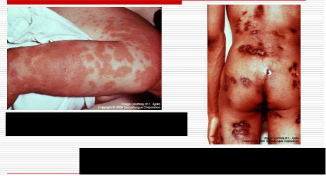
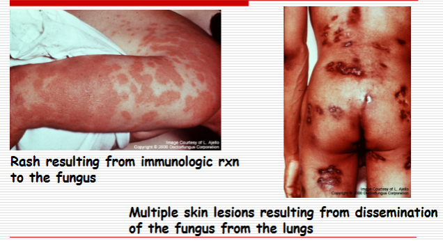
List the Consequences of CNS dissemination of coccidioidomycosis
Consequences of CNS dissemination of coccidioidomycosis
Chronic granulomatous meningitis
Basilar meninges
Cerebral and cerebellar abscesses
Clinical symptoms
Headaches, nausea, vomiting
Altered mental status