Chapter 9 , Chapter 8 Anatomy JCCC
1/149
There's no tags or description
Looks like no tags are added yet.
Name | Mastery | Learn | Test | Matching | Spaced | Call with Kai |
|---|
No analytics yet
Send a link to your students to track their progress
150 Terms
Joints
Joints occur where two bones meet (articulations).
Joint Classification
The functional classification of joints includes Synarthrosis (immovable joint, no movement), Amphiarthrosis (a slightly movable joint), and Diarthrosis (a freely movable joint).
Fibrous Joints
Bones held together by dense collagen fibers, with no joint/synovial cavity.
Cartilaginous Joints
Cartilaginous joints are bones held together by cartilage, with no joint/synovial cavity.
Synovial Joints
Bones held together by ligaments, fluid, capsules, and have a joint cavity.
Sutures
Synarthrotic joints only between bones of the skull, with short CT fibers and irregular, interlocking edges that increase strength.
Syndesmoses
Amphiarthrotic joints that permit more or less movement depending on fiber length, such as the tibiofibular ligament and interosseous membrane.
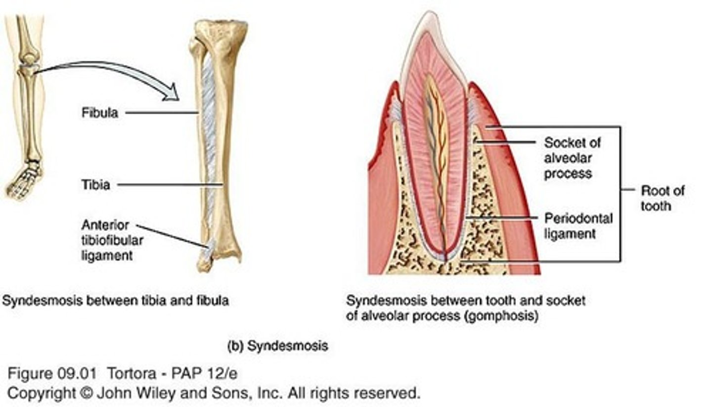
Gomphoses
Synarthrotic peg-in-socket joints, such as the articulations of the teeth with the sockets of the maxillae and mandible.
Synchondroses
Synarthrotic joints where connecting tissue is hyaline cartilage, such as the epiphyseal plate and the 1st rib and manubrium of sternum.
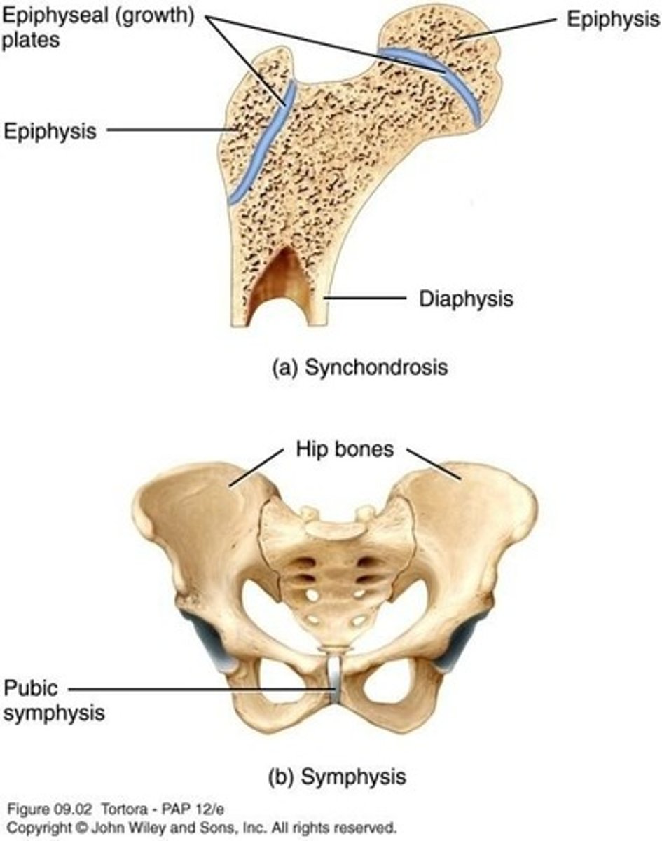
Symphyses
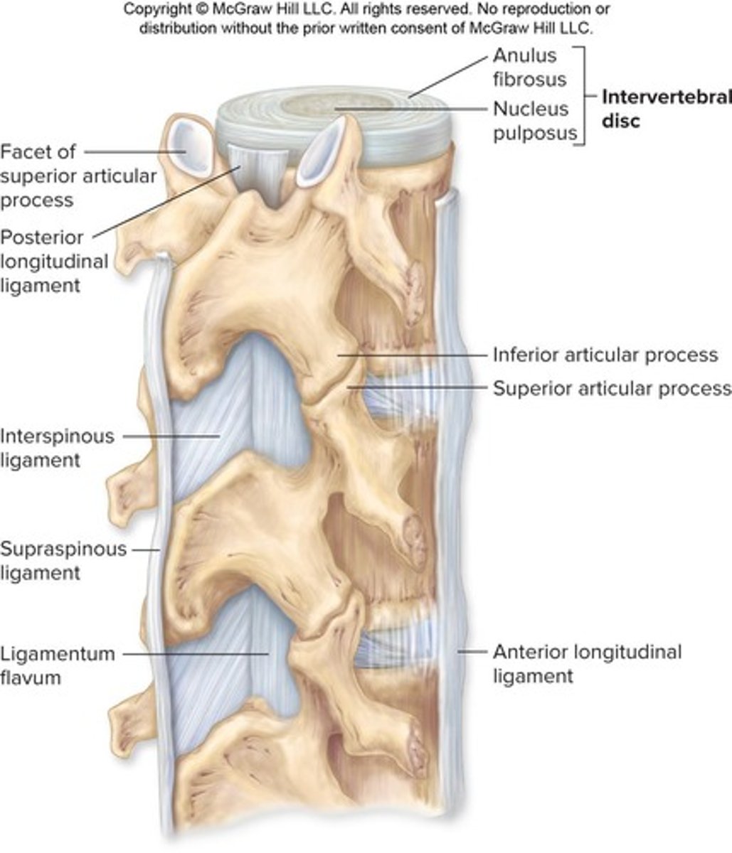
Amphiarthrotic joints where the ends of the articulating bones are covered with hyaline cartilage, connected by a disc of fibrocartilage, such as the pubic symphysis and intervertebral disks.
Articular Cartilage
Hyaline cartilage that covers the ends of bones in synovial joints.
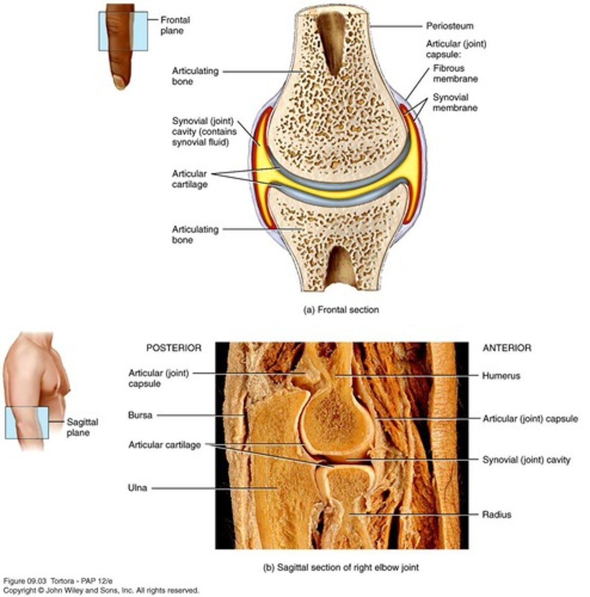
Synovial Cavity
Allows free movement and is characteristic of diarthrotic joints.
Articular Capsule
Sleeve-like capsule that encloses the joint/synovial cavity, consisting of an outer fibrous capsule and an inner synovial membrane.
Synovial Fluid
Secreted by the synovial membrane, lubricates the joint, prevents erosion, reduces friction, acts as a shock absorber, and supplies oxygen/nutrients while removing CO2/metabolic wastes.
Reinforcing Ligaments
Several layers that stabilize the joint.
Nerves and Blood Vessels
Detect pain and monitor joint position and stretch, while capillary beds supply filtrate for synovial fluid.
Fatty Pads
Cushion between the capsule fibrous layer and synovial membrane or bone.
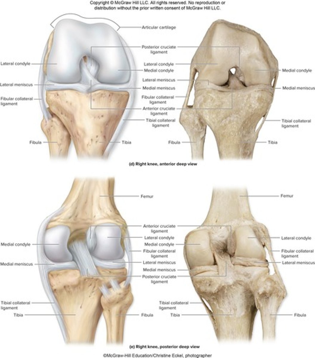
Articular Discs (Menisci)
Fibrocartilage structures that separate articular surfaces to improve the fit of bone ends, stabilize the joint, and reduce wear and tear.
Bursae
Bags of synovial fluid that reduce friction where ligaments, muscles, skin, tendons, or bones rub together.
Tendon Sheaths
Elongated bursae wrapped completely around tendons subjected to friction.
Factors Influencing Synovial Joint Stability
Stability of joints to prevent dislocations: shape of articular surface, ligament number and location.
Shape of Articular Surface
Shallow surfaces are less stable than ball-and-socket joints.
Ligament Number and Location
The more ligaments, the stronger the joint.
Muscle tone
Keeps tendons taut as they cross joints, extremely important in reinforcing shoulder and knee joints and arches of the foot.
Origin
Attachment to immovable bone.
Insertion
Attachment to movable bone.
Muscle contraction
Causes insertion to move toward origin.
Transverse plane
One of the planes along which movements occur.
Frontal plane
One of the planes along which movements occur.
Sagittal plane
One of the planes along which movements occur.
Planar/Gliding Joints
Allow back-and-forth and side-to-side movements; examples include intercarpal and intertarsal joints.
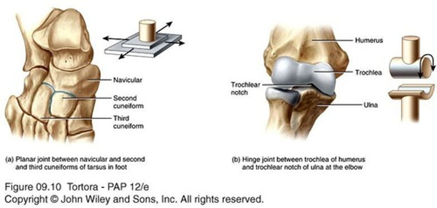
Hinge Joints
Produce an opening and closing motion like that of a hinged door; examples include knee (femur & tibia) and elbow (humerus & ulna).
Flexion
Bending movement that decreases angle of joint and brings 2 articulating bones closer together.
Extension
Bending movement that increases angle of joint and takes 2 articulating bones further away.
Pivot Joints
Allow rotation; examples include head of radius on ulna and atlas/axis.
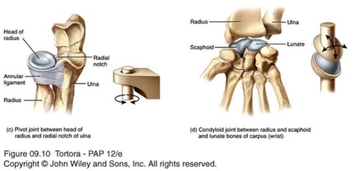
Condyloid Joints
Permit angular movements such as circumduction, adduction, and abduction; examples include wrist and metacarpophalangeal joints.
Circumduction
Combination of flexion/extension and abduction/adduction, resembling a circular motion.
Abduction
Movement along frontal plane away from midline of body.
Adduction
Movement along frontal plane toward midline of body.
Saddle Joints
Articular surface of one bone is saddle-shaped, fitting into the saddle of another; example includes the thumb.
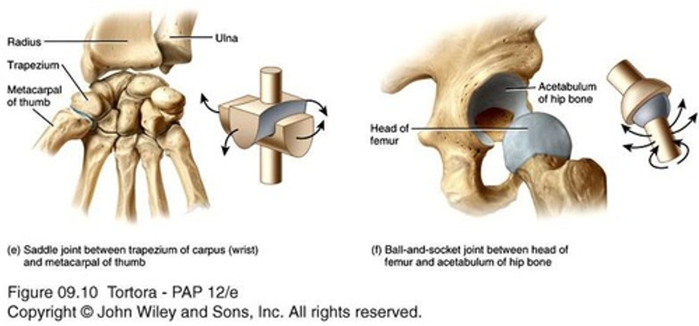
Ball-and-Socket Joints
ball-like surface of one bone fits into a cuplike depression of another; most freely moveable; examples include shoulder (humerus, scapula, & clavicle) & hip (pelvis & femur).
Elevation
upward movement of a part of the body; example includes closing the mouth; its opposing movement is depression.
Depression
downward movement of a part of the body; example includes opening the mouth.
Protraction
movement of a part of the body anteriorly; example includes thrusting the mandible forward; its opposing movement is retraction.
Retraction
movement of a protracted part of the body back to normal.
Inversion
movement of the sole medially; its opposing movement is eversion.
Eversion
movement of the sole laterally.
Dorsiflexion
bending of the foot at the ankle in an upward direction; its opposing movement is plantar flexion.
Plantar flexion
bending of the foot at the ankle in a downward direction.
Pronation
movement of the forearm so that the palm is turned downward; its opposing movement is supination.
Supination
movement of the forearm so that the palm is turned upward.
Opposition
movement of the thumb across the palm to touch the tips of the fingers on the same hand.
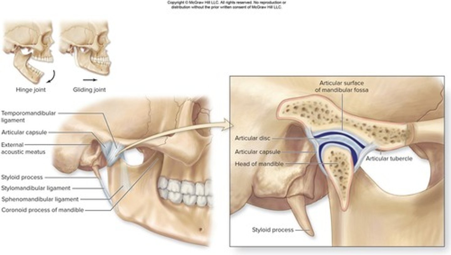
Temporomandibular Joint
the articulation between the head of the mandible and the articular tubercle of the temporal bone anteriorly and mandibular fossa posteriorly; it is a diarthrotic, synovial hinge joint.
Intervertebral Articulations
amphiarthroses between vertebral bodies; diarthroses between articular processes; vertebral bodies separated by intervertebral discs.
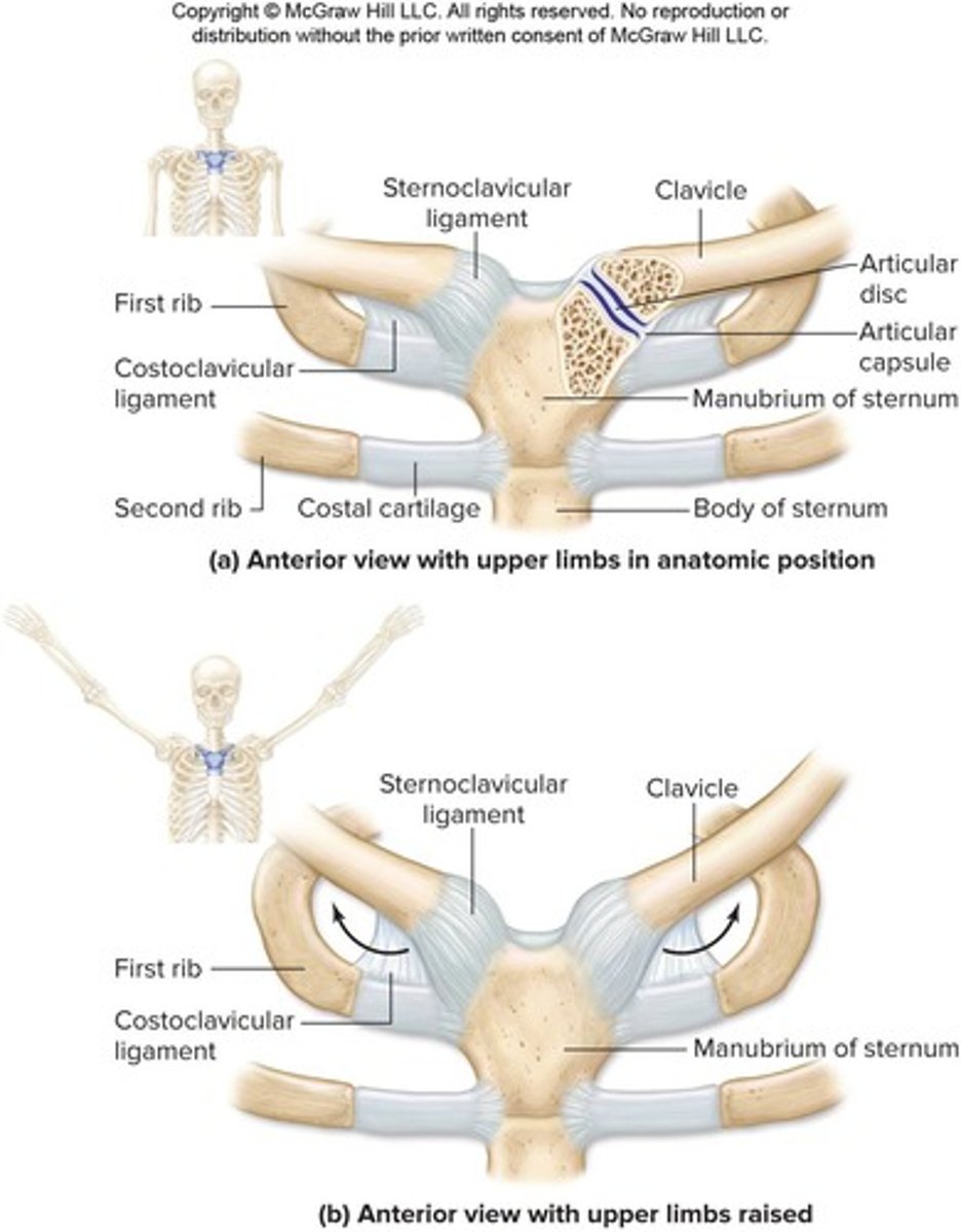
Sternoclavicular Joint
diarthrotic saddle joint between manubrium of sternum and sternal end of the clavicle; articular disc separates two joint cavities.
Acromioclavicular Joint
diarthrotic plane joint between acromial end of clavicle and acromion of scapula; articular disc within joint cavity.
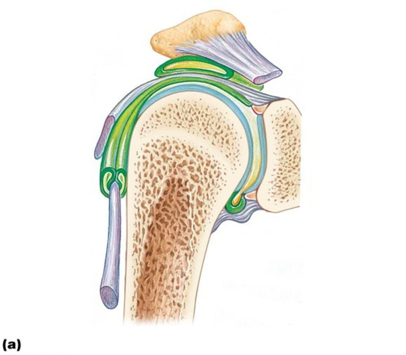
Glenohumeral (Shoulder) Joint
diarthrotic ball-and-socket joint between head of humerus and glenoid cavity of scapula; greatest range of movement but also the most unstable.
Elbow Joint
diarthrotic hinge joint composed of humeroulnar and humeroradial joints; both joints enclosed in a single articular capsule.
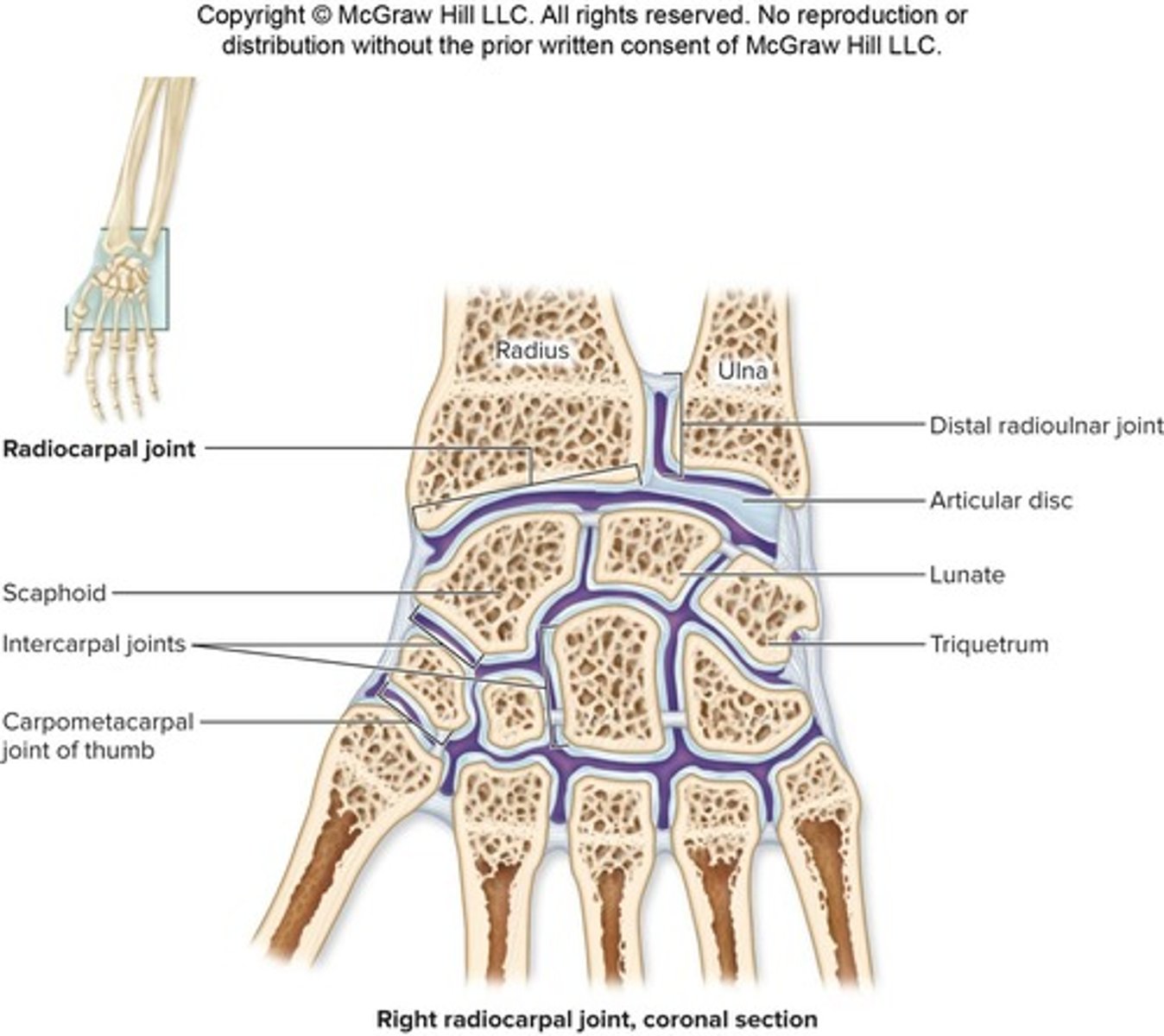
Radiocarpal (Wrist) Joint
diarthrotic condylar joint between the three proximal carpal bones (scaphoid, lunate, triquetrum), the distal articular surface of radius, and an articular disc.
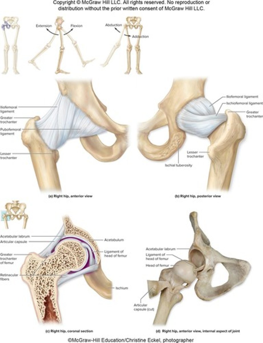
Hip (Coxal) Joint
diarthrotic ball-and-socket joint between head of femur and acetabulum of os coxae; features include fibrocartilaginous acetabular labrum.
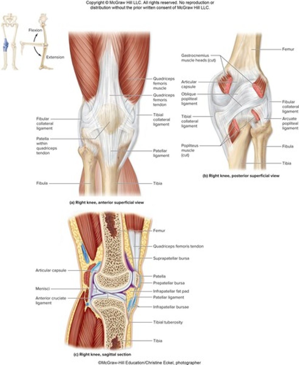
Knee Joint
diarthrotic hinge joint containing two articulations: tibiofemoral joint and patellofemoral joint; largest and most complex diarthrosis of body.
Talocrural (Ankle) Joint
diarthrotic hinge joint composed of two articulations between distal end of tibia and the talus, and between distal end of fibula and the lateral aspect of the talus.
Intertarsal Joint
plane joint between tarsals.
Tarsometatarsal Joint
plane joint between distal tarsal bones and metatarsals.
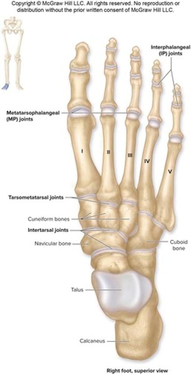
Metatarsophalangeal (MP) Joint
condylar joint between metatarsal and proximal phalanges.
Interphalangeal (IP) Joint
hinge joint between phalanges.
Arthritis
a common inflammatory or degenerative disease that comes in many forms, including gouty arthritis, osteoarthritis, and rheumatoid arthritis.
Appendicular Skeleton
Includes bones of the limbs and the girdles of bones that attach limbs to axial skeleton.
Pectoral Girdle
Articulates with the trunk and supports the upper limbs; consists of the clavicles and scapulae.
Clavicle
S-shaped bone with a sternal end that articulates medially with the manubrium of sternum and an acromial end that articulates laterally with the acromion of the scapula.
Scapula
Shaped like a broad, flat triangle with three borders and three angles; glenoid cavity articulates with head of humerus.
Upper Limb
Consists of 30 bones including the humerus, radius, ulna, carpal bones, metacarpals, and phalanges.
Humerus
Proximal features include a head that articulates with scapula and greater and lesser tubercles for muscle attachment.
Radius
Bone of the forearm that is lateral to the ulna in the anatomic position.
Ulna
Bone of the forearm that is medial to the radius in the anatomic position.
Carpals
Eight wrist bones arranged in two rows of four.
Metacarpals
Five bones in the palm of the hand.
Phalanges
Fourteen bones in the fingers.
Glenoid Cavity
Articulates with the head of the humerus.
Intertubercular Sulcus
Passage for biceps brachii tendon and site for muscle attachment.
Capitulum
Round, lateral projection of the humerus for articulation with the radius.
Trochlea
Pulley-shaped, medial projection of the humerus for articulation with the ulna.
Olecranon Fossa
Large basin on the posterior humerus that accommodates the olecranon of ulna.
Styloid Process (Radius)
Lateral 'wrist bump' at the distal end of the radius.
Trochlear Notch
Accommodates the trochlea of the humerus.
Olecranon
Projection that forms the posterior 'bump' of the elbow; attachment site for triceps brachii.
Coronoid Process
Inferior lip of the trochlear notch of the ulna.
Scaphoid
First bone in the proximal row of carpals.
Lunate
Second bone in the proximal row of carpals.
Triquetrum
Third bone in the proximal row of carpals.
Pisiform
Fourth bone in the proximal row of carpals.
Trapezium
First bone in the distal row of carpals.
Trapezoid
Second bone in the distal row of carpals.
Capitate
Third bone in the distal row of carpals.
Proximal Phalanx
The first phalanx in a finger, closest to the hand.
Middle Phalanx
The second phalanx in a finger, located between the proximal and distal phalanges.
Distal Phalanx
The last phalanx in a finger, furthest from the hand.
Pollex
The thumb, which has only two phalanges (proximal and distal).
Pelvic Girdle
The pelvis is composed of left and right ossa coxae, as well as the sacrum and coccyx.
Os Coxae
The 'hip bone', a fusion of ilium, ischium, and pubis between 13 and 15 years of age.