Neuroscience Final
1/281
There's no tags or description
Looks like no tags are added yet.
Name | Mastery | Learn | Test | Matching | Spaced |
|---|
No study sessions yet.
282 Terms
Individual neurons form neural circuits (PYR, PV+, SST+, VIP+)
PYR: pyramidal neuron
PV+: Paravalbumin-expressing interneuron
SST+: Somatostatin-expressing interneuron
VIP+: Vasointestinal peptide-expressing interneuron
Why study neural circuits?
The postsynaptic effect of a single action potential is small
Population coding is a common theme in neural information processing
Neural Coding
Action Potential Frequency (rate code)
The firing rate (frequency of APs) code for the strength of a sensory signal
Coordinated activity (synchrony code)
Precision of spikes among different neurons
Graded Potential
The amplitude of depolarization codes for the strength of the sensory signal; the more depolarization, the more transmitter is released
Examples of Neural Systems
Visual, Somatosensory, auditory, motor, reward, learning/memory
Example study of synchrony code
EEG (Electroencephalography) recording during “moony face recognition” in human subjects
Higher spectral power: stronger synchrony
Properties of Sensory Systems: Central Pathway
Sensory signal is transmitted from peripheral neurons to “central” neurons in the brain
Periphery → Subcortical → Cortical
Properties of Sensory Systems: Sensory Receptors (Cells)
Modality specific
Transduce energy of different forms to electrical activity so that can be passed on to other neurons
Even within a single sensory system, different populations of neurons are specialized to detect different features of the same input
Properties of Sensory Systems: Topographic Map
Nearby neruons have nearby receptive fields → orderly representation of the sensory space in the nervous system
The Visual Pathway
Retina →
1. Suprachiasmatic Nucleus (SCN)
Superior Colliculus (SC)
Lateral Geniculate Nucleus → Visual Cortex: V1 → V2→ V3, V4, V5
Light starts visual processing
Electromagnetic radiation that is visible to our (human) eyes: 400 (higher energy) to 700nm (lower energy)
Light Energy to Neural Activities is called
Phototransduction; occurs in the retina
how light energy leads to a change in membrane potential
Iris
Surrounds pupil/eye color/contains muscles that can change the size of pupil (controls amount of light reaching the eye)
Sclera
continous with the cornea/”white of the eye”; is a tough outer wall
Extraocular muscles
move the eye
Retinal Information Processing: 1 pathway
Linear Pathway (Direct):
Photoreceptor Cells (P): (only cell type directly affected by light) → Bipolar Cells (B) → Retinal Ganglion Cells (R) : The retina’s only output cell type
Retinal Information Processing: 2nd pathway
Lateral Pathway (Indirect):
Retinal Ganglion Cells ←> Amarcine cells <→ Bipolar
Bipolar ←> Horizontal Cells ←> Photoreceptor cells
Cones detect light of specific wavelength
Blue Cones: 430nm
Green cones: 530nm
Red cones: 560nm
Distance across retina
Temporal Periphery Central Retina Nasal Periphery
Photoreceptor cells are constantly depolarized in the dark
Intracellular cyclic guanosine monophosphate (cGMP) binds to cGMP gated Na+ channel → cGMP keeps the channel open and allows Na+ influx → Photoreceptor cell membrane is kept depolarized (-30mv)
Dark Current → NT glutamate is constantly released at the terminal
Photoreceptor cells are hyperpolarized by the light
Light stimulation reduce cGMP → The Na+ channel close, allows the membrane hyperpolarization (-65mv) → glutamate release stops at the terminal
Neural Codes
the meaning of activity (action potentials) by a neuron depends on the system it’s part of
AP by neurons in a pain system, would yield the perception of pain
AP by neurons in visual system would yield the perception of vision
APs by a neuron in motor systems would cause movement
APs by a neuron in system underlying emotion might cause feeling of fear or another emotion
Systems have _ and neurons in _ code for _
Systems have subsystems and neurons in different subsystems code for different functions
Visual subsystem example
In visual system different areas and groups of neurons perform different functions
Some neurons responsible for detecting the form of a visual object, others responsible for detecting color, others for movement, others for location (code to location of an object in visual field)
What is a neuron coding? What attribute of the visual world is a neuron in the visual system encoding?
refers to how a neuron represents information through its electrical activity (like firing rate or pattern of action potentials)
encodes attributes such as orientation, color, motion, depth or location of visual stimuli in the visual field
How do neuroscientists determine the function of a neuron or of a system of neurons?
By recording their activity (using electrodes, imaging, or fMRI) while presenting specific stimuli or tasks, then analyzing how firing patterns change
They may also manipulate the neurons (lesions, electrical stimulation, optogenetics) to see how behavior or perception is affected
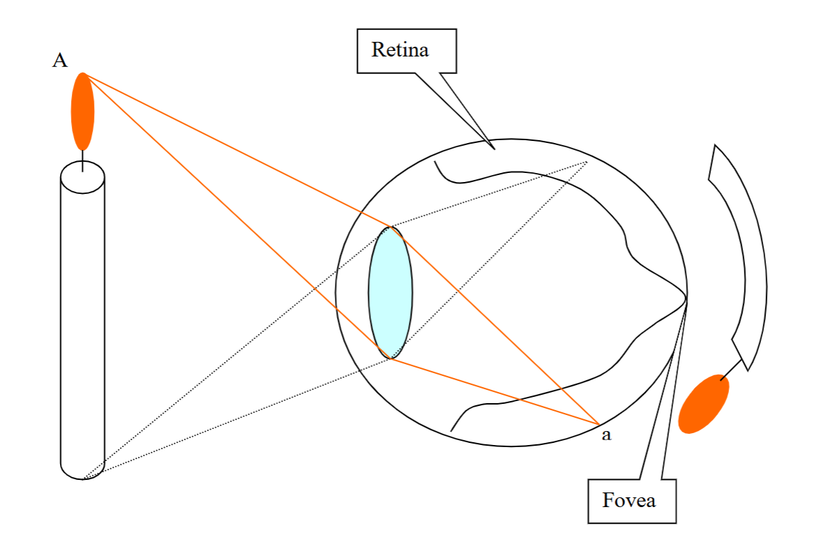
Retina
Consists of several thin layers of cells distributed across the inside of the eye
Contains photoreceptors that convert light into neural signals (light → images sent to the brain)
Fovea
The center of the visual field, provides color vision
Light directly reaches to the photoreceptor cells here
Less distortion, less blur → clear vision
It is the portion of the retina with the highest acuity, the ability to resolve fine detail and patterns of light
Visual Acuity
The ability of the eye to distinguish two points near eachother
Optic Disk
is the retinal location where axons from a type of retinal cell collect and exit the eye and form the optic nerve
What is the blind spot and why
The optic disk because there are no photoreceptors
Lens
Fine tunes focus by helping the eye focus light onto the retina and adjust refraction for near and far vision
Cornea
Covers pupil and iris
Bends (refracts) light toward retina (main refractive surface of the eye)
Pupil
Controls how much light enters the eye (goes to retina)
Optic Nerve
Sends visual info to the brain
Properties of Sensory Systems: receptive field
the location in the environment (or the surface of the body, i.e, sensory space) from which an appropriate stimulus will change that cell’s activity
Photoreceptor Receptive Field
is circular
of a given photoreceptor is determined by its location in the retina
responds to changes of light intensity in its field
RGC A (peripheral) has la arger field than RGC B (Central)
Divergence of single PR cells onto multiple RGC → overlapping fields
Example of neuron’s receptive field
Light at point a (near the fovea) will affect the activity of retinal cell in location “a” in the retinal”
Cells in different locations in the retina have receptive fields in different locations in the visual fields
The cells activated by
Retinal Ganglion cells, Amacrine, Bipolar and Horizontal cells: activated by signal from photoreceptor cells
Photoreceptor cells: directly activated by light
Receptive Field Size Difference in Distinct RGC types
In perpiheral: convergence of synaptic input: large field; magno (m)-type
In central: no or less convergence of synaptic input: small field; parvo (p)-type
RGC Receptive Field: Center-Surround
On-center: increase spiking when light is on the center receptive fieldl increases APs
Off-center: increase spiking when light is off the center receptive field; decreases Aps
Two Different Glutamate Receptors in Bipolar Cells: Inhibitory Glutamate Receptor
Glutamate release from pre-synapse in PR cell → Received by inhibitory receptor at post-synapse in BP cell → Decrease membrane potential in BP cell → No NT release
Two Different Glutamate Receptors in Bipolar Cells: Excitatory Glutamate Receptor
Glutamate release from pre-synapse in PR cell → Recieved by excitatory receptor at post-synapse in BP cell → increase membrane potential in BP cell → NT release
Mechanism of BP cell response to receptive field Surround
Horizontal cell depolarization → photoreceptor cell inhibition + bipolar cell excitation
Visual Preception Does NOT depend on _ and why?
Illumination level
Contrasts in light intensity are more informative than the overall illumination. so that our perceptions of what we see are not dramatically affected by the level of ambient illumination
5 cells types in the retina
Photoreceptors
Bipolar cells
Retinal ganglion cells
Horizontal cells
Amacrine cells
Photoreceptors
a specialized neuron in retina that detects light and converts it into electrical signals
only cell type in visual system that’s directly sensitive to light
two types: rods & cones
project to the bipolar cells
Rods
Highly sensitive to light, responsible for vision in very dim light
Bleached in bright light and thus unresponsive in bright light
Not responsible for high acuity vision (not good for fine detail)
Achromatic (insensitive to colors/ so black and white vision)
Only exist outside of the fovea
120 million rods in human retina
Cones
Low sensitivity: Less sensitive to light intensity and are inoperative in dim light
Needs a lot of light
High resolution
Sensitive to color, three subtypes: selectively sensitive to red, blue, and green wavelengths light
Most concentrated in the fovea
6 million
Bipolar cells
relay signals from photoreceptors to ganglion cells
Retinal ganglion cell
sends action potentials to the brain via the optic nerve
Horizontal cell
integrates signals across photoreceptors
excited by increased glutamate and release GABA (inhibitory) which inhibits nearby photoreceptors, sharpening the contrast (lateral inhibition)
Amacrine cell
regulate bipolar and ganglion cell communication
located btwn bipolar & ganglion cells and used input from bipolar cells to modify the input using GABA or glycin to inhibit some ganglion cells in order to help detect motion, edges, direction
Emmetropia
Normal vision
Hyperopia
refractive error
farsighted
Hyopia
refractive error
nearsighted
Why is it that only retinal ganglion cells have axons?
axons are needed for long-distance transfer of info
in retina the cells are close together so they don’t need APs or axons
also, communication by PSPs may be able to convey info that is more subtle than can be conveyed by the AP frequency code
Why do only RGCs and Amacrine cells generate action potentials?
The rest of the cell types use graded depolarization to release neurotransmitter to the next cell
A depolarization increases NT release
Small depolarizations cause small release of NT; large depolarizations cause large release of NT
Relationship between diff cell types in retina
retina is “inside out” w photoreceptors furthest away from light (very back of ete), all light must pass thru other cell types to reach photoreceptors
works bc all cells in eye (except photoreceptors) are translucents
also at the foveal pit all cell types (except photoreceptors) are pushed out of the way
resting membrane potential of photoreceptors
-30mv in the dark
maximum hyperpolarization
-65mv and produced by bright light
Glutamate
neurotransmitter used by photoreceptors. the greater the intensity of light, the less NT released
Visual Pathway route
Eye → optic nerve → optic chiasm → optic tract → Lateral Geniculate Nucleus → Optic Radiation → primary visual cortex
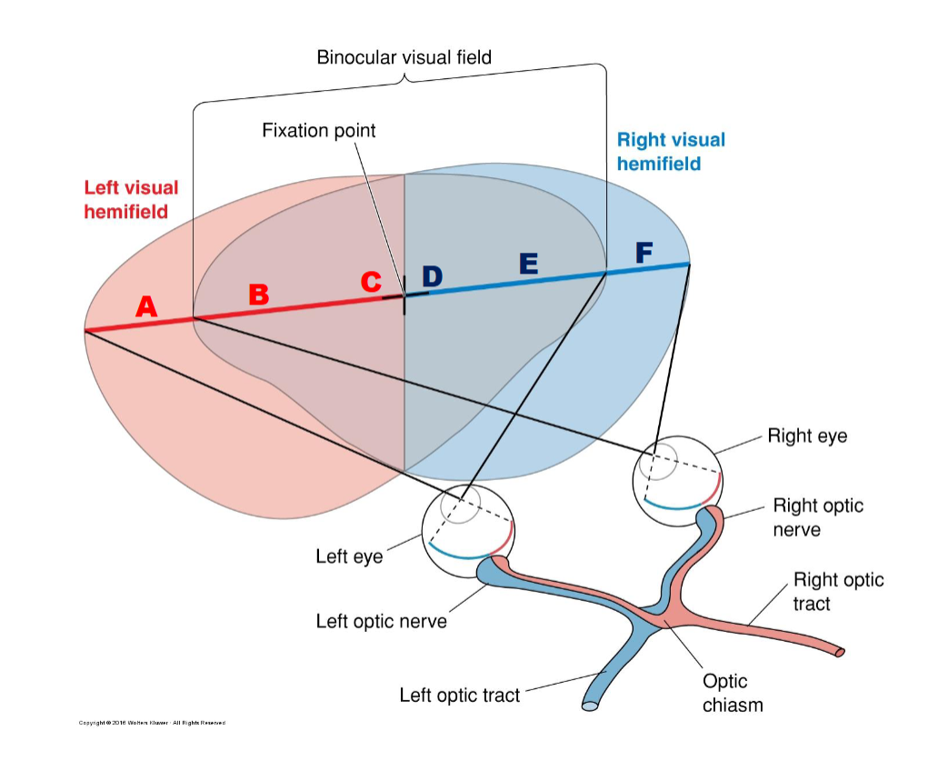
A-F & right vs left visual hemifields
Right eye, optic nerve, optic tract will see left visual hemifield: A, B, C
Left eye, optic nerve, optic tract will see right visual hemifield: D, E, F
Bioncular visual field: B, C, D, E
Fixation point is between C and D
Lateral Geniculate Nucleus (LGN)
six distinct layers
Layers 1 & 2 contain larger cells: Magnocellular layer
Layers 3-6 contain smaller cells: Parvocellular layer
Each layer recieves inputs from one eye
Parallel Processing of Different Visual Properties
Layer 1: contralateral, magnocellular
Layer 2: ipsilateral, magnocellular
Layer 3: ipsilateral, parvocellular
Layer 4: contralateral, parvocellular
Layer 5: ipsilateral, parvocellular
Layer 6: contralateral, parvocellular
CIICIC: chloe is intelligent cool inspiring cute
Ventral to each principal layer: nonM-nonP (Koniocellular LGN cell type), contra/ips it’s same as overlying prinicpal layer

Topographic Map in V1
Visual Field:
Left top: 1 & A
Left bottom: 2 & B
Right top: 3 & C
right bottom: 4 & D
V1 cortex:
Right hemibrain posterior: 1 & A Anterior: 2 & B
Left hemibrain posterior: 3 & C Anteriror: 4 & D
Suprachiasmatic Nucelus
Circadian rhythm: 24 hr fluctuations of biological processes
Recieves inputs from intrinsically photosensitive RGC (ipRGC)
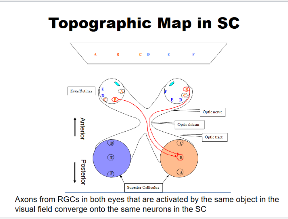
Topographic map in Superior Colliculus
Axons from RGCs in both eyes that are activated by the same object in the visual field converge onto the same neurons in the SC
Right eye sees: BC, DEF → right superior colliculus (posterior): ABC
Left eye: ABC, DE → left superior colliculus (posterior): DEF
Layered Structure in SC
← Anterior Posterior →
C B A
Superficial layers Visual input
Intermediate and deep layers Visual, auditory, somatosensory input, motor outpu
Sensory neurons directly synapse onto motor neurons to control movements, such as gaze and saccades
Cortical Visual Subsystems
Retina → LGN → visual coretex V1 → V2 → V3, V4, V5
Blindsight pathway
Retina → superior colliculus (can track/avoid objects subconsciously) → Suprachiasmatic Nucleus (circadian rhythm intact)
(no LGN/Visual cortex)
Parallel Processing Streams in the Perceptual Visual Pathway
Dorsal stream: movement; V1 → V3 & V5
Ventral Stream: color; V1 → V2 → V4 → Inferotemporal cortex (IT): complex objects
Rhodopsin
photopigment; a light sensitive receptor protein in rod photoreceptors
made of 1. the protein opsin (GPCR) and 2. retinal (11-cis-retinal or trans-retinal)
How does light produce the graded hyperpolarization?
The ligand-gated Na+ channels in the outer segment membrane are open in the dark, causing depolarization (to the resting membrane potential of -30mv)
These ligand-gated channels r like receptors, but “inside-out", meaning they bind their ligand cGMP to a binding site on the intracellular face of the Na+ channel and this opens the channel
How does light decrease the concentration of cGMP?
photopigment (highly concentrated in membrane of disks in outer segement of photoreceptors) is purple in dark, when it absorbs light its bleached to pale yellow
Photopigment is rhodopsin
The steps of cGMP activation
Rhodopsin: retinal
the only light sensitive molecule in visual system
precursor of retinal is vitamin A
exists in 2 confromations:
in the dark: 11-cis-retinal
in light will switch it to: all trans-retinal
Steps of cGMP activation
Opsin passes thru membrane 7 times (it’s a metabotropic or G-protein-coupled receptor)
release of retinal from opsin allows opsin to change shape and this activates a G-protein (transducin)
The G-protein (G) dissociates and travels along the membrane and activates an enzyme (cGMP phosphodiesterase)
cGMP phosphodiesterase converts cGMP to GMP, and thus lowers the concentration of cGMP
In the dark, cGMP bound to Na+ channel. light decreases concentration of cGMP, causing cGMP to disassociate from the channel, consquently, channels close and photoreceptors hyperpolarize
Why do we have this type of system (what’s the advantage)? 1st answer
Increased surface area + increased photopigment produced by having the photopigment molecules on the stacked disks, instead of on the Na+ channels, increases the chance of light being detected by a rod
system so sensitive that a single photon can produce a detectable change in the membrane potential of a rod-type photoreceptor
Why do we have this type of system (what’s the advantage)? 2nd answer
The use of G-proteins allows for amplification
each molecule of opsin can activate many G-proteins, each of which can activate many enzymes of cGMP phosphodiesterase, which then can convert many molecules of cGMP into GMP
depolarized photoreceptor
Dark= glutamate released= ON bipolar inhibited OFF bipolar excited
hyperpolarized photoreceptor
Light= glutamate decreased= ON bipolar excited OFF bipolar inhibited
Excited
more likely to send signal to ganglion cell → action potential
Inhibited
less likely to pass signal to ganglion cell → no action potential
OFF bipolar cell
sends signals to OFF ganglion cells → OFF pathway → detects when light turns OFF
ON bipolar cell
sends signals to ON ganglion cells → ON pathway → detects when light turns ON
3 Types of Retinal Ganglion Cells
Parvocellular (P-type)
Mangnocellular (M-type)
NonM-NonP
Parvocellular (P-type)
small receptive fields, sustained response involved in color and detail
Mangnocellular (M-type)
large receptive fields, transient responses, sensitive to motion and low contrast
Each ganglion cell responds to light in a specific area:
its receptive field
Center: direct input from photoreceptors
Surround: indirect input via horizontal cells
Metabotropic Receptors (how they differ from Ionotropic receptors)
Linked to G-proteins not ion channels
slower but long-lasting effects
acts thru secondary messangers
How they work: NT binds → activates G-protein → starts secondary cascade → affects ion channels indirectly
Ionotropic Receptors (how they differ from Metabotropic receptors)
ion channels themselves
fast-acting
How they work: NT binds → channel opens → ion flow in or out → direct change in membrane potential
ON bipolar cells are _
Metabotropic since they express mGluR6 receptors (GRCRs)
OFF bipolar cells are _
Ionotropic since they express AMPA or Kainate receptors
The three visual pathways
Retinofugal
Retinotecal
Retinohypothalamic
Retinofugal Pathway
Main conscious vision pathways
Retina → optic nerve → optic chiasm → optic tract → LGN (thalamus) → primary visual cortex
Retinotecal Pathway
involved in eye movement and reflexes
Retina → Superior Colliculus
Retinohypothalamic Pathway
regulates circadian rhythms
Retina → Suprachiasmic Nucleus (SCN)
Each eye see both visual fields but:
The nasal retina crosses at the optic chiasm
The temporal retina stays on the same side
Left Eye sees
left visual field → hits nasal retina → crosses at optic chiasm → goes to right hempisphere
right visual field → hits temporal retina → does NOT cross → goes to right hemisphere
Right Eye sees
right visual field → hits nasal retina → crosses at optic chiasm → goes to left hemisphere
left visual field → hits temporal retina → does NOT cross → goes to right hemisphere