Med Micro Readings
 Call Kai
 Learn
Learn Practice Test
 Spaced Repetition
 Match
Match Flashcards
 Knowt Play
1/74

There's no tags or description
Looks like no tags are added yet.
Name | Mastery | Learn | Test | Matching | Spaced | Call with Kai | Chat |
|---|
No analytics yet
Send a link to your students to track their progress
75 Terms
Darkfield microscopy
Light objects are visible against a dark background
Opaque disk placed in condenser
Only light reflected off the specimen enters the objective lens
Used to visualize Treponema palldum
phase contact microscope
Splits a beam of light into 2 types of light,
- direct
- reflected
brings them together to form an image of the specimen; it allows the detailed observation of living organisms, especially the internal structures

Hooke's contribution
First to describe cells using a microscope.
Coined the term "cell"
Idenfitied cell as the basici unit
Scientific Nomenclature
Two-name system for naming organisms
The first name is the Genus; it is italicized and capitalized
The second name is the specific epithet (species name); it is italicized, but no cap
Types of microorganisms
Bacteria
Archaea
Fungi
Protozoa
Algae
Viruses
Multicellular animal parasites
Bacteria characteristics
- unicellular and prokaryotic
- 3 shapes: Cocci (circular), Bacillus( Rod), and Spiral
- enclosed in carbohydrate wall & petidoglycan
- Replicate via binnary fission
* bacteria obtain nutrience through mostly organic matter, but some can photosynthesis
Archea characteristics
unicellular, are often extremophiles, living in harsh environments (high temp, high salinity, no light). and often using alternative sources of energy such as chemosynthesis.
- prokaryotic but lack peptidoglycan wall
- these are not known to cause disease to humans
Fungi
An organism that absorbs nutrients from the environment.
- can be singel celled or multicellular
- eukaryotic
- most cannot carry out photosynthesis
-
Mold
Forms visible masses known as mycelia, which are composed of long filaments called hyphae)
Protozoa characteristics
-eukaryotic
- unicellular
- lack a cell wall
- sexual or asexual reproduction
they can either live as free entities or as parasites
- If parastitic will absorb nutrients from host
- some can utlize photosynthesis
Algae
a very simple plant without stems or leaves that grows in or near water.
- Photosynthetic Euks
- Asexual or sexual reproduction
- cell walls are made of cellulose
viruses characteristics
-acellular
-nucleic acids with protein coat
-has capsid
-can only be seen with mircoscope
- Viruses require a host to reproduce
Spontaneous Generation Theory
Hypothesis stating that life could arise from nonliving matter.
Supported by John Needham Experiment
- Boiled nutrients broth and poured it into covered flask
- Microbes grew
Conclusion:
Claimed a mircobes arose spontaneously from a "vital force" in the broth. What really happened was the jar was not sterilzed prior
Van Leeuwenhoek
First to see organisms under a microscope
- This contributed to the cell theory
Biogenesis Theory
Theory that states living things can only come from other living things
Supporting experiments include:
- Redi's Meat Jars
- Spallazoni's sterialized jars
- Pastuers S-shaped flasks
Francesco Redi
This scientist disproved spontaneous generation by showing that maggots do not spontaneously arise from decaying meat.
Placed decaying meat under 3 conditions:
- Open jar, tightly sealed jar, gauze covered jar
- the maggots only appeared in the open jar
This showed that large organisms do not arsie spontaneously
Lazzaro Spallanzani (1765)
repeated Needham's experiments to show that organisms entered the broth from air
- The nutrience broth was boiled in the flask and then it was sealed
- one set allowed air in and the other set didn't
Demonstrated that no mircoorganisms grew in the sealed thoroughly boiled flask
Louis Pasteur
A French chemist, this man discovered that heat could kill bacteria that otherwise spoiled liquids including milk, wine, and beer.
His biogenesis experiment: Boiled nutrience broth in a S-shaped neck flask which allowed fresh air in but prohibited microbes
The broth remained sterile as long as the neck remained intact.
Conclusion:
Disproved spontaneous generation definitively for microorganisms, establishing that living cells arise only from pre-existing cells
Germ Theory of Disease
the principle that microorganisms cause disease
Experiments
- Pasteur: Microbes cause the " spoilage of wine"
- Se
Compound Light Microscope
microscope that allows light to pass through a specimen and uses two lenses to form an image
condenser lens
located immediately below the specimen, focuses light from the light source onto the specimen
objective lens
the part of a compound light microscope that is located directly above the specimen and that magnifies the image of the specimen
ocular lens
Eyepiece of a microscope
this adds a 10x magnification
Path of light through a compound microscope
light source; condenser; specimen; objective lens; ocular lens
Total Magnification
objective lens x ocular lens
- Remember to start at the lowest objective lens and work up
Resolution of a microscope
measures direct clarity or ability to distinguish small but close objects
This is the ability of the microscope to distingish fine + small details.
Specifcally it is the ability of the lens to distingish two points that are a specific distance apart.
For example, a mircoscope w/ a resolving power of 0.4nm can tell points at least 0.4nm apart
refractive index
a measure of the light-bending ability of a medium
- If we can change the refractive index of specimens it can help get a clearier more sharp image
- we can stain the speciman to obtain a sharper contrast between the medium and speciman. When the ray changes direction the angles change increasing the contrast
Also remember to preserve the direction of the light rays, we use immersion oil at high resolution
Differential Interference Contrast Microscopy
Uses two beams of polarized light.
The combined images look as if the cell is casting a shadow on one side.
This adds contrasting colors to the speciman
The resolution of DIC microscope is higher than a standard phase microscope

Fluorescence Microscopy
Uses UV light
Fluorescent substances absorb UV light and emit visible light
Cells may be stained with fluorescent dyes (fluorochromes)
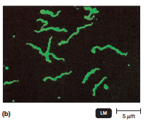
Immunofluorescence
a method of tagging antibodies with a fluorescent dye to detect or localize antigen-antibody combinations
confocal microscopy
a light microscope that uses fluorescent stains and laser to make two- and three-dimensional images

Transmission Electron Microscopy (TEM)
An electron microscope in which a beam of electrons is transmitted through a specimen and focused to produce an image.

Use for Dark-Field microscope
to examin living microorganisms that are invisable in brightfield microscopy and do not stain easily or are distorted by frequent staining
- Live, unstained microorganisms
- View ones typically distorted by staining
Phase Contrast Microscopy Use
To facilitate detailed examination of the internal structure of living specimens
Detailed examination of the internal structures of living organisms
Use of differential interference constract microscope
To provide three-dimensional images
- generating brightly colored psuedo-3D images of lving cells & internal structures
Use of a fluorescence microscope
For flourscent antibody techiniques to rapidly detect and identify microbes in tissues or clinical specimens
use of a transmission electron microscope
Observing the internal ultrastructure of cells under high magnification and resolution
To examine viruses or the internal ultrastructure in thin sections of cells
- Very high magnification!
- produces structure 2D of internal features
Use of scanning electron microscope
Observing the surface of a sample
To study the surface feature of cells and viruses
- This is used for the external surface
- produces 3D image of external structures like Cilia or Flagella
Use of Confocal Microscope
To obtain a two or three dimensional image of a cell for biomedical application
Glycocalyx functions
- in cell adhesion
- recognition and immunity; it prevents phagocytosis
It can be capsule form or slime form
Gram negative flagella
It contains two pairs of rings
- outer pair of rings ancored to various portions of the cell wall
- inner pair of rings anchored to plasma membrane
gram postive flagella
Only the inner ring is present
Bacteria Flagella
contain a filament composed of flagellin, a basal body that anchors and rotates the flagellum, and a hook that connects the two
The movement is generated by the rotation of the basal body. It can go clockwise or counter clockwise
Runs and tumbles
Atrichous flagella
bacteria that lack flagella
peritrichous flagella
flagella that cover the surface of a cell
monotrichous flagella
single flagellum at one end
lophotrichous flagella
multiple flagella at one end
amphitrichous flagella
flagella at both poles of the cell
Archaella
•Archaeal motility structure
•Made of glycoproteins archaellins
•Anchored to the cell
•Archaella (singular: archlaellum) rotate like flagella
axial filaments
-also called endoflagella
-in spirochetes. -anchored at one end of a cell. -rotation causes cell to move
Brightfield characteristics
- Speciman is darker against white, bright background
- Not good for objects less than 0.2 µm
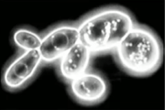
Darkfield characteristics
Specimen: appears bright and luminous against an entirely black background
- Thin glowing outlines will be against pitch black
Phase contrast characteristics
Background: Flat, neutral gray.
Specimen: Sharply defined internal structures with characteristic bright or dark halos around the cell perimeter.
Dead giveaway: Unstained, living cells with distinct glowing fringes or rings around the edges.
Differential interference contrast (DIC) characteristics
Background: Often gray or soft pastel colored.
Specimen: Looks like an embossed, 3D relief sculpture with distinct shadows cast across the cell surface, often with prism-split colors.
Dead giveaway: Appears pseudo-3D and brightly colored despite being unstained.
Transmission electron microscopy characteristics
Format: Flat, 2D slice.
Specimen: High-magnification internal ultrastructure (ribosomes, organelle membranes, viral envelopes, DNA filaments).
Dead giveaway: Looking inside a cut cell or viewing tiny viruses in black and white (or artificially tinted).
scanning electron microscopy characteristics
Format: Dramatic 3D perspective.
Specimen: Intact outer surfaces, showing external textures like cilia, flagella, cellular grooves, or biofilms with realistic shadows.
Dead giveaway: Looks like a photograph of an intact object taken with a camera, showing full external depth.
Diplococci
Spherical bacteria that grow in pairs and cause diseases such as pneumonia.

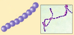
Streptococci
bacteria that form a chain
Tetrads
4 cocci in a square

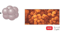
Sacrine
Cube like Cocci
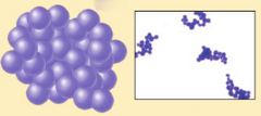
Staphylococci
a group of about 30 species of bacteria that form irregular groups or clusters resembling grapes
Diplobacilli
A double bacillus, two being linked end to end to each other

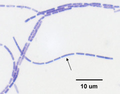
Streptobacilli
rod-shaped bacteria occurring in chains
Coccobacilli
very short rods

Semmelwies
-Brilliant individual in the 19th century
-Stressed the importance of washing hands before conducting surgery
- Advocated for handwashing bc of observatiions
Robert Koch
German doctor who identified the bacterium that caused tuberculosis
Proved bactereium cause disease
Koch's Postulates steps
1. The same pathogen must be present in every case of the disease.
2. The pathogen must be isolated from the diseased host and grown in pure culture.
3. The pathogen from the pure culture must cause the disease when it is inoculated into a healthy, susceptible laboratory animal.
4. The pathogen must be isolated from the inoculated animal and must be shown to be the original organism.
basic dyes
cationic, positively charged chromophore
Bind negative molecules; nucleic acids
- crystal violet
- methylene blue
- malachite green
- safranin
This will be used in postive staining
acidic dyes
anionic, negatively charged chromophore
- eosin
- acid fuchsin
- nigrosin
This is used in negative staining
normal mircobiota
permanently colonize the host and do not cause disease under normal conditions
Simple stains
one dye is used; reveals shape, size, and arrangement
- typically a basic dye
- mordant can be used to increase affinity of stain to speciman and coat structure like flagellum
acid fast stain
carbolfuchsin used to stain Mycobacterium species
A staining procedure for identifying bacteria that have a waxy cell wall.
A differential stain used to identify bacteria that are not decolorized by acid-alcohol
Negative staining
microbe repels dye, the dye stains the background
- Capsule can be identified as clear, colorless halo
Bacterial capsules are composed water-soluble polysaccharides to they repel stains and wash off in water
flagella staining
mordant applied to increase thickness of flagella
Mordant on flagella
Carbolfuchsin simple stain
endospore stain
A differential stain used to detect the presence and location of spores in bacterial cells.
Malachite green will be used to stain endospores, and safrin will be applied to rest