liver Quiz
 Call Kai
 Learn
Learn Practice Test
 Spaced Repetition
 Match
Match Flashcards
 Knowt Play
1/49

Earn XP
Description and Tags
Ovel
Name | Mastery | Learn | Test | Matching | Spaced | Call with Kai | Chat |
|---|
No analytics yet
Send a link to your students to track their progress
50 Terms
The adult liver is considered enlarged after the anteroposterior diameter exceeds
15 cm
Sonographic findings commonly associated with portal hypertension include which of the following?
splenomegaly and hepatofugal flow in the main
portal vein
Gain settings should be placed to demonstrate the normal liver:
as a medium shade of gray
A hepatic cavernous hemangioma most commonly appears on ultrasound as a(n):
hyperechoic mass
Which of the following ligaments separates the left lobe from the caudate lobe of the liver?
venosum
The most common cause of cirrhosis in the United States is:
alcohol abuse
Which of the following symptoms in not associ- ated with hepatocellular carcinoma?
elevated serum albumin
In the United States, a hepatic abscess is most likely to develop in which of the following conditions?
ascending cholangitis
Which of the following structures separate the left lobe of the liver from the right lobe?
middle hepatic vein and main lobar fissure
The right lobe of the liver is divided into anterior and posterior segments by the:
right hepatic vein
A patient presents with a history of right upper quadrant pain, fever, and leukocytosis. On further questioning, the patient discloses recent travel abroad. A complex mass is identified in the right lobe of the liver. This mass most likely represents a(n)
hepatic abscess
Patients with a history of hepatitis B have a predisposing risk factor for developing:
a hepatoma
“Daughter cysts” are associated with which of the following pathologies?
echinococcal cyst
Which of the following hepatic structures is interlobar in location?
hepatic vein
The normal blood flow pattern in the main portal vein is described as:
phasic
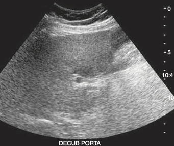
An asymptomatic obese adult patient presents
with elevated aspartate aminotransferase (AST) and alanine aminotransferase (ALT) levels discovered during a life insurance physical. The hypoechoic area documented in the sonogram most likely represents
normal liver tissue
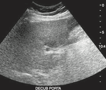
The hepatic pathology demonstrated in this sonogram most likely represents:
fatty infiltration
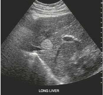
A postmenopausal patient presents with a history of right upper quadrant pain and normal liver function tests. She denies hormone replacement therapy or previous abdominal surgery. A solitary mass is identified in the right lobe of the liver. This mass most likely represents a(n):
cavernous hemangioma
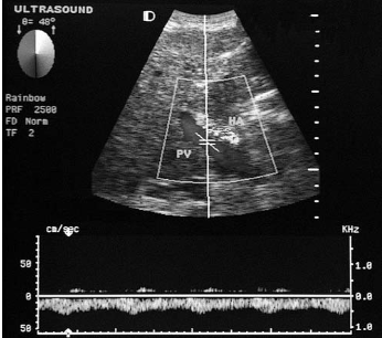
A patient presents with a history of hepatitis C and abdominal distention. The finding in this duplex image of the porta hepatis is most com- monly associated with which of the following conditions?
portal hypertension
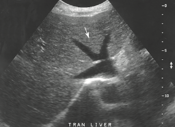
The arrow identifies which of the following he- patic lobes?
medial left lobe
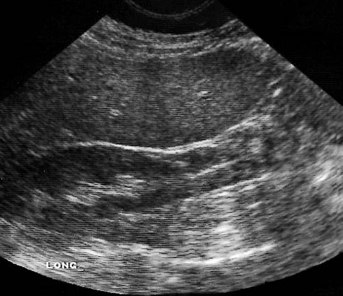
A female patient presents with a history of post- prandial pain. The left lobe of the liver does not extend across the midline. This image of the right lobe is most likely demonstrating which of the following conditions?
Riedel lobe
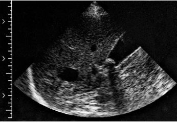
An asymptomatic patient presents to the ultrasound department for evaluation of a solitary hepatic mass documented on a recent computed tomography (CT) examination. A nonvascular anechoic structure is identified in the area in question. Based on this clinical history and sonogram, the mass identified is most consistent with a(n):
simple cyst
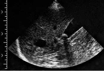
Which of the following pathologies is also dem- onstrated in the sonogram?
cholelithiasis
Which of the following liver pathologies is associ- ated with immune-suppressed patients?
candidiasis
Metastatic lesions involving the liver most com- monly originate from a primary malignancy of the:
colon
Which of the following ligaments serves as a barrier between the subphrenic space and Morison pouch?
coronary
An abnormally enlarged or dilated vein is most commonly termed a(n)
varix
Traditional lobar anatomy divides the liver into:
four lobes
Severe insult to the liver cells leading to subse- quent necrosis describes:
cirrhosis
Von Gierke disease is most commonly associated with:
glycogen storage disease
Prominence of the portal veins is most commonly associated with which of the following pathologies?
hepatitis
A transjugular intrahepatic portosystemic shunt (TIPS) is commonly placed between the
right hepatic vein and the right portal vein
paraumbilical vein courses from the umbilicus to the:
left portal vein
Which of the following conditions describes a congenital extension of the liver anterior and inferior to the right kidney
Riedel lobe
Which of the following spaces is located superior to the liver and inferior to the diaphragm?
subphrenic space
Enlargement of the caudate lobe is most commonly associated with which of the following pathologies?
cirrhosis
On spectral Doppler, the hepatic veins are characterized by which of the following flow types?
multiphasic
Which of the following ligaments attaches the liver to the anterior abdominal wall?
falciform
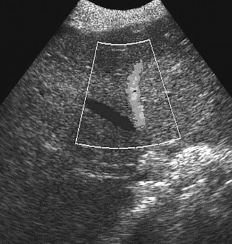
A transverse duplex image of the liver displays:
normal and abnormal hepatic venous flow
Which of the following most accurately describes the location of the caudate lobe?
posterior to the porta hepatis
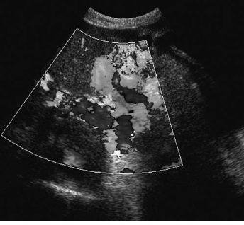
A patient presents with a history of cirrhosis. Based on this clinical history, the duplex image of the left upper quadrant is most suspicious for:
a. flash artifact
gastric varices
Decreases in prothrombin time are associated with which of the following?
acute cholecystitis
The most common symptom associated with acute thrombosis of the portal veins is:
severe abdominal pain
The diameter of a transjugular portosystemic shunt (TIPS) should measure a minimum of:
8 mm
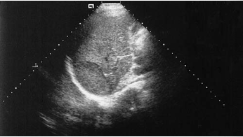
A 30-year-old female patient presents to the ultrasound department with postprandial pain. Gallstones are identified along with a mass in the right lobe of the liver. The patient has been taking oral contraceptives for 10 years. Based on this clinical history and sonogram, the mass is most suspicious for:
an adenoma
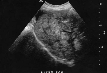
A 70-year-old patient presents with elevated alkaline phosphatase, right upper quadrant pain, and rectal bleeding. A sagittal sonogram of the right upper quadrant is documented. Based on this clinical history, the sonographic findings
are most consistent with which of the following pathologies?
liver metastasis
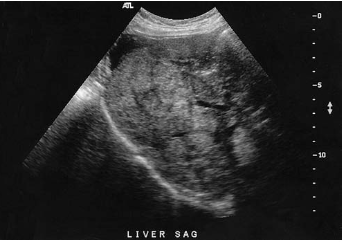
The fluid collection identified in this image is located in which of the following spaces?
right subphrenic space
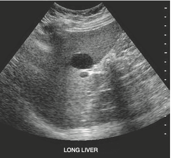
A patient who is complaining of postprandial pain presents for an abdominal ultrasound. Laboratory values are within normal limits. Based on this clinical history and sonogram, the mass is most suspicious for a (an):
simple hepatic cyst
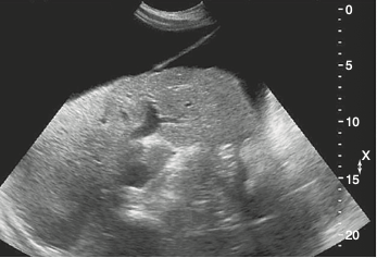
Which of the following ligaments is demonstrated in this sagittal image of the liver?
falciform
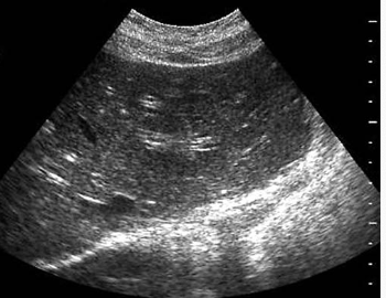
A 40-year-old patient arrives for an abdominal ultrasound to rule out gallstones. The patient complains of fatigue, fever, chills, and a loss of appetite for the previous week. Laboratory tests show marked elevation in AST, ALT, and bilirubin levels. Based on this clinical history, the sonogram is most suspicious for which of the following pathologies?
acute hepatitis