ANAT 5010 - unit 11
1/65
There's no tags or description
Looks like no tags are added yet.
Name | Mastery | Learn | Test | Matching | Spaced | Call with Kai | Chat |
|---|
No analytics yet
Send a link to your students to track their progress
66 Terms
- superciliary arches
- lower border of the mandible
- ear to ear
what are the boundaries of the face?
- superciliary arches
- EOP and super nuchal lines
- zygomatic arches
what are the boundaries of the scalp?
glabella
smooth, hairless region between eyebrows
superciliary arches
"eyebrows"; bony ridge above the supraorbital margin
supraorbital margin and notch/foramen
sharp edge of the upper rim of the orbit; notch allows the nerves, arteries, and veins to pass to and from the orbit and face
medial and lateral canthi
corners of the eyes/palpebral fissure
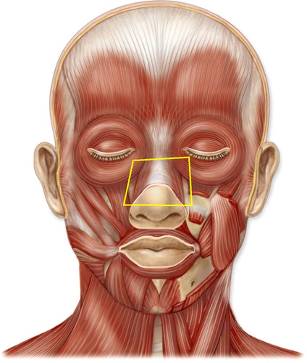
nasal bones
forms bridge of the nose on the face
dorsum of the nose
long, sloping superior surface of the nose
apex of the nose
anterior end/tip of the dorsum of the nose
external nares
nostrils
septal and major alar cartilages
cartilage support for dorsum and apex of nose
infraorbital foramen
inferior to lower orbital margin; contains nerves, arteries, and veins of the same name
vermillion border
where skin of the face meets skin of the lips
labial commissure/angle
corners of the mouth
mental protuberance
midline anterior-projecting bump on the external mandible
mental foramen
on the anterior surface of the body of the mandible; contains nerve, artery, and vein of the same name
philtrum
vertical indentation between nose and mouth
- skin
- connective tissue
- aponeurosis (musculotendinous)
- loose areolar tissue
- pericranium
what are the layers of the scalp?
- occipital artery
- posterior auricular artery
- superficial temporal artery
what arteries to the scalp come from the external carotid artery?
ophthalmic artery in the orbit branches into the...
- supratrochlear artery
- supraorbital artery
what arteries to the scalp come from the internal carotid artery?
CN V (trigeminal nerve)
what provides sensory innervation to the scalp in front of the ears?
- VPR of C2 and C3 from cervical plexus
- DPR of C2 and C3 directly
what provides sensory innervation to the scalp behind the ears?
supraorbital nerves
CN V1 location on face
zygomaticotemporal nerves
CN V2 location on face
auriculotemporal nerves
CN V3 location on face
- ophthalmic (CN V1)
- maxillary (CN V2)
- mandibular (CN V3)
what are the three parts of the trigeminal nerve and what numbers correlate?
supraorbital foramen
where do the CN V1 fibers exit the skull?
infraorbital foramen
where do the CN V2 fibers exit the skull?
mental foramen
where do the CN V3 fibers exit the skull?
ophthalmic nerve (CN V1)
exits cranial cavity through the superior orbital fissure; moves through the orbit to reach the upper face; has five terminal branches
- external nasal
- supratrochlear
- infratrochlear
- supraorbital
- lacrimal
what are the terminal branches of CN V1 on the face?
maxillary nerve (CN V2)
exits cranial cavity via the foramen rotundum; moves through the pterygopalatine fossa; has three terminal branches
- zygomatic-facial nerve
- zygomatic-temporal nerve
- infraorbital nerve
what are the terminal branches of CN V2 on the face?
mandibular nerve (CN V3)
exits cranial cavity via foramen ovale; courses through infratemporal fossa; has three terminal branches
- auriculotemporal nerve
- buccal nerve
- mental nerve
what are the terminal branches of CN V3 on the face?
corneal reflex
- protects and moistens the eye
- afferent limb - nasociliary nerve from CN V1
- efferent limb - blinks to close eye - facial nerve (CN V||)
how does the corneal reflex work?
ciliary ganglion
inside the orbit along the lateral side of CN 2; receives CN 3 and some branches of CN V1; target is pupillary constrictor muscle
pterygopalatine ganglion
housed in the pterygopalatine fossa in the deep midface; receives CN V||'s greater petrosal nerve; ANS fibers travel with CN V2 branches; target is the glandular mucosa of the nasal and oral cavities
otic ganglion
supported by CN V3 just inferior to foramen ovale; receives CN IX's lesser petrosal nerve; ANS fibers travel with CN V3's auriculotemporal nerve; target is parotid gland
submandibular ganglion
supported by CN V3's lingual nerve near back of mouth; receives CN V||'s chorda tympani nerve; target is submandibular and sublingual glands
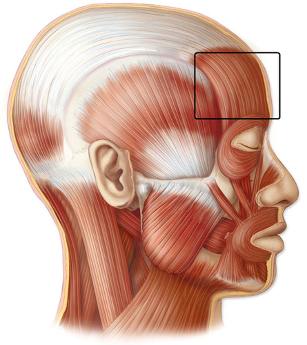
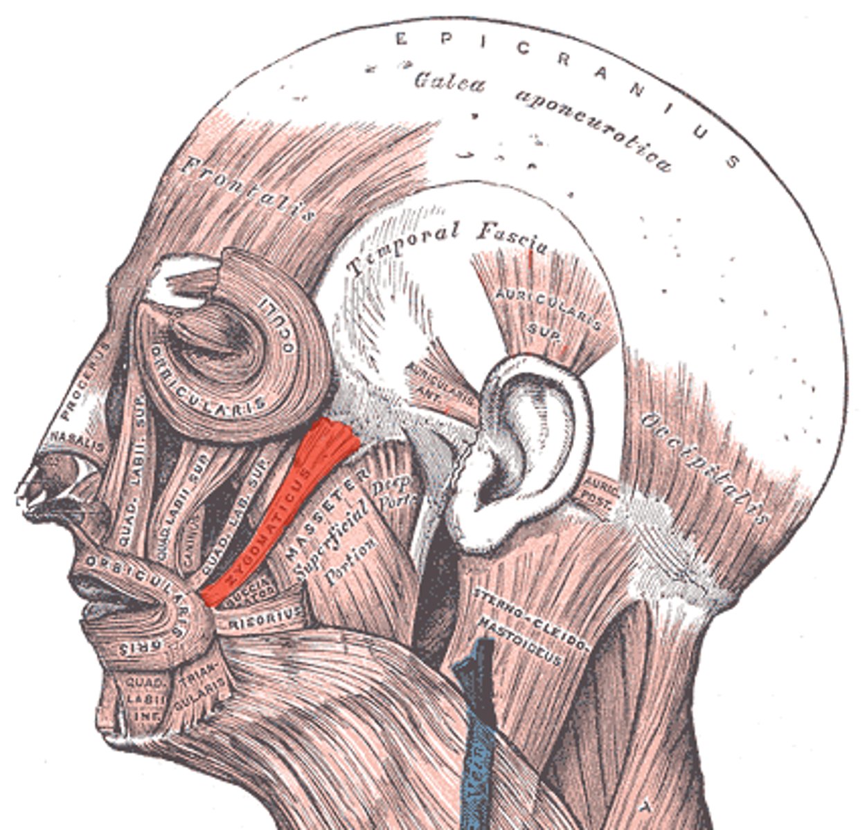
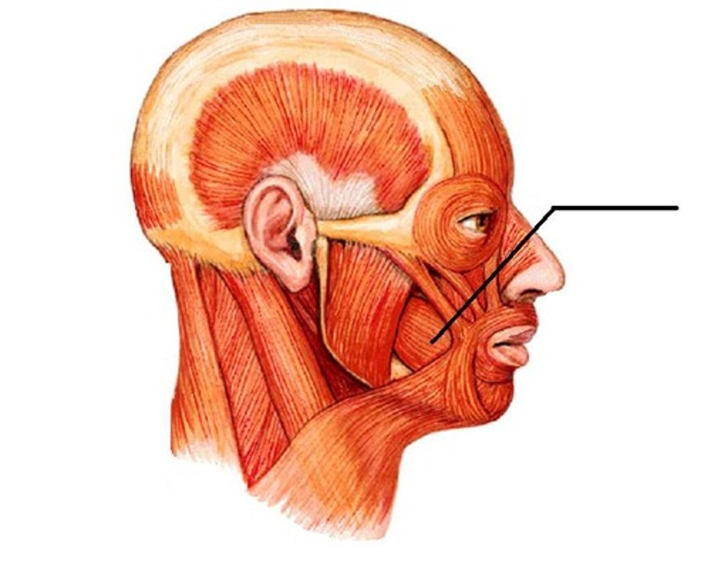
insertion: skin near eyebrows; aponeurosis of scalp
action: elevates eyebrows, wrinkles forehead
what is the insertion and action of the occipitofrontalis?
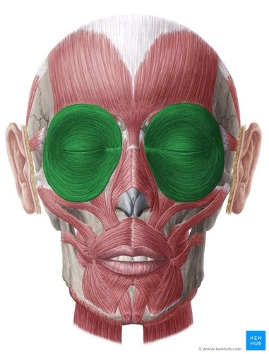
insertion: skin around orbit margins, tarsal plates
action: closes eyelids
what is the insertion and action of the orbicularis oculi?
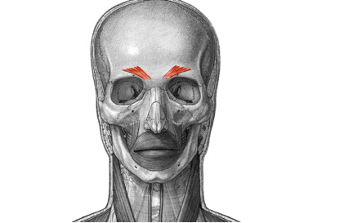
insertion: skin between supraorbital margins
action: draws eyebrow inferomedial, creates vertical wrinkles above nose
what is the insertion and action of the corrugator supercilli?
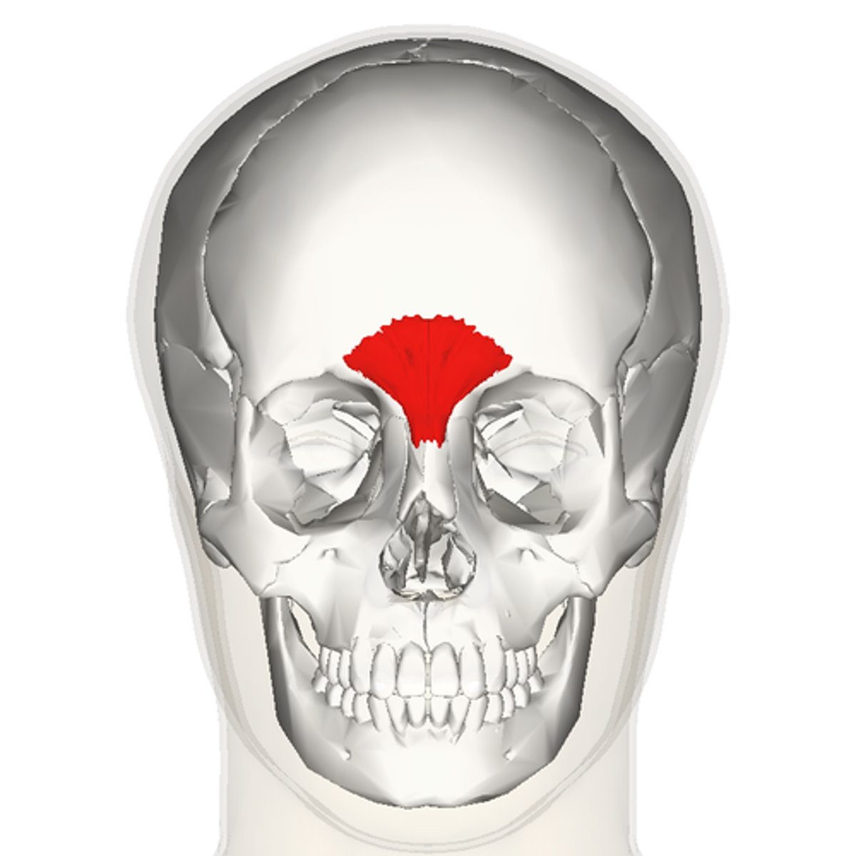
insertion: skin of lower forehead between eyebrows
action: depresses medial end of eyebrow, wrinkles skin over dorsum of nose
what is the insertion and action of the procerus?
insertion: major alar cartilage of the nose
action: depresses ala laterally, dilating anterior nasal aperture
what is the insertion and action of the nasalis?
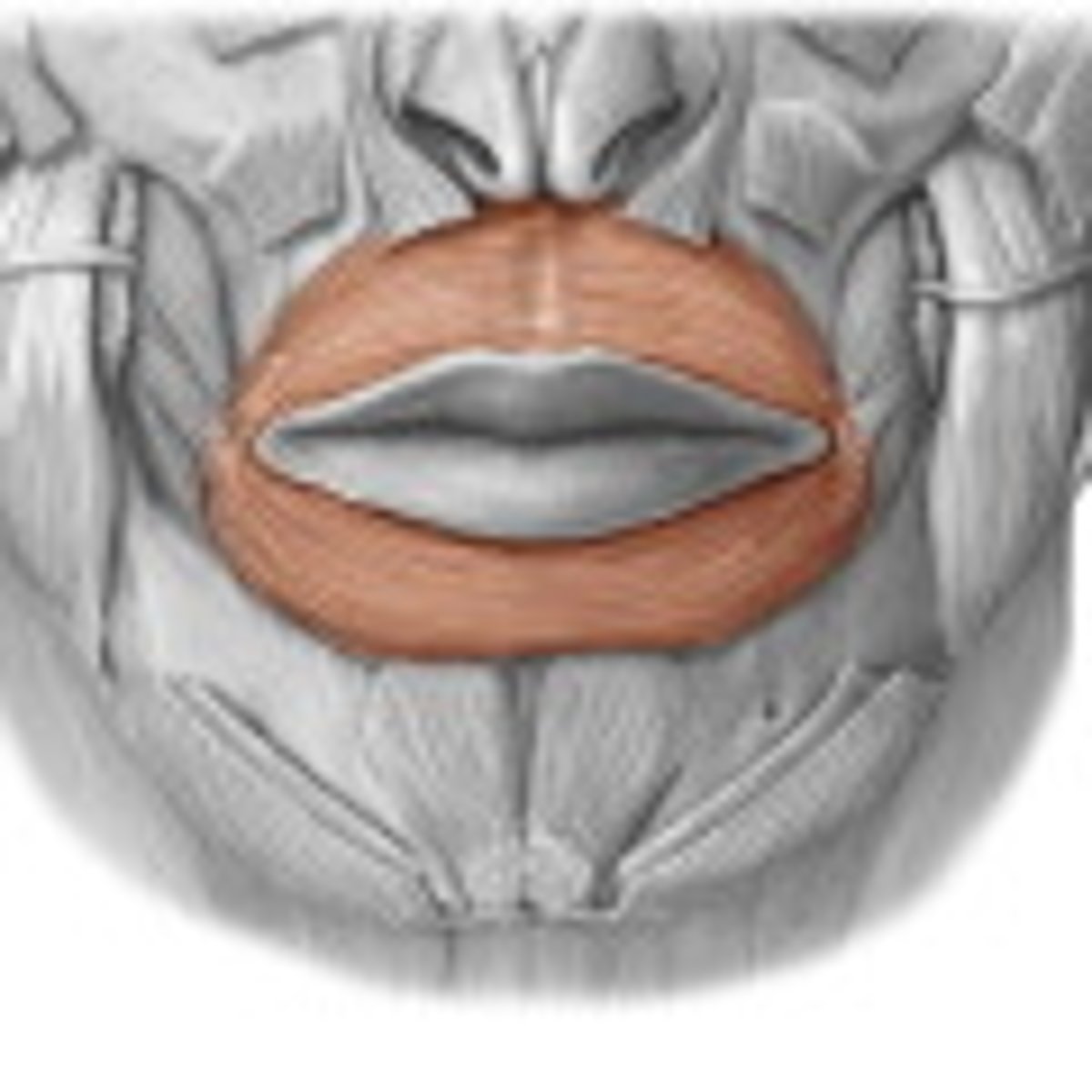
insertion: mucous membrane of both lips
action: closes oral fissure; compresses and protrudes lips; resists distention of cheeks
what is the insertion and action of the orbicularis oris?
insertion: skin of upper lip
action: elevate upper lip; deepen nasolabial sulcus; alaque nasia portion aids in dilating nostrils
what is the insertion and action of the levator labii superioris (and alaque nasi part)?
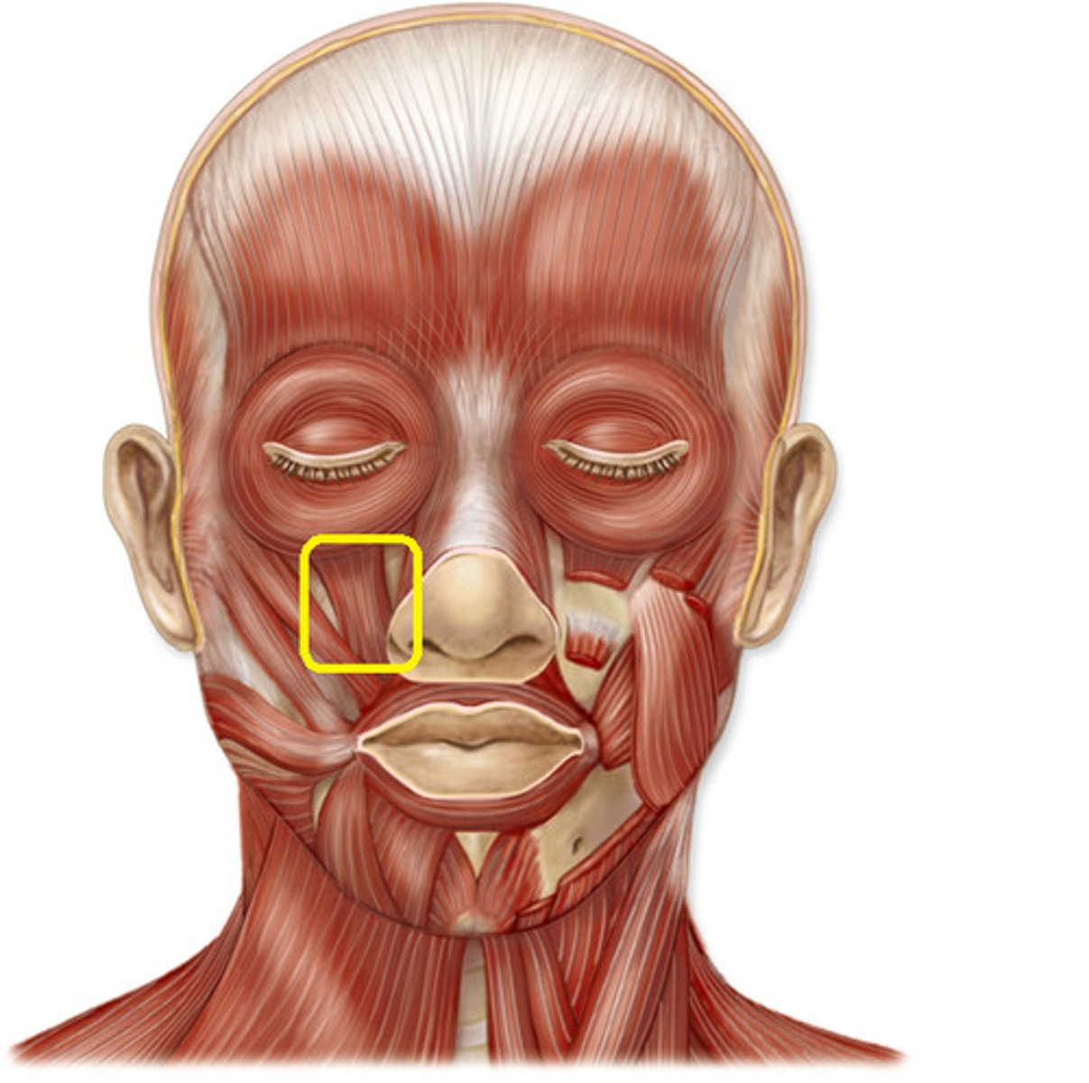
insertion: skin of upper lip
action: elevate upper lip; deepen nasolabial sulcus; alaque nasia portion aids in dilating nostrils
what is the insertion and action of the zygomaticus minor?
insertion: angle of the mouth
action: elevate/retract upper lip
what is the insertion and action of the zygomaticus major?
insertion: angle of the mouth
action: presses cheek against teeth; aids in keeping food between teeth; resists distention
what is the insertion and action of the buccinator?
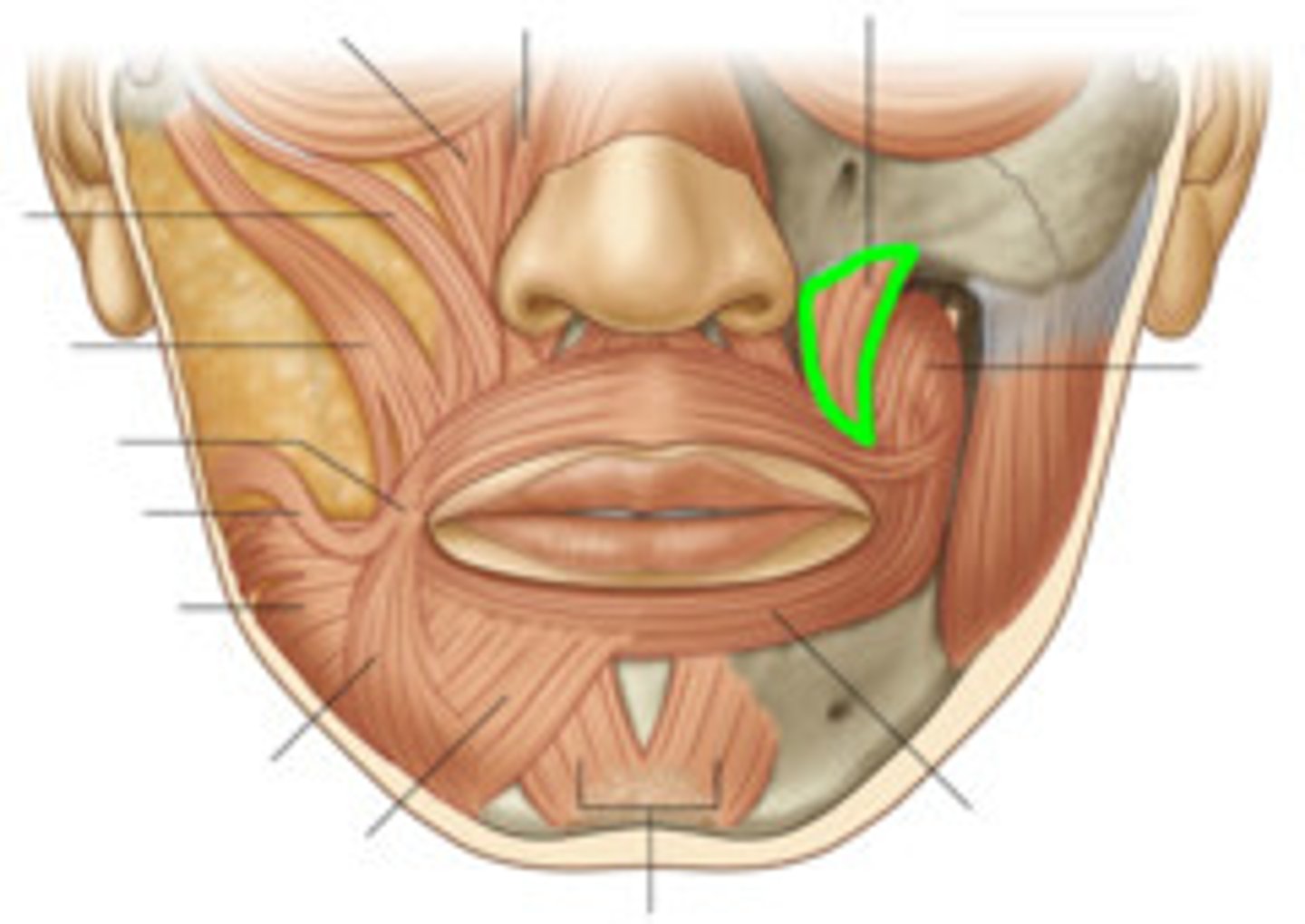
insertion: angle of the mouth
action: elevates corner of the mouth; widens oral fissure
what is the insertion and action of the levator anguli oris?
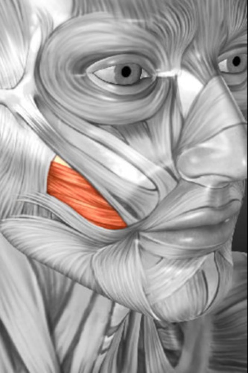
insertion: angle of the mouth
action: depresses/retracts angle of mouth
what is the insertion and action of the risorius?
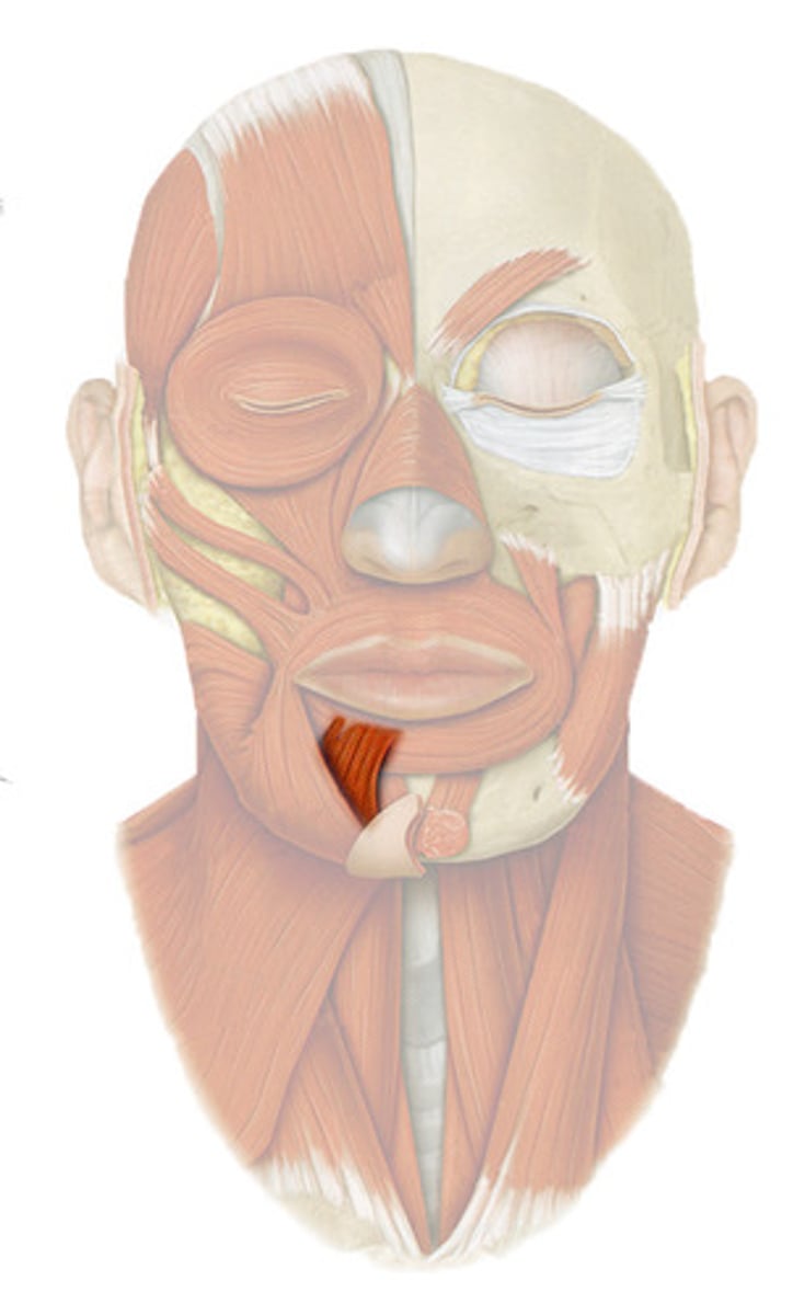
insertion: angle of the mouth
action: depresses lower lip
what is the insertion and action of the depressor anguli oris?
insertion: skin of lower lip
action: depresses/everts lower lip
what is the insertion and action of the depressor labii inferioris?
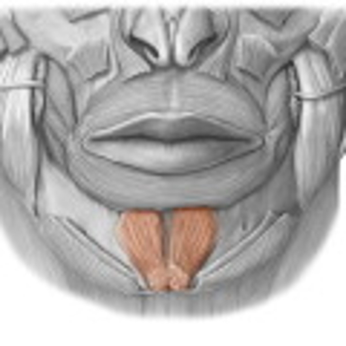
insertion: skin of chin
action: elevates/protrudes lower lip; wrinkles skin of chin
what is the insertion and action of the mentalis?
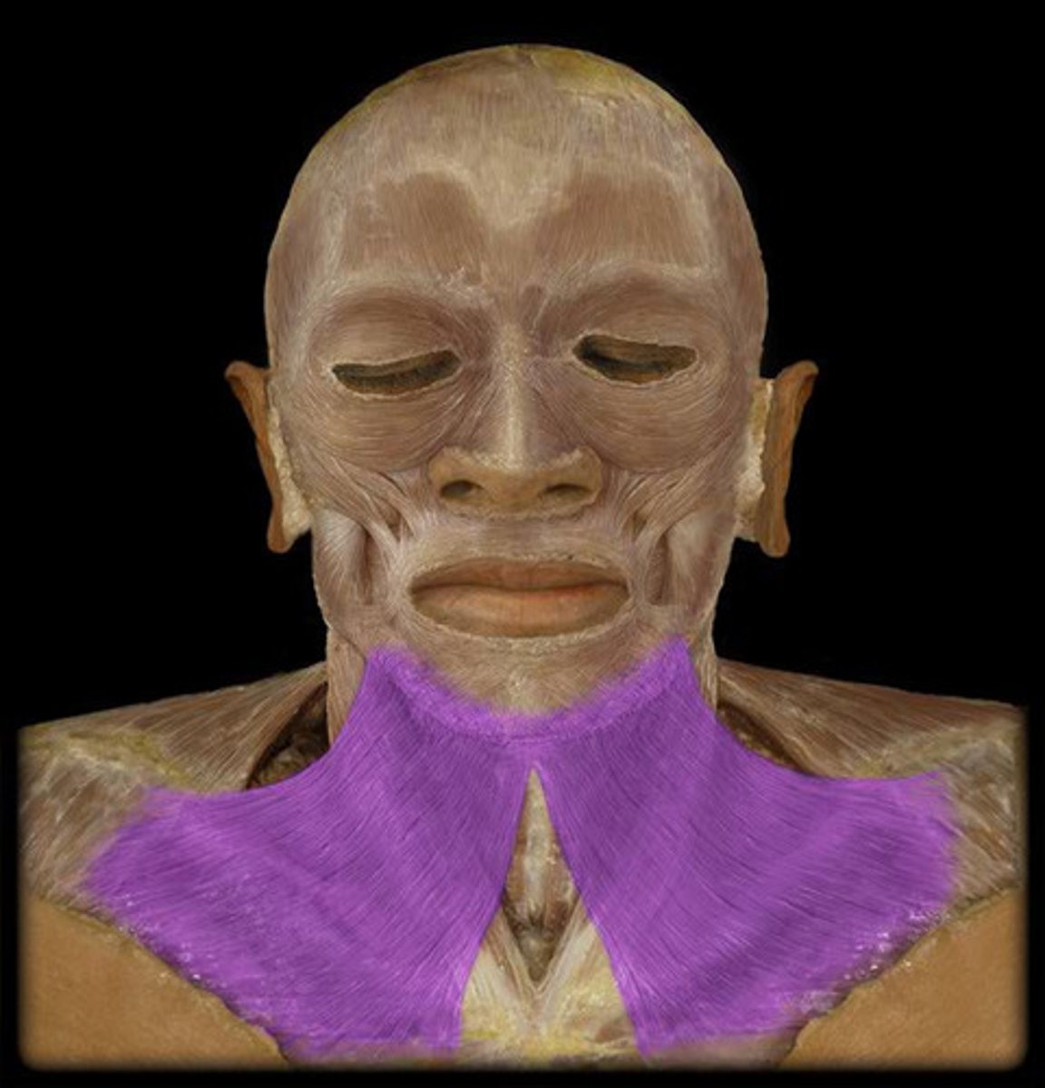
insertion: inferior border of mandible; skin of the lower lip and cheeks
action: tenses skin of inferior face and neck
what is the insertion and action of the platysma?
internal auditory meatus; motor fibers exit through the stylomastoid foramen
where does CN V|| exit the cranial cavity? where do the motor fibers exit?
- temporal branch
- zygomatic branch
- buccal branches
- marginal mandibular branch
- cervical branch
what are the five terminal branches of the facial nerve?
- inferior labial artery
- superior labial artery
- lateral nasal artery
- angular artery
what are the branches of the facial artery on the superficial face?
- starts at medial canthus
- angular veins forms at medial canthi
- runs posterior to the facial artery branches and receives the external nasal vein, superior labial vein, deep facial vein, and inferior labial vein to create the facial vein
- facial vein then crosses the mandible and forms the common facial vein by the union of the facial vein, submental vein, lingual vein, and anterior division of retromandibular vein
- then empties into the IJV
explain the route of venous return from the face
parotid fascia
thick, strong extension of the investing layer of DCF
buccopharyngeal fascia
covers buccinator muscle and extends posteriorly to cover the pharyngeal constrictor muscles
temporal fascia
strong fascia over the temporal fossa and muscle, attached to the superior temporal line above and to zygomatic arch below
parotid gland (and duct)
largest salivary gland; contained within the parotid sheath; emerges from the anterior border of the gland to cross the cheek superficial to the masseter; duct pierces the buccinator muscle to enter the mouth at the parotid papilla located near the second maxillary molars
- glossopharyngeal nerve contains parasympathetic motor fibers
- sympathetic fibers can reduce blood flow
- sensory innervation from CN V3 and C2-3 VPR
what innervates the parotid gland?