Speech Anatomy Module 1
 Call Kai
 Learn
Learn Practice Test
 Spaced Repetition
 Match
Match Flashcards
 Knowt Play
1/61

There's no tags or description
Looks like no tags are added yet.
Name | Mastery | Learn | Test | Matching | Spaced | Call with Kai | Chat |
|---|
No analytics yet
Send a link to your students to track their progress
62 Terms
Non-patholgoical structural anomalies
• Different from “normal” or “typical” structure
• Tons of variation across individuals
• Differences do not always indicate presence of a disorder
• Differences in structure do not always mean differences
Standard Anatomical position
Body is standing erect with the feet together
or slightly apart
• face is directed forward
• arms are at the side and rotated outward
• palms facing forward
• thumbs are pointed away from the body
Axial Skeleton
comprises the bones of the trunk and the head
• Consists of 80 bones: vertebral column, hyoid bone, skull bones, rib
cage, sternum, and the ossicles of the middle ear

Appendicular skeleton
comprises bones of the limbs, pelvic girdles, and pectoral girdles
• Consists of 126 bones of the arms, legs, hips, and
shoulders

Sagittal / Vertical
divides the body into right and left sides vertically.
• Median or midsagittal: if this plane courses down the middle of the body
• Longitudinal or parasagittal: if it divides the body into unequal
right and left sides
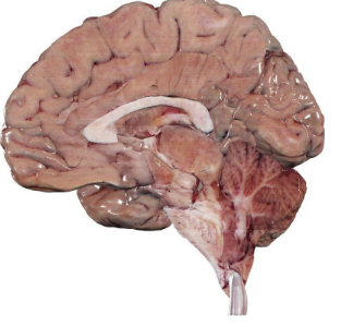
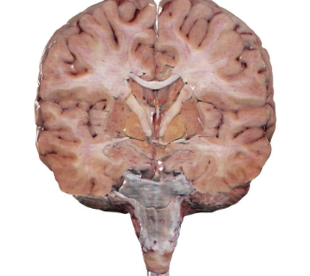
Frontal/coronal
divides the body intoventral/anterior and dorsal/posterior
• longitudinal plane perpendicular to thetransverse plane
Transverse /
horizontal
divides the body into upper and lower parts
• Perpendicular to the sagittal and coronal planes
• Often used to describe the location of parts of the body
in relation to one another

Superior or Rostral
Upper – toward head
Inferior or Caudal
lower— towards tail
Anterior or ventral
towards front—belly
posterior or dorsal
towards back
Supine
Lying flat on back
• Arms at sides
• Typically palms down
prone
Lying flat on stomach
• Arms at sides
• Typically palms up
DEEPER / TOWARD MIDLINE
Medial
• Internal
• Deep
• Central
• Endo
• Intrinsic
CLOSER TO SURFACE / AWAY
FROM MIDLINE
Lateral
• External
• Superficial
• Peripheral
• Ecto
• Extrinsic
proximal
Toward body or
root of extremity
Distal
Away from body or
root of extremity
bilateral
paired structures having a right
and left members
Unilateral
structure occurs only on one
side of the body.
Ipsilateral
refers to something occurring
on the same side of the body in relation to
another structure
Contralateral
refers to something
occurring on the opposite side of the body
in relation to something else
The respiratory system
responsible for breath support during speech
Organs: Pharynx, nasal cavity, larynx, trachea, bronchi,
lungs, and diaphragm
phonatory system
is responsible for voicing during
speech
articulatory/resonance
system changes the voice and
converts it to words that we
identify as speech
Skeletal system
provides structural
support and protection via bones,
ligaments, & cartilage
muscular system
is involved with
movement via the muscles, tendons,
and fascia that cover them
Circulatory or Cardiovascular system:
pumps blood and lymph through the body
• Moves oxygen and nutrients to cells and
removes carbon dioxide from the body
endocrine system
communicates by using hormones produced by the
thyroid, parathyroid, adrenal glands,
and endocrine glands
nervous
obtains, transfers, and processes information
Connective: cartilage
Smooth elastic tissue
• Function is to protect the bones at the joints
• It also forms the structure of the ear, nose, rib
cage, and bronchial tubes
• Can be hyaline (gas-like), elastic (easily bent), or
fibrous
connective tissue: bone
Part of thevertebrate skeleton
The hardeststructure of the body
Enables the body to have structure and provide the ability for the body to move
Covered by a fibrous membrane called periosteum
Deep layers of periosteum comprise osteoblasts that help with forming new bones when there are fracture
muscular tissue types
Skeletal, Smooth, Cardia
Muscles that permit movements are called
prime movers (agonists) and muscles that
provide stability are called fixators
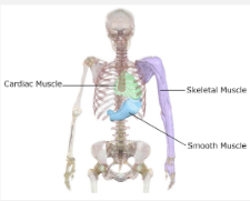
M U S C U L A R T I S S U E : S K E L E TA L
( S T R I AT E D ) M U S C L E S
Contract voluntarily because of messages that are received from the CNS
Connected to the bones by tendons
Groups of muscle fibers are called fasciculi (singular: fasciculus)
Each muscle, in turn, is covered with fascia, which is dense connective tissue
Type I (slow twitch) muscle fibers are used for endurance activities
Type II (fast twitch) muscles are used for sprint and intermediate activities
neurons
send and receive information
Glial cells
transmit nerve impulses, provide nutrients, support the
neuron, electrically insulate neurons, and remove dead neuron
ogliodendrocytes
(in the CNS)
create myelin (layers of a fatty substance that speeds signal transmissio
Schwann Cell
PNS
create myelin (layers of a fatty substance that speeds signal transmissio
gray matter
is found in the
brain, cerebellum, brainstem,
and spinal cord and is made of
neuron cell bodies
white matter
is made up of
myelinated axons that are
called tracts (CNS) or nerves
(PNS)
PNS
Cranial nerves – 12 pairs, arise from
the brain and brainstem
Spinal nerves – 31 pairs emerge from
the spinal cord
CNS
brain
spinal cord
Flexion:
Extension:
Abduction:
Adduction:
Protraction:
Retraction:
Flexion: Decreasing the angle between two bones (e.g., bending your elbow).
Extension: Increasing the angle between two bones (e.g., straightening your elbow).
Abduction: Moving a limb away from the midline of your body (e.g., raising your arm out to the side).
Adduction: Moving a limb toward the midline of your body (e.g., lowering your arm back down).
Protraction: Moving a body part forward or anteriorly (e.g., pushing your jaw or shoulders forward).
Retraction: Moving a body part backward or posteriorly (e.g., pulling your shoulder blades together).
Cranial nerves
send and receive messages from the head and neck
12 pairs of cranial nerves (meaning that there is
one for the right side of the body and an analogous one for
the left side of the body)
spinal nerves
send and receive messages from the rest of the body
how are cranial nerves ordered?
Numbered by Roman numerals in the
order they synapse or connect to the
CNS superiorly to inferiorly; order they
exit the brainstem
OLFACTORY
(AFFERENT)
I
• Sense of smell in the mucous
membranes of the nasal cavity
• Has a close relationship to
taste, eating, and swallowing
• Damage can affect a person’s
perception of taste

OPTIC
II
Relays information about vision from
retinas toward brain
• The optic nerve leaves the orbit (eye socket) and proceeds medially and posteriorly to the optic chiasm and then the occipital lobe
• All visual information on the left side of each eye is sent to be processed in the right occipital lobe and vice versa
• Damage to the optic nerve or the optic tract negatively affects vision
• The visual fields that are lost depend on the location of the lesion along the optic tract or optic nerve

• Optic chiasm
the point at which the left and right optic nerves
come together
OCULOMOTOR
III
efferent
Controls the movements of the eyes
Controls how the eye reacts to light and adducting/abducting the eyelids
• general somatic efferent (GSE): innervates extrinsic ocular muscles
• Controls moving eyes up & out; down & out; or inward
• general visceral efferent (GVE): innervates intrinsic ocular muscles
• Controls focus and reflexes of pupils
TROCHLEAR
IV
(EFFERENT)
moves eyes down
lesion could affect persons ability to turn eyes down and out
TRIGEMINAL
V
both (afferent and efferent)
One of the most important cranial nerves for speech
• Has three branches: ophthalmic, maxillary, mandibular
• Sensory: controls sense of touch on the
face
• Motor: innervates most of the muscles for mastication
(chewing)
2/3 of tongue

Abducens
VI
efferent
innervates muscles that abduct the eye
Damage here could cause diplopia (double vision) because the eyes would rotate medially
facial
VII
both (efferent and afferent
• Important for speech
production
• Communicates with other
cranial nerves such as the
acoustic, trigeminal, vagus,
glossopharyngeal, and even
some cervical nerves
• Has four branches
• Sensory: transmits taste
from the anterior two-thirds
of the tongue to the CNS
• Motor: innervates muscles of
the face

VESTIBULOCOCHLEAR
VIII
afferent
Also known as the auditory
nerve
• Has two branches
• Cochlear branch: Transmits
sound from the cochlea to the
brainstem
• Vestibular branch: Transmits
information from the vestibular
system for spatial orientation
and balance
GLOSSOPHARYNGEAL
IX
(AFFERENT – EFFERENT
• Has multiple branches
• Some branches important
for speech
• Sensory: transmits taste,
temperature, touch & pain from
the posterior one-third of the
tongue and from a portion of the
soft palate
• Motor: Innervates the superior
muscles of the pharynx
(swallowing) and the parotid gland
(saliva production)
• Damage to this nerve would lead a
person to lose all sensation on
the posterior 1/3 of the tongu
Vagus
X
both (efferent and afferent)
Largest cranial nerve
• Most important cranial nerve for SLPs
• A long and complex nerve
• Has many branches, not all relevant to speech
• Sensory: info from the inside of the larynx
• Motor: innervates the
muscles of the soft palate,
pharynx, and larynx

Accessory nerve
XI
efferent
Spinal component responsible for innervating muscles of the shoulders and neck
Trapezius
Sternocleidomastoid
Cranial component works alongside the vagus (as an
accessory) and shares functions with the vagus
• Innervates muscles used for raising the velum
• Innervates the uvula CN XI’s cranial component is functionally distinct from the vagus but still poorly understood.
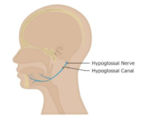
Hypoglossal
XII
efferent
Innervates all intrinsic and most extrinsic muscles of the tongue
• Intrinsic muscles (body of tongue) used for fine motor movements of articulation
• Extrinsic muscles used for gross motor movements for mastication and swallowing
• Innervates some muscles of the neck
Phrenic
bonus
spinal nerve
Responsible for
innervating many of the
muscles of respiration
• Damage to the phrenic
nerve can cause issues
with breath support for
speech