Elbow Complex Bony Injury & Dislocation
 Call Kai
 Learn
Learn Practice Test
 Spaced Repetition
 Match
Match Flashcards
 Knowt Play
1/43

There's no tags or description
Looks like no tags are added yet.
Name | Mastery | Learn | Test | Matching | Spaced | Call with Kai | Chat |
|---|
No analytics yet
Send a link to your students to track their progress
44 Terms
elbow dislocation has no
specific classification system:
Timing
articulations involved
direction
degree of placement
presence/absence of fx
Simple Elbow Dislocations involve surgical interventions such as Closed Reduction due to
Possible neural injury
Ligamentous injury likely
Non-op tx for Simple Elbow Dislocations
Period of immobilization in hinged brace
3-4 weeks
Complex Elbow Dislocations/Instability involves
fracture + extensive tissue damage
Radial Head/neck most common
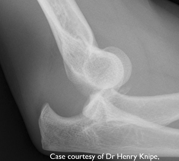
The Terrible Triad involves
Elbow dislocation (ulna moves posteriorly)
Radial head fracture
Coronoid Fracture
MOI for elbow terrible triad
FOOSH
Male > Female
Concurrent Dislocation
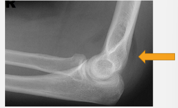
an elbow fracture results in a
Fat pad Sign
Inability to fully extend elbow ***
History of trauma
Elbow Extension Test to rule out
fracture; inability to straighten elbow after trauma
Olecranon fx are most common in elderly, displaced fx is most common type, complications include
Disruptions of triceps function
Displaced intra-articular joint fracture
surgical intervention for Olecranon Fracture
ORIF
Bone excision w/ triceps repair
complications post olecranon fracture
Loss of ROM (Extension)
Ulnar Neuropathy
Post-traumatic arthritis
Instability
Non-union, Complications w/ fixation device
Radial Head Fracture accounts for 1/3 of all elbow fractures. it occurs more often in
females than males (20-60 y/o)
Occur in 10% of all elbow dislocations
Radial Head Fracture MOI
Foosh
Direct Blow to Elbow
Hyperflexion Injury (crash into humerus)
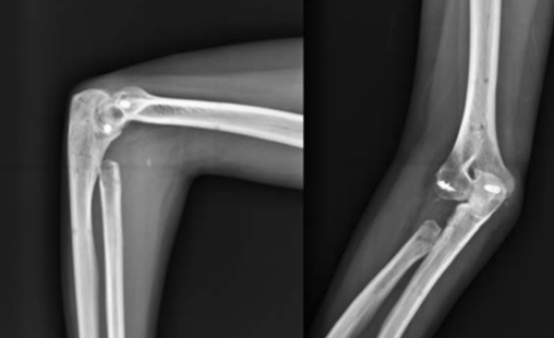
Indications for radial head excision includes substantial fracture of radial head but UCL Intact, why?
radial side gets compressed against capitulum, so need UCL on medial side to resist against valgus stress
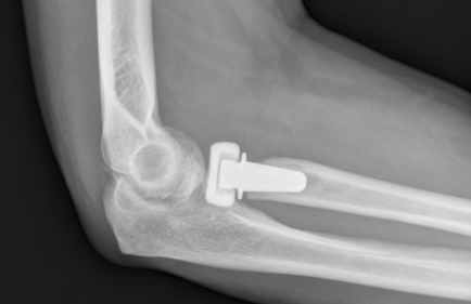
Indications for Radial Head Replacement
Severe fractures w/ UCL or RCL injury leading to instability
benefits of radial head replacement include a normalization of jt articulation, which restores
intrinsic stability & physiologic loading of distal ulna
↓ risk of radial head migration proximally (if not, under load radius would migrate toward ulna)
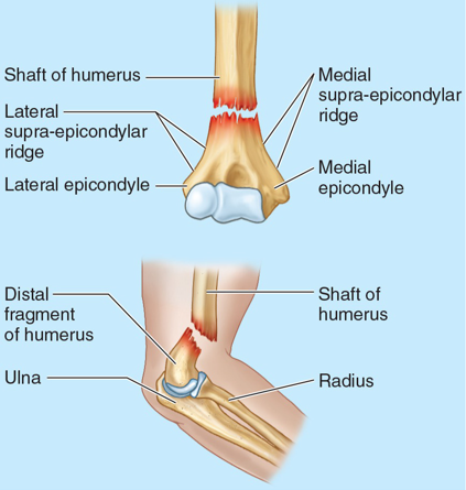
Supracondylar Fracture is common in children under the age of 10, Boys>girls; MOI
Fall on extended Elbow (growth plate)
Most common types of Supracondylar Fractures
Extension >95% vs Flexion
Olecranon acts as fulcrum
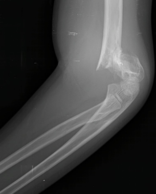
complications w/ Supracondylar Ridge Fracture include Non/Malunion → Cubitus Varus; additionally
Brachial Artery injury: Volkmann's Contracture (hand no longer functions), Compartment Syndrome
Neural Injury: Medial, Ulnar, or Radial Nerve Injuries
Complex Elbow Dislocation rehab w/ surgical fixation is immobilization for 4 weeks. Forearm position based on ligamentous instability:
Supinated to protect MCL, Pronated to protect LCL
(PT’s must try to avoid elbow stiffness)
Most Complex Elbow Dislocations will have some degree of limitation in ROM. Successful outcome after low-energy fracture shows
15-140° of ROM
More complex & articular involvement will have greater ROM limitations
Complex Elbow Dislocation rehab involves establishing controlled motion protocols early w/in
1st few days using gravity to assist
Strengthening exercises around 8-12 wks post-op
Complications of Elbow Fracture
Elbow Stiffness
Heterotopic Ossificans
Instability
Non-Union
Post traumatic arthrosis
intrinsic elbow stiffness is
tightness of capsule
extrinsic Elbow Stiffness is
tightness of muscles (elbow flexors, brachialis)
Elbow Stiffness is the most common complication of Surgical Intervention &/or Fracture. Factors impacting elbow stiffness are
thickening of anterior & posterior capsule which can lead to loss of Elbow extension & supination
elbow Stiffness Management is Prevention & Early Recognition in order to perform
early AROM/PROM
Elbow stiffness mgmt involves MT, modalities/meds for pain (HEAT) but mostly
Sustained stretching programs: Low low prolonged stretches (TERT)
high pain use AAROM
Surgical mgmt for Elbow stiffness is necessary when a contracture is present for
12 months
Lack functional ROM
Elbow OA is not generally characterized by loss of joint space (NWB) but will have appearance of
osteophytes & joint contracture usually related to past injury or trauma
Pain primary complaint: dull/achy/stiff
TEA dependent on functional expectation & other pathology (instability)
Elbow rheumatoid Arthritis involved in 50% of cases, common populations are
Females 35-45
Loss of extension ROM, jt instability
Conservative: Pain control & functional training/strengthening
Surgical: TEA
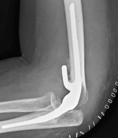
indications for Total Elbow Arthroplasty
Advanced age
Low physical demand
Chronic instability
Advanced RA, Posttraumatic OA
Ankylosis of elbow
Elbow stiffness, Functional ROM Loss
Pain
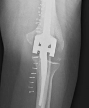
Total Elbow Arthroplasty (TEA) Constrained vs Semiconstrained outcomes
20 year survivorship: 61%
Complication rates: 20-40% (hardware loosening, infection)
Functional ranges 30-115° (Lower Functional demands)
Total Elbow Arthroplasty (TEA) rehab ensures what early?
ROM
Strengthening may be delayed depending on muscular resection for visualization
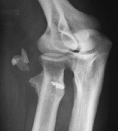
Heterotopic Ossificans (bone growth in muscle) is Ectopic bone formation in para-articular soft tissues, it has unknown etiology but high risk how long after trauma?
2 months after trauma (56% of elbow fracture/dislocation)
common location for Heterotopic Ossificans
Between brachialis & anterior capsule
Between triceps & posterior capsule
Heterotopic Ossificans is associated w/
aggressive passive mobilization of jt
Heterotopic Ossificans presentation includes pain w/ progressive
loss of ROM
Hyperemia→Swelling/Warmth
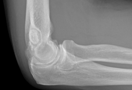
Osteochondritis Dissecans is a Lateral Compression injury in adolescents of articular surface of the capitulum due to
Repetitive activities, WBing or OH activities (adolescent baseball)
Osteochondritis Dissecans Presentation
Vague lateral elbow pain, ext ROM loss
Swelling, dull, tender to palpation at radiohumeral jt
Radiographs→radiolucency of capitulum
Panner’s Disease is loss of blood supply to growth plate of capitulum, leads to
flat capitulum
Panner’s Disease occurs in
Children 7-10 y/o
Non-traumatic: repetitive stress
Panner’s Disease signs & sxs
Dull lateral ache, joint swelling, 5-20° of ext lost
Panner’s Disease treatment
Rest, avoidance of valgus stresses
May take years to resolve
Self-limiting