Ch. 4 Tissue: Lecture 5-6
 Call Kai
 Learn
Learn Practice Test
 Spaced Repetition
 Match
Match Flashcards
 Knowt Play
1/98

There's no tags or description
Looks like no tags are added yet.
Name | Mastery | Learn | Test | Matching | Spaced | Call with Kai | Chat |
|---|
No analytics yet
Send a link to your students to track their progress
99 Terms
Epithelial tissue
Lines body surfaces, forms boundaries between body spaces, and forms glands
Connective tissue
Supports, protects, and connects other structures together
Muscle tissue
Responsible for movement in the body
Nervous tissue
Transmits electrochemical signals throughout the body to coordinate functions
Epithelium
Sheet or membrane of cells covering body surfaces and cavities or forming glands
Lining epithelium
Epithelium that covers body surfaces and cavities
Glandular epithelium
Epithelium that forms glands
Function of epithelium
Protection of deeper tissue, absorption and filtration, secretion (glands), sensory reception (nerve endings embedded within epithelia)
Polarity of epithelium
The organization of epithelial cells into distinct apical and basal sides
Apical side
Side of an epithelial cell facing the external environment or lumen
Lumen
Space within a tube duct or gland
Basal side
Side of an epithelial cell facing deeper layers of connective tissue
Cell-to-cell adhesions of epithelium
Adjacent cells are joined mainly by tight junctions and desosome
Basement membrane
Structure that separates epithelium from deeper connective tissue, made up of basal lamina and reticular lamina
Basal lamina
Very thin sheet-like part of the basement membrane
Reticular lamina
Web-like network of fibers in the basement membrane
Characteristics of epithelium
polarity, cell-to-cell adhesions, supported by basement membrane, avascular but highly innervated, regenerative (stimulated by loss of polarity of contact with adjacent cells
Microvilli
Finger-like extensions of the apical cell membrane that increase surface area for absorption, non motile
Cilia
Extensions of the apical cell membrane that use whip-like motion to propel material along the cell surface. Typically longer than microvilli, motile
Simple epithelium
Epithelium consisting of one cell layer
Stratified epithelium
Epithelium consisting of more than one cell layer
Squamous
Flat cell shape

Cuboidal
Square-shaped cell shape

Columnar
Tall rectangular cell shape

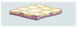
Simple squamous epithelium
Single layer of flat cells specialized for rapid diffusion. Found in lungs, blood vessels, kidneys
Simple cuboidal epithelium
Single layer of square or rounded cells involved mainly in secretion and absorption. Found mainly lining glands and ducts

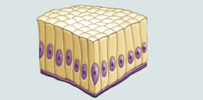
Simple columnar epithelium
Single layer of tall elongated cells (often with microvilli or cilia) involved in absorption, secretion, and mucus propulsion when ciliated. Found in digestive tract, smaller airways in lungs, uterus
Stratified squamous epithelium
Multiple cell layers with flat squamous cells at the apical surface
Non-keratinized stratified squamous epithelium
Stratified squamous epithelium forming a wet protective lining against abrasion. Apical: flat, squamous cells; basal: cuboidal or columnar in shape, actively undergoing mitosis. Found in oral cavity, esophagus, vagina, anal canal
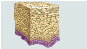
Keratinized stratified squamous epithelium
Stratified squamous epithelium with a superficial layer of dead keratinized cells forming a dry water-repellant barrier. No nuclei on apical surface, most superficial layer of skin
Stratified cuboidal epithelium
Usually only two cell layers, rare and usually found in ducts of glands such as sweat and salivary glands
Stratified columnar epithelium
Apical layer: columnar; basal layer: cuboidal. Rare epithelium found in areas including the conjunctiva of eye, glands ,and male urethra and vas deferens
Transitional epithelium
Made up of cuboidal and columnar cell shapes, found in urinary structures that stretch and relax (bladder). Apical layer of cells is bulging, club-shaped when unstretched, but flattened out when stretched.

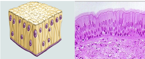
Pseudostratified columnar epithelium
Single layer of columnar cells of different heights that appears stratified because nuclei are at different levels. Nuclei at different levels, but all cells in contact with basal membrane. Function in absorption, secretion, propels mucus (if ciliated). Ciliated type found in airways, non-ciliated type found in epidydimis and vas deferens
Endocrine gland
Gland that releases substances directly into the bloodstream
Exocrine gland
Gland that releases substances into body cavities or onto the surface of the skin
Duct
Structure through which most exocrine glands release their products
Simple exocrine gland
Exocrine gland with an unbranched duct
Compound exocrine gland
Exocrine gland with a branched duct
Tubular gland
Exocrine gland with an elongated tubular secretory portion
Alveolar gland
Exocrine gland with a round grape-like secretory portion
Merocrine secretion
Secretion of products by exocytosis
Holocrine secretion
Secretion in which cells die and disintegrate to release their products
Apocrine secretion
Secretion in which the apical portion of the cell pinches off and the damaged portion regenerates
Goblet cell
Unicellular mucin-producing exocrine gland
Mucin
Substance produced by goblet cells that combines with water to form mucus
Mucus
Mucin combined with water
Connective tissue
Tissue derived from mesenchyme with cells embedded in extracellular matrix
Mesenchyme
Embryonic tissue composed of gel-like ground substance fibers and undifferentiated stem cells
Extracellular matrix
Material surrounding connective tissue cells consisting of ground substance and fibers
Ground substance
Gel-like material occupying the interstitial space of connective tissue
Collagen fibers
Fibers that provide tensile strength
Elastic fibers
Fibers that allow tissues to stretch and recoil
Reticular fibers
Fibers that form a structural network
Blast cells
Immature connective tissue cells that produce ground substance and fibers
Cyte cells
Mature connective tissue cells that maintain the extracellular matrix
Adipocyte
Cell specialized for storing fat
Fibroblast
Connective tissue cell that produces ground substance and fibers
Areolar connective tissue
Loose connective tissue that provides support and binds other tissues together
Lamina propria
Layer of loose areolar connective tissue just deep to epithelium
Adipose connective tissue
Loose connective tissue with closely packed lipid-filled adipocytes
Adipose tissue functions
Energy storage insulation heat generation and shock absorption
Reticular connective tissue
Connective tissue made primarily of reticular fibers that forms a structural network
Stroma
Web-like structural network that supports other cells
Dense regular connective tissue
Connective tissue with closely packed parallel collagen fibers providing high strength under tension
Dense irregular connective tissue
Connective tissue with collagen fibers arranged in multiple directions to resist tension in multiple directions
Elastic connective tissue
Connective tissue with densely packed elastic fibers that provides stretch and recoil
Cartilage
Firm but flexible connective tissue containing chondrocytes in lacunae
Chondrocyte
Mature cartilage cell located in a lacuna
Lacuna
Space containing a chondrocyte within cartilage
Hyaline cartilage
Most abundant cartilage type found at ends of bones nose and walls of airways
Elastic cartilage
Cartilage with a higher proportion of elastic fibers that allows stretching while maintaining original shape
Fibrocartilage
Cartilage with rows of chondrocytes between collagen fibers that withstands both tension and compression
Bone
Connective tissue with calcified matrix that supports and protects body tissues
Osteoblast
Cell that produces and calcifies bone matrix
Osteocyte
Cell that maintains bone matrix
Osteoclast
Cell that removes bone matrix
Osteon
Structural unit of bone
Blood
Connective tissue with plasma as ground substance and blood cells and platelets as cellular components
Plasma
Fluid ground substance of blood
Cutaneous membrane
Dry body membrane forming the outer surface of the skin
Epidermis
Epithelial layer of the cutaneous membrane consisting of keratinized stratified squamous epithelium
Dermis
Connective tissue layer beneath the epidermis
Mucous membrane
Wet membrane lining cavities that open to the external environment
Mesothelium
Simple squamous epithelium lining serous membranes
Serous membrane
Wet membrane lining closed ventral body cavities
Myofilaments
Protein filaments made of actin and myosin that produce muscle contraction
Actin
Contractile protein found in myofilaments
Myosin
Contractile protein found in myofilaments
Skeletal muscle
Voluntary striated muscle made of multinucleated cylindrical cells with peripheral nuclei
Smooth muscle
Involuntary non-striated muscle made of uninucleate spindle-shaped cells
Cardiac muscle
Involuntary striated muscle made of uninucleate branching cells found in the heart
Tissue repair
Process by which damaged tissue is repaired following injury
Inflammation
Initial response to tissue damage involving inflammatory signals and increased blood vessel permeability
Clot
Structure that initially seals a damaged site during tissue repair
Granulation tissue
Tissue containing an ingrowing capillary network that restores blood supply to a damaged area
Fibroblast activity during tissue repair
Fibroblasts secrete growth factors and collagen fibers near the damaged area
Fibrotic scar tissue
Scar tissue that remains deep to the epithelium depending on the extent of the wound