Mediastinum and Heart
 Call Kai
 Learn
Learn Practice Test
 Spaced Repetition
 Match
Match Flashcards
 Knowt Play
1/39

There's no tags or description
Looks like no tags are added yet.
Name | Mastery | Learn | Test | Matching | Spaced | Call with Kai | Chat |
|---|
No analytics yet
Send a link to your students to track their progress
40 Terms
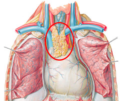
thymus
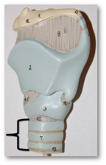
trachea
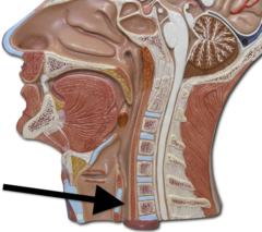
esophagus
mediastinum
area between the lungs containing the heart, aorta, venae cavae, esophagus, and trachea
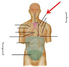
superior mediastinum
Upper compartment of the thoracic cavity between thoracic inlet and T4
inferior mediastinum
lower compartment of the thoracic cavity between T5 and T9
aortic arch
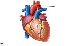
brachiocephalic trunk
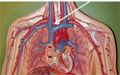
left common carotid artery
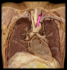
left subclavian artery
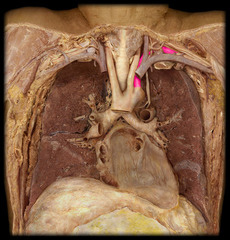
brachiocephalic veins
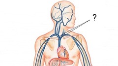
superior vena cava
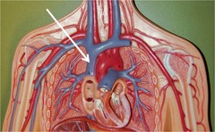
phrenic nerve
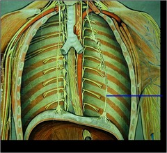
vagus nerve
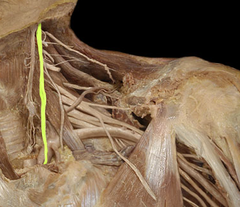
thoracic duct
major lymphatic vessel of the body
fibrous pericardium
outter pericardial layer; holds heart in the mediastinum and attached to to vessels off the heart and diaphragm
serous pericardium, parietal layer
outter layer of serous pericardium, attached to underside of fibrous pericardium
serous pericardium, visceral layer
inner layer of serous pericardium, attached to surface of heart
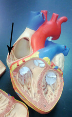
pectinate muscles
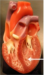
trabeculae carnae
pulmonary/semilunar valve
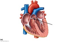
aortic/semilunar valve
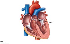
left atrioventricular/bicuspid/mitral valve
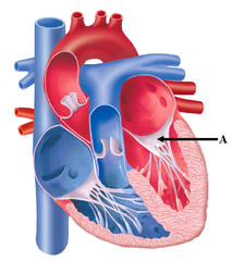
right atrioventricular/tricuspid valve
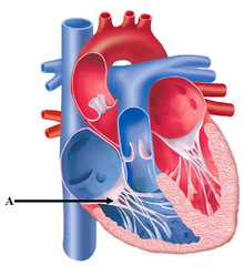
chordae tendineae
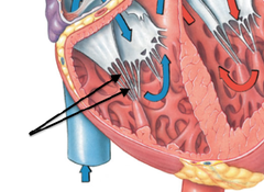
papillary muscles
only found on the AV valves; contact during heart contraction to AV valves from opening
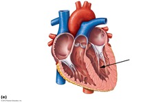
how semilunar valves close
back flow of blood down the pulmonary trunk and aorta
how AV valves open
blood flowing from the atriums into the ventricles
right coronary artery
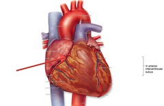
right marginal artery/vein
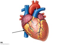
Circumflex branch of left coronary artery
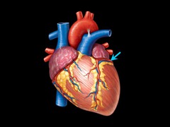
anterior interventricular branch
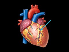
posterior interventricular branch
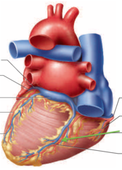
small cardiac vein
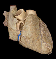
great cardiac vein
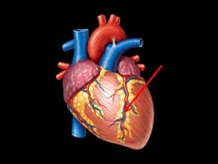
middle cardiac vein
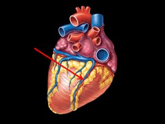
coronary sinus
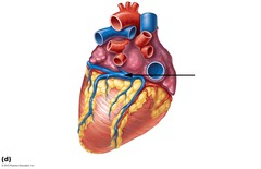
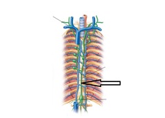
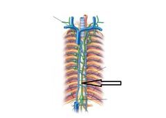
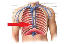
azygous vein
drains blood from thoracic region
sympathetic trunk
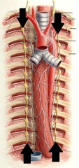
thoracic duct