Neck and Upper Limb Osteology
1/120
There's no tags or description
Looks like no tags are added yet.
Name | Mastery | Learn | Test | Matching | Spaced | Call with Kai |
|---|
No analytics yet
Send a link to your students to track their progress
121 Terms
cervical plexus
Formed by anterior rami of the first four cervical nerves
covered by the prevertebral layer of deep cervical fascia and related to internal jugular vein within the carotid sheath
Supplies skin and muscles of head, neck and shoulder • Cervical nerve • C1 - C4
segmental
prevertebral muscles, levator scapulae
ansa cervicalis c1,2,3
omohyoid, sternohyoid, sternothyroid
c1 fibers via hypoglossal nerve
thyrohyoid, genohyoid
phrenic nerve c3,4,5
nerve to diaphragm, important for contraction
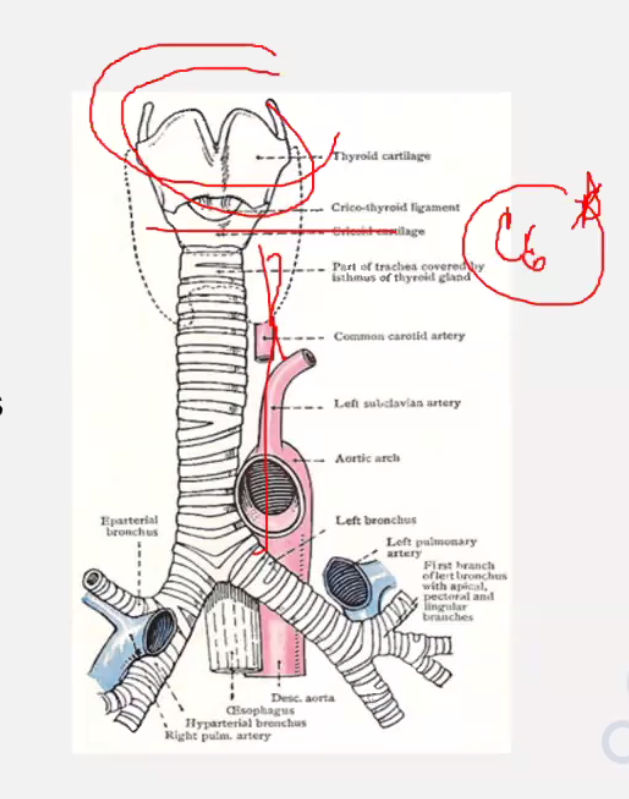
Thyroid Gland
Consists of right and left lobes and isthmus (level of 2 nd to 4th tracheal ring)
hyoid
c3
thyroid cartilage
c4-5
cricoid
c6
Pyramidal lobe
found in the isthmus more on left
Levator glandulae thyroideae
muscular band connecting the pyramidal lobe to the hyoid
anterolateral Thyroid Gland
Sternothyroid, superior belly of omohyoid, sternohyoid, anterior 2/3 of SCM
medial Thyroid Gland
Larynx, trachea, pharynx, esophagus
Posterolateral Thyroid Gland
Carotid sheath with common carotid artery, internal jugular vein and vagus n
Posterior Thyroid Gland
Superior and inferior parathyroid gland
Anastomosis of inferior and superior thyroid artery
thyroxine and triiodothyronime
increases metabolic activity of most cell in body
Thyrocalcitonin
produced by parafollicular cells that lower the level of blood calcium
Parathyroid
Small ovoid, parathyroid glands lie external to the fibrous thyroid capsul
superior parathyroid gland
lie middle of the posterior border of the thyroid gland and inferior parathyroid glands lie close to the inferior pole of thyroid gland
Trachea
Mobile cartilaginous and membranous tube
Starts as a continuation from larynx ( at level of cricoid, lower border of C 6) and ends at carina (division of the tranchea to principal bronchus) at the level of sternal angle of Louis (between 4th and 5th thoracic vertebrae)

anterior trachea
• Skin, fascia • jugular arch • Isthmus of thyroid (2nd to 4th tracheal rings) • thyroidea ima • Inferior thyroid vein • L bracheo cephalic v • Sternothyroid and sternohyid muscles
posterior trachea
• Esophaus • Right and left recurrent laryngeal nerves
lateral trachea
• Lobes of thyroid gland • Carotid sheath and contents
trachea
Made of U shaped cartilaginous bars of hyaline
Posterior is free
Trachealis muscle
connects free end
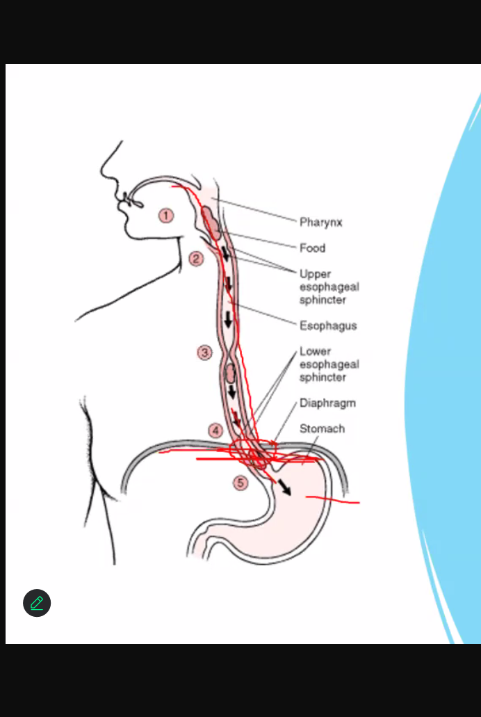
Esophagus
Tubular structure 10 inches long
From pharynx to stomach
Continuous above with the laryngeal part of the pharynx opposite 6th cervical vertebra and passes diaphragm at level of 10th thoracic vertebra to join stomach (c6 to t10)
anterior esophagus
• Trachea • Recurrent laryngeal nerve
posterior esophagus
• Prevertebral layer of the cervical fascia; vertebral column and longus colli
lateral esophagus
• Thyroid gland (lobe) and carotid sheath
parasympathetic and sympathetic efferent and afferent fibers, esophageal nerve plexus
nerve of esophagus
root of the neck
Area immediately above the inlet to the thorax
Contents: ■ Subclavian artery ■ Subclavian vein ■ Thoracic duc
Interscalene triangle
gap between scalenus anterior and medius muscle and first rib
Where subclavian artery and roots fo brachial plexus pass through
clavicle
Collar bone • Between sternum and scapula; base of neck • Connected medially to the sternum and laterally to the scapula • S shape
First bone to ossify

strut
__ connecting the upper limb to the thorax and together with scapula, it allows the limb to move freely from the trunk

functions of clavicle
Transmits forces from the upper extremity o Holds arm away from trunk o Provides attachment for muscles o Absorbs force from upper extremity

clavicle
Articulates with costal cartilage and sternum medially and acromion scapula laterally
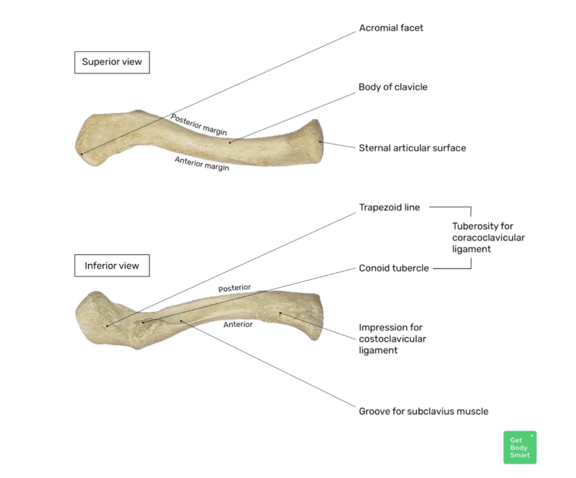
Medial (sternal extremity) end
Proximal end, blunt, thickened o Attaches to the sternum through Clavicular notch o Forms the Sternoclavicular joint o Convex

Lateral (acromial extremity) end
o Flat o Attached to acromion process of scapula o Concave

clavicle
Origin of deltoid, pectoralis major, SCM
Insertion of subclavius m., upper fibers of trapezius

clavicle
most commonly fractured bone in the body because it absorbs force from upper extremity

anterior clavicle
more smooth
posterior clavicle
rough because this is where ligaments attach
body
Conoid tubercle is found; this is for the attachment of the conoid ligament

Subclavian groove
Where the subclavian vessels will pass through

Costal tuberosity
Attaches to the first rib

scapula
Shoulder bone or shoulder blade • Flat, triangular in shape
• Between 2nd to 7th ribs (ex: 2nd rib attached to T2 and so on)

ventral or costal surface
Concave o Forms shallow subscapular fossa (entire thing) o Nothing divides it

spine
– divides posterior side to supraspinous and infraspinous fossa

Acromion
- free lateral end of spine; articulates with clavicle
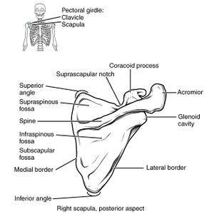
Coracoid process
- found above glenoid cavity, for attachment of muscles and ligaments

Suprascapular notch
medial to base of coracoid process

Glenoid cavity
superolateral angle of the scapula; pear shape, articulates with the humeral head (and its attachment site); shallow cavity

Supraglenoid tubercle
Long head of biceps

Infraglenoid tubercle
Long head of triceps

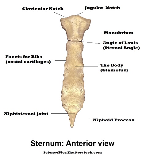
sternum
Flat bone also known as breastbone • Connected to shoulder girdle • Medial area where clavicle attaches

clavicular facets
connects the clavicle to sternum; where clavicle is attached

costal facets
where 1st rib is attached o Jugular notch/ Suprasternal notch

body
Where 2nd to 7th rib attaches to
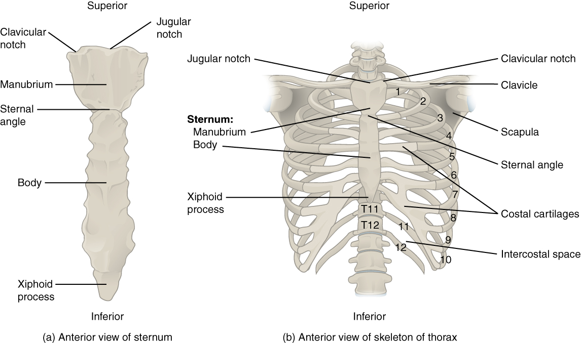
Xiphoid Process
8th to 10th will join to be the angle and will attach to the 7th rib

Sternal angle of Louis
Junction of body and manubrium o Cartilaginous joint o Between Intervertebral disc of T4-T5

Sternal angle of Louis
marks the superior level of the pericardium, the sac enclosing the heart, and the superior limit of the pulmonary trunk
Shows the level of the beginning and end of the arch of the aorta
is the level at which the trachea bifurcates into right and left main bronchi

Sternal angle of Louis
o marks the site of articulation of rib 2 with the sternum o Boundary between the superior and inferior portion of the mediastinum o Passage of the thoracic duct from right to left behind esophagus o End of the azygos system into SVC
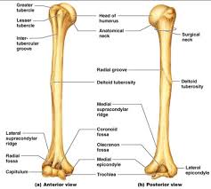
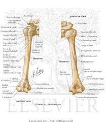
humerus
Articulates with the scapula (glenoid fossa) to form the shoulder joint and radius and ulna at the elbow joint • Forms glenohumerus joint/main shoulder joint • Longest bone in the UE

head
1/3 of a sphere, articulates with glenoid fossa (because glenoid is very shallow and why dislocated shoulders are common); contains the greater and lesser tuberosities which are divided by an intertubercular notch/groove
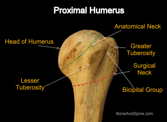
surgical neck
- area where fracture is most common

Deltoid tuberosity
- attachment/insertion of deltoid

spiral groove
- accommodates the radial nerve

Deltoid tuberosity
rough triangular elevation for attachment of deltoid

Radial groove or spiral groove
posterior shallow depression; between deltoid tuberosity and lateral supracondylar ridge o Radial nerve and profunda brachii vessels
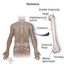
medial epicondyles
sometimes called the funny bone; more prominent than lateral

lateral epicondyle
where radial bone will be attached

Capitulum
round, articulates with radial head; caput
radial notch
above capitulum; where elbow bends, where head of radius goes

trochlea
pulley shape, articulates with trochlear notch of ulna

Coronoid fossa
above trochlea; for the articulation of the coronoid process

Olecranon fossa
receives olecranon process of ulna when elbow is extended
radius
• Lateral bone in the forearm • Articulates proximally with humerus (capitulum) and ulna at (to form proximal radius ulnar joint) • distally, articulates with the scaphoid and lunate bone (to form ellipsoid/wrist joint) and distal ulna (distal radius ulnar joint) • Becomes wider distally

head
small, circular ▪ Concave upper surface articulates with the capitulum of humerus ▪ Circumference articulates with notch of ulna

Bicipital tuberosity
- insertion of biceps

shaft
wider below than above ▪ Interosseous border ▪ Pronator tubercle (insertion of pronator teres muscle)

Interosseous border
where there is an interosseous membrane

Interosseous membrane
forms a fibrous joint connecting the radius to the ulna
fibrous joint
Middle radioulnar joint
pivot joint
Proximal and Distal radioulnar joint
Radial tuberosity
attachment of biceps
Styloid process
projects distally from the lateral margin

Ulnar notch
medial surface for attachment of ulna

Inferior surface
articulates with scaphoid and lunate
Dorsal tubercle (Lister’s tubercle)
- grooved on its medial side by the tendon of the extensor pollicis longus; tendons pass through here

ulna
Medial bone • Articulates with trochlea of humerus at the elbow joint and with head of radius at the proximal radioulnar joint • Becomes narrower distally • Distal end articulates with radius at distal radioulnar joint
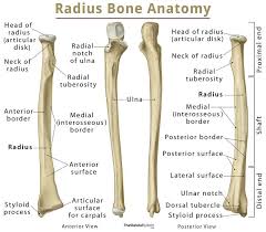
Olecranon
forming the prominence of the elbow posteriorly

Trochlear notch
articulates with the trochlea of the humerus

Coronoid process
triangular end below the trochlear notch

Radial notch
articulation of radial head

Supinator crest
– found below the radial notch

8
how many carpal bones
Cartilaginous
__ at birth (carpal bone)
Capitate
__ first to ossify
scaphoid
Most commonly fracture carpal bone is