Anemia Biochem-Folate/B12, Iron, HMP shunt
 Call Kai
 Learn
Learn Practice Test
 Spaced Repetition
 Match
Match Flashcards
 Knowt Play
1/55

There's no tags or description
Looks like no tags are added yet.
Name | Mastery | Learn | Test | Matching | Spaced | Call with Kai | Chat |
|---|
No analytics yet
Send a link to your students to track their progress
56 Terms
Nutritional anemias are classified to three groups:
Microcytic (small red blood cell = RBC) defined by MCV smaller than 80 fentliters
-def in iron (major), copper, pyridoxine
Normocytic (normal size RBC) defined by MCV from 80-100
-protein-calorie malnutrition (kwashiorkor)
Macrocytic (large RBC) defined by MCV more than 100
-def in vitamin B12, folate (more common)
Megaloblastic anemia
folate and B12 essential for nucleotide synthesis
deficiency->megaloblastic
makes cell larger (S) phase takes longer time, stem cells
oval and larger in mature RBC
Folate metabolism
Dihydrofolate (FH2) + DHFR (dihydrofolate reductase)-->Tetrahydrofolate (FH4) (one carbon unit) (active form)
FH4->nucleotide biosynth or AA metab or B12-CH3 Meth metab
folic acid->convert to active form in two step rxn above. requires 2 NADPH (from Niacin/B3) and (DHFR). Each step adds 2 hydrogens to folate
FH4 pathways
Nucleotide synthesis
-de novo purine syn
-thymidine syn
AA metab
-serine synthesis (Gly->Ser or N-meythl Gly)
Meth metab
-homocysteine (hc) -> methionine
Folate components
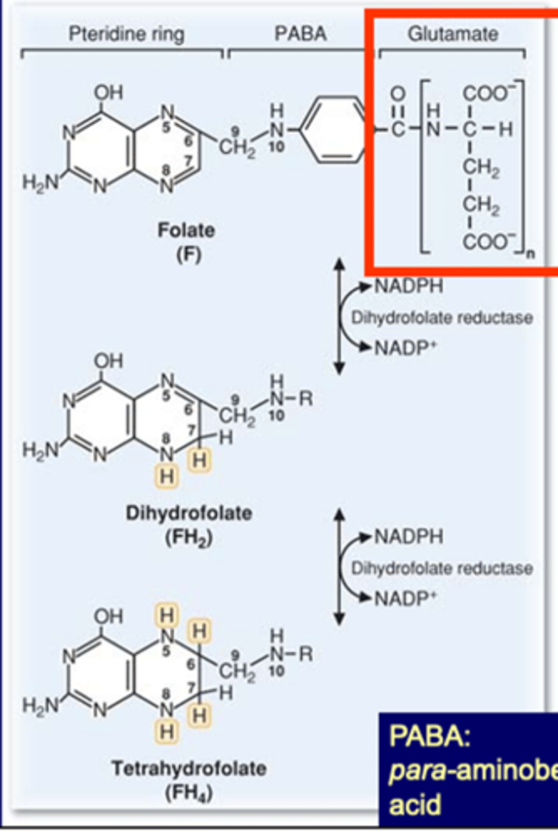
absorbed as polyglutamate
needs to be reduced using NADPH (from niacin/B3) (uses dihydrofolate reductase)
3 parts of folate
1. ring
2. PABA
3. glutamate
dihydrofolate reductase is the target of methotrexate, inhibits folic acid
Folate function
transports one carbon units
used in DNA, AA and meth metab
Forms of FH4
FH4
FH4-C
-Formyl (N10-formyl FH4)->de novo purine, -CHO
-Methylene (N5, N10-methylene-FH4)->Thymidine (dTMP), Serine, -CH2-
-Methyl (N5-methyl-FH4)->B12-CH3, Methionine, -CH3
N10-formyl FH4 (Formyl = CHO) is used in:
De novo purine synthesis
N5, N10-methylene-FH4 (Methylene = CH2) is used in:
Thymidine (dTMP) synthesis
Serine synthesis (from glycine)
N5-methyl-FH4 (Methyl = CH3) is used in:
Methionine synthesis (from Homocysteine) via B12-CH3
The methylene form of folate can be converted into N5-methyl-FH4 via the enzymatic activity of _________________________.
Methylenetetrahydrofolate reductase (MTHFR)
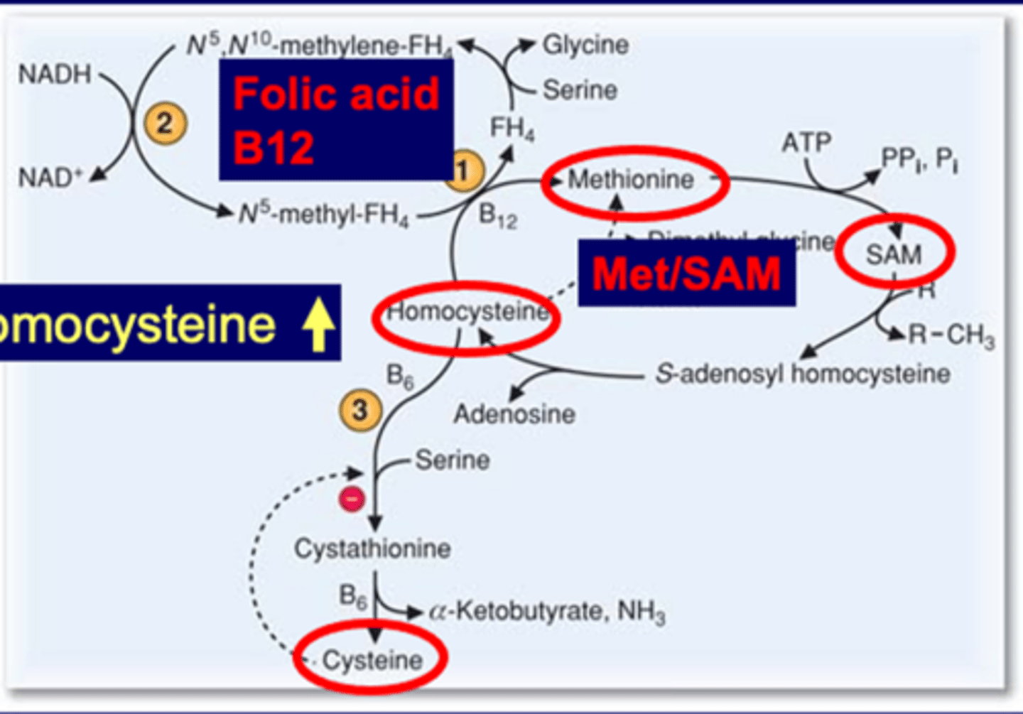
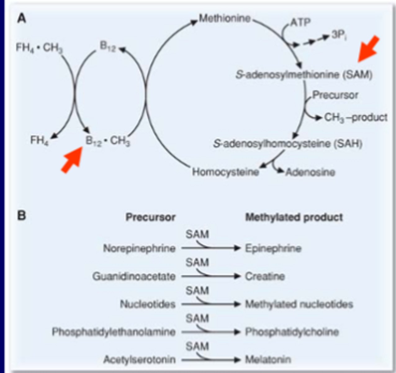
Folic acid/B12-CH3 Methionine Metabolism
Meth->SAM->SAH->homocys
homocys is toxic->Meth (requires B12 and folic acid)
homocys (alt path)->cystathrionine->cysteine (dep on B6)
when hc goes up try to push to cys but has neg feedback (inhib)
will keep circling between meth, but hc will still build up
high hc is nemia in blood, uria in the urine
initially give folic acid, B12 and B6 for treatment
if B6 responsive or not, binds to enzyme to make cysteine. sometimes enzyme mutates and not binds
severe form of high hc
Hyperhomocysteinemia
Severe form (homocystinuria) is a rare (enzyme dep)
-CBS def, two types->B6 responsive and non-responsive (not based on activity, based on mutation site, bind or not)
less severe form is more common (5-7% total pop) (nutritional or genetic)
-most common is MTHFR def
-Folate/B12/B6 def
-Folate/B12 pathway def in other genes (MTR, MTRR, MMADHC)
Hyperhomocysteinemia lab conditions
Lab diagnosis:
-normal (5-15)
-moderate (15-20)
-intermediate (30-100)
-severe (>100)
normal value always given, if 10x going to be severe
in between is moderate or intermed
severe always genetic problem
less severe induced or dietary problem
Methyl-trap hypothesis
B12 deficiency blocks the reaction from FH4-CH3 (N5-methyl-FH4) to FH4
Folate is "trapped" as a form of FH4-CH3
This results in appearance of folate deficiency (but actually a B12 deficiency)
increases homocysteine
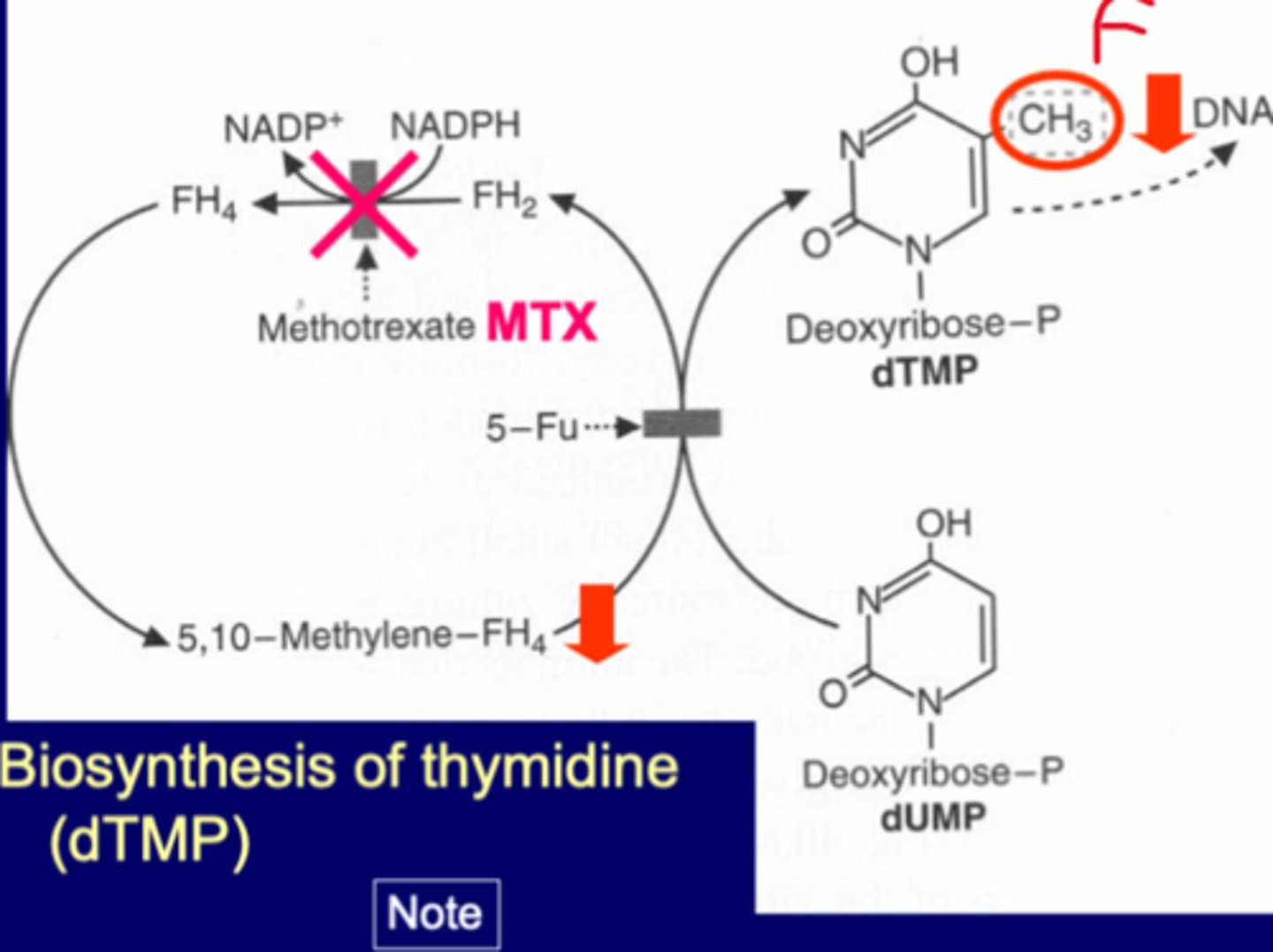
Biosynthesis of thymidine (dTMP)
thymidine synthase enzyme
-5-FU
-if knock out 5FU inhib both dTMP and FH4 pathway
MTX-inhibitor of FH2-FH4
MTX (methotrexate)
-used as therapy for rheumatoid arthritis
-side effects-inhib enzyme dihydrofolate reductase
-reduce immune cells, and rxns
also used in chemotherapy (inhibits cancer)
inhibits proliferation of lymphocytes and other cells responsible for inflammation in the join
MTX side effects (how to decrease)
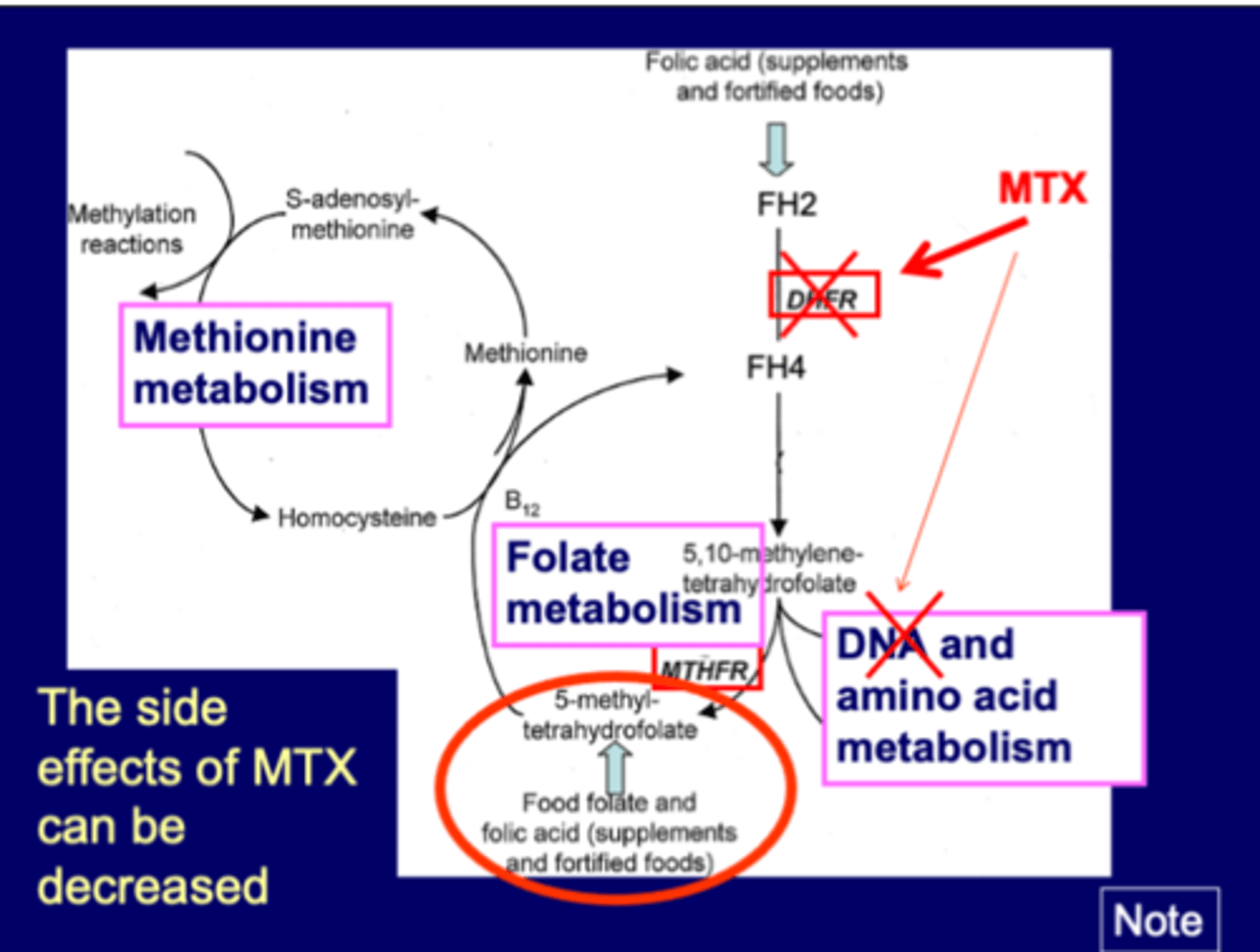
DHFR-makes active form FH4 (from FH2)
coupled with B12
MTX inhibits DHFR, blocks up and downstream
goal is to knock out DNA nucleotide biosynthesis
knocks out many others too
can provide alternative form of tetrahydrofolate
-from food or supplement, alternative form of folic acid
-5-meythl form
Nutrition: B12 produced by bacteria
stored for 2 years in the liver
meat, eggs, dairy products good sources
not salad/veggie
also made by bacteria
Key role of B12 in stomach
B12 binds to intrinsic factor (IF) in stomach
-IF comes from parietal cells (IF is glycoprotein)
Transcobalamin (TC) then binds to B12 and carries to liver
1st issue-issue with parietal cell, if doesn't make IF, can't absorb B12, leads to B12 deficiency
-lose parietal cells due to damage or removal by surgery
-common autoimmune problem, gastric arthritis
2nd issue-stomach cancer, remove part of stomach that has parietal cells
Pernicious anemia
B12 deficiency due to lack of intrinsic factor (NOT RELATED TO FOLATE)
Current understanding of pernicious anemia (PA) is an _____________________ in which autoantibodies prevent the formation of the vitamin B12-IF complex, which in turn dramatically decreases vitamin B12 absorption.
autoimmune condition
The followings result in severe B12 deficiencies (lack of intrinsic factor) in the order of (from most common to rare):
Autoimmune gastritis (against parietal cells) (most common)
Surgery to remove stomach
Inherited disorders which prevent intrinsic factor production
Carbon (methyl) donors (how affect HC)
-folic acid->runs cycle to make HC->Meth, more or less good
-B12->same way as above
-SAM->if give SAM, increase SAM, increases HC/everything
makes condition worse, if HC worse, HCnuria/nemia worse
-Betaine (trimethylglycine)->alter pathway, HC->Meth
minor pathway, 3 methyl group makes Meth
1 carbon unit donors
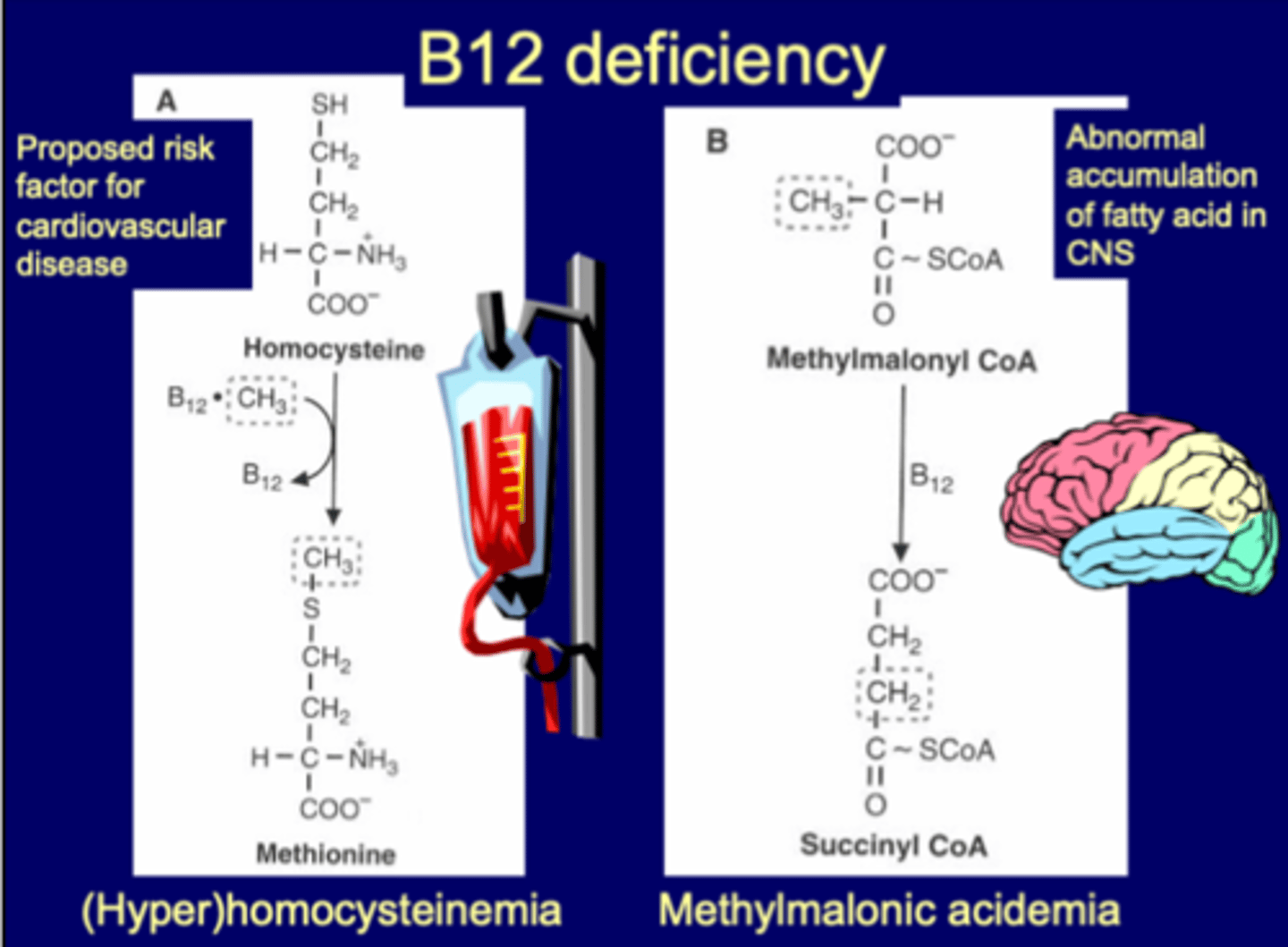
Vitamin B12 deficiency
risk factor for cardiovascular disease
-(hyper)homocysteinemia
abnormal accumulation of FA in CNS
-leads to accumulation of methylmalonyl CoA (methylmalonic acidemia- MMA)
-MMA up specific to B12 (NOT FOLIC ACID)
-acidemia is the condition of high MMA (also called MMA)
-if MMA going up, pathway is blocked, FA not degrading, accumulate in body->typically in brain or CNS
Folate and B12 deficiencies
Folate and B12
-megaloblastic anemia
-high HC (total plasma)
--concen>13 mcmol/L abnormal, including cystathionine-B-synthase (CBS) deficiencies
--if concen 10x more then its genetic not nutrition
-neural tube defects (NTDs)
B12 deficiencies
-methylmalonic acidemia (MMA)
--measurement of methylmalonic acid (or methylmalonyl CoA) (MMA), distinguishes between B12 (cobalamin) and folate deficiencies.
--also can be caused by genetics, methylmalonyl CoA mutase deficiencies
if MMA HIGH->B12 ONLY
if MMA NORMAL->folate/B12 (probably both)
Cobalamin (Vitamin B12)
B12 has cobalt inside
original name cobalamin
different enzyme name
Free iron vs heme
anemia associated with iron
common health issue
easiest way to get iron is heme
not possible to absorb ionized free iron
can absorb some iron forms
Iron deficiency anemia
microcytic anemia
hypochromic-clear inside, can't make heme
MCV < 80
Iron distribution in a human
male, female distribution amount is the same
Hemoglobin heme component
ferritin-storage form iron
transferrin-small 1% form
Iron deficiency anemia causes
males-mostly absorption problem, inability to absorb iron, lack of iron in diet
females-blood loss, use of blood components, can be pregnancy
Iron is mainly absorbed in the duodenum and upper jejunum through either one of the following:
heme transporter --Heme and Fe2+ (using DMNT1 to to help absorb), heme best form for absorption
endosomes--Fe2+ (stored in ferritin)->once goes in as Fe2+ turns into Fe3+ (storage form) then when leaves converts back
FP1 transporter-takes Fe2+ out of ferritin, transports to TF (transferrin) to leave
-regulated by hepcidin (from liver), will block FP1/stop iron transport
TF-has two arms, can bind 2 irons
The major diet form of free iron Fe3+ needs to be converted to Fe2+ by:
Duodenal Cytochrome B (ferric reductase)
-gives an e-
-reduces so can use DMT1 transporter
Iron transport summary:
- Fe2+ (in the cells) is transported out from the cell through ferroportin (iron transporter).
- Fe2+ (outside of the cells) is immediately converted to Fe3+
- Fe3+ (outside of the cells) binds to transferrin
Iron absorption summary:
-Iron more readily absorbed in ferrous state (Fe2+)
-Most dietary iron is in ferric form (Fe3+)
- Fe3+ (a dietary form of free iron) is converted to Fe2+ by Duodenal Cytochrome B (= ferric reductase)
-gastric secretions: dissolve and form soluble complexes with ascorbic acid, reduction to the Fe2+ form
Inhibitors of iron absorbtion:
phytates, tannins, soil clay, laundry starch, iron overload, antacids
Competitors of iron absorption:
lead, cobalt, strontium, manganese, zinc
Facilitators of iron absorption:
ascorbate, citrate, amino acids, iron deficiency
Iron absorption, storage and transport
Iron Transport: Transferrin
Absorption: Free iron, heme
Iron Storage Ferritin
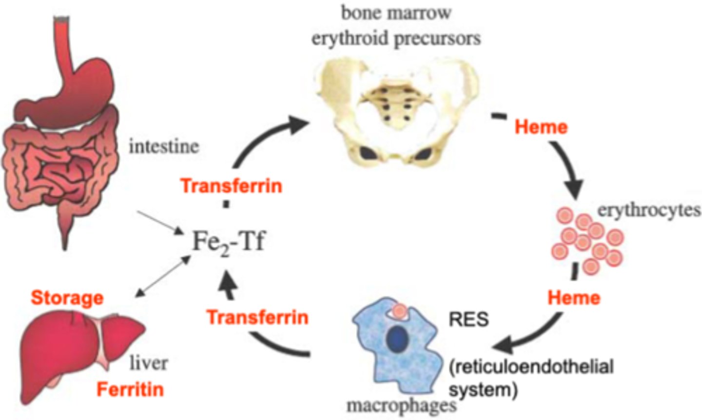
Iron Cycle
Transferrin takes iron from storage/ferritin (Fe2+ form) in the liver/instestine
TF1 binds to bone marrow to make heme
Heme packages RBC (life of 120 days)
Heme->recycled, free iron to make transferrin
Heme portion goes to heme degredation cycle->
RES (redticuloendothelial system) in macrophages
makes biliverdin->bilirubin (B)
B->BG->Bile
Iron storage
Ferritin stores the 3+ form
Fe2+ enters ferritin and is bonded with (OOH) to store as Fe3+
converted back to Fe2+ when leaves
Iron stores: sex susceptibility
Ferritin good marker of serum iron levels
normal male-goes up after about 20 years
normal female-goes up around 50 years due to menopause
females can maintain iron better than males, but can go down due to store depletion
Iron deficiency 3 phases:
-gradual, start
-middle-iron stores deplete, show deficiency, not anemia
-final-show phenotype, impact RBC, anemia
Iron absorption disease
Hemochromatosis, type 1
-HFE gene deficiency
-Too much iron absorbed from GI system
-preventable complications
-1/3 not show phenotype (reduced penetrance)
Rec for testing: phenotypic screening->genetic testing
HMP (hexose monophosphate shunt)
attached to upstream of glycolysis
2 functions:
NADPH (oxidative/irreversible)
DNA/RNA synthesis (non-oxidative/reversible)
G6P 2nd product after glucose->HMP, makes NADPH
other path back and forth with F6P, final destination is ribose 5P-sugar for nucleotide->makes purines and pyrimidines (can make gout worse)
G6PD deficiency-enzyme in first path, can't make NADPH (deficient in G6P dehydrogenase that makes 6-P gluconolactone from G6P). NADPH needed for GSH (antioxidant) leads to oxidative stress damage RBC
Von Gierke-can't make glucose from G6P, accumulates G6P->overproduce ribose 5-P, too many purines (hyperuricemia and gout)
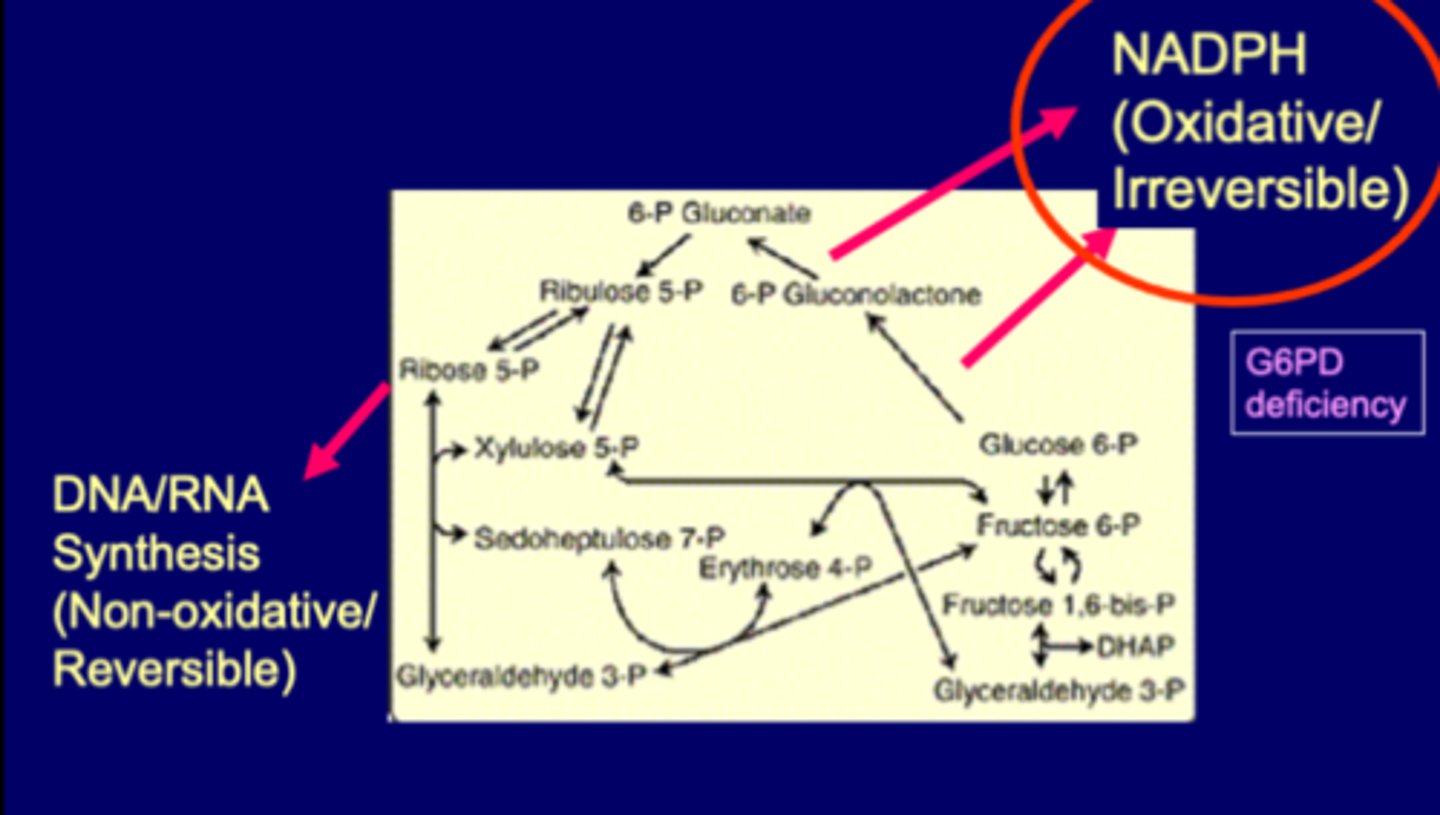
Summary HMP shunt
Oxidative (irreversible)
-Glucose 6-P->>>2NADPH, Ribulose-5-P
-Enzyme: Glucose 6-P dehydrogenase (rate limiting)
Non-oxidative (reversible) (for nucleotides)
-ribulose-5-P->>>Ribose-5-P, G3P, F6P
-Enzyme: transketolases (cofactor vitamin B1) (thiamine)
thiamine impacts ribose5P, NOT NADPH
The five biochemical uses of NADPH are:
Scavenging hydrogen peroxide (H202) (protection from oxidative damage)
Phagocytic respiratory burst
Synthesis of nitric oxide (NO)
Reductive biosynthesis
Cytochrome P450 monooxygenase system
Uses of NADPH and GSH
Reduction of H2O2
H2O2 detoxed by GSH + enzyme GPx to make water
similar to catalase
GSH/GPx->cell membrane
GSH-made by glycine, cysteine, glutamate
GSH-attached form, can be recycled as GSSG
NADPH used to recycle
if NADPH knocked out->GSH stuck in inactive form (GSSG)
leads to accumulation H2O2, trouble in RBCs
Uses of NADPH and RBC
HMT shunt pathway generates NADPH to protect cell membrane lipids and proteins from oxidation, through regeneration of reduced glutatione
G6PD (glucose-6-phosphate dehydrogenase) deficiency
-blocks HMT shunt
-reduced supply of NADPH
-reduced GSH/increased oxidative stress (H2O2)
-causes hemolysis
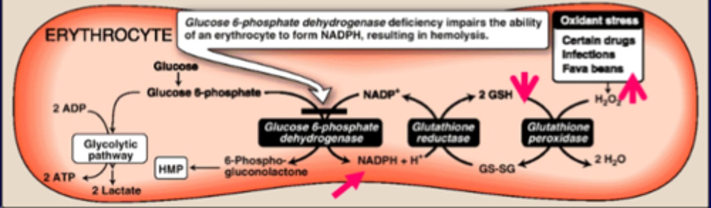
Glucose 6-P dehydrogenase (G6PD) deficiency genetics
-X-linked, most common disease enzyme abnormality
-no clinical manifestations in most individ with def mutation
-hemolytic anemia with precipitating factors, including oxidants (oxidative stress)
--oxidative stress can cause the phenotype to express in individuals with mutation that didn't express before
-shows heinz bodies in RBCs
Precipitating factors in G6PH deficiency:
Ingestion fava beans (favism)
-all patients with favism have G6PD deficiency
-NOT all G6PD patients show favism
-creates oxidative stress
Use of an oxidant drug
Infection
Neonatal jaundice
NADPH for NOS
From Arginine to Citrulline
-nNOS (neuronal)
-eNOS (endothelial)
-iNOS (Inducible)->manny immune cells, macrophages, brain astroglia
NOS in OMM
NO:
-Endothelial cells (eNOS), which need NO for vascular smooth muscle relaxation (Ca2+ cGMP pathway)
-known as endothelium derived relaxation factor (EDRF)
OMT:
-can induce NO, leading to its analgesic, anti-inflam and relaxation effects
NAPDH for ROS respiratory burst
hydroxyl radical via iron-dependent pathway (fenton reaction)
peroxynitrite (or peroxynitrile) via iNOS pathway
hypolchlorus acid (HOCL) via myeloperoxidase-dependent pathway
Defects in phagocytic respiratory burst
Chronic granulomatous disease (CGD):
-immunodef, caused by defects in respiratory burst by immune cells
-formation of granulomas (nodular areas of inflammation)
-NADPH oxidase deficiences
--low (or no) superoxide (neutrophil function tests)
--can use hydrogen peroxide (H2O2) generated by invading organisms
-At high risk of infection by catalse-positive organisms (S. aureus and aspergillus)
Chronic granulomatous disease (CGD)
NADPH oxidase deficiency
-cant kill catalase positive bacteria (the bacteria will detox H2O2 and live and cause infection)
-can kill catalase negative bacteria (bacteria has hydrogen peroxide which is used to kill them)