MED 204: Visualization of Cells and Tissues
 Call Kai
 Learn
Learn Practice Test
 Spaced Repetition
 Match
Match Flashcards
 Knowt Play
1/39

There's no tags or description
Looks like no tags are added yet.
Name | Mastery | Learn | Test | Matching | Spaced | Call with Kai | Chat |
|---|
No analytics yet
Send a link to your students to track their progress
40 Terms
what is it: Contrast
diff in light intensity between specimen and background
Generate through stains, dyes, fluorescent dyes
what is Magnification?
microscope make image larger than actual
Useful ONLY when see details that u can't with your unaided eye
objective lens ENLARGE image
What is Resolution?
distinguish detail
min distance where 2 points of specimen can be seen
Objective lens, imaging media, wavelength of light determine fine detail
X-ray
x-ray radiation to detect tissue
what is CT?
computed tomography)- compilation of x rays (greater resolution than X-ray)
What is MRI?
- use radio waves and magnets to detect tissue
What is PET scan?
positron emission tomography)- radioactive tracers to track blood flow, O2 intake, metabolic activity (ex: detect change like cancer)
Microscopes use (blank) or (blank) to make image
Microscopes use light or electron to make image !
Light Microscope
light pass through specimen is altered to see contrast
Transmission Electron Microscopy
What is it?
accelerated electron beam through specimen to phosphor screen
(dense obj= scatter electrons = dark)
Scanning Electron Microscopy
What is it?
focused electron beam. Electron interation with atoms on sample makes scatter that generates image
Formula: Total Magnification
multiple magnification of each component
Ex: (10x eyepiece) x (40x objective) = 400x
Visible light ?
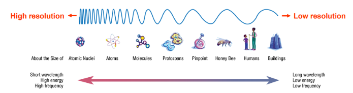
What is relationship between wavelength and resolution?
type of electromagnetic radiation (no mass only photons)
Shorter wavelength= higher resolution = more damage to tissue
Refractive Index (RI)
ratio of speed of light in Air to speed of light in Material

2 types of specimens
Phase specimen and Amplitude specimen
Phase specimen
colorless, retarded slows down, amplitude don’t change
Microscope components
Amplitude specimen
stained visible dye, amplitude changes, looks darker , light is being absorbed
Stains and dyes
Brightfield microscopy
Diffraction
Tendency of waves to bend around edges
Magnification
make image bigger with objective lenses (4x, 10x, 40x, 100x)
Smaller angle, fewer diffraction
Resolution
shortest distance between 2 points on a specimen that can still be distinguished by observer or camera as separate entities – u need space between them to resolve the image
RESOLUTION IS INVESERLY PROPORTIONAL TO WAVELENGTH
Abbe diffraction limit
resolution in light microscopy is Limited by light's physical properties
Rayleigh Criterion
2 objects can be resolved if space between (valley) between overlapping brightness is reduced by 20% between two maxima to show 2 diff objects
Point spread function
3d representations of diffraction pattern
Prepping Samples :
Light microscopy?
Electron microscopy?
Light microscopy- LIVING or FIXED
Electron microscopy- MUST BE FIXED
Contrast
stains and dyes- which specimen?
Microscope components- which specimen?
made by stains, dyes, enzymatic color change
Stains and dyes- amplitude specimens
Microscope components: Phase specimens
Phase optics, DIC optics,
Brightfield microscopy
What is it and which specimen?
light DAMPENED when passed through cell that have been stained with dyes
=Amplitude specimens- amplitude of light waves change when passed through samples
Generate contrast with dyes and stains
Fixed or live samples?
must be first FIXED
Generate contrast in light microscopy with optical components
Fixed or live samples?
live or fixed
Phase contrast microscopy
physical impediments to shift light waves as they pass through specimen= destructive interference
Study internal cell structure without need to strain and kill cells
primarily used on live samples, unstained
Differential Interference Contrast Microscopy (DIC)
use polarized light to generate contrast/details as light pass through specimen
(google says both live and fixed)
Generate contrast with Fluorophores
Fixed or live samples?
cells living or fixed
Fluorescent moles must be excited by light at 1 wavelength to emit light at a second wavelength
Advantages of Flourophores?
Advantages: sensitivity, resolution
Specificity
Sensitivity (50 m/mm3)
Fluorophores Sensitive to physical-chemical environment
Temporal resolution (10-9 sec)
Spatial resolution (can detect single molecules)
Greater resolving power than staining with visible dyes or color enzyme assay
Generating contrast in light microscopy with immunological reagents
Fixed or live samples?
- usually FIXED
Immunolabeling to visualize specific compenents of cells –
Fixed or live samples?
usually FIXED
Direct labeling of a molecule of interest to see its cell components
What is it? Fixed or live samples?
– LIVING or FIXED cells = molcularly manipulating gene of interest to add a detectable tag
Introduce Plasmids--> tagged protein
Fluorescent protein tag (GFP), visualized by shining wavelength of light on sample
Non-Flourescent epitope tage (myc, His, GSt, FLAG, HA), visualized by immunostaining & indirect flourscence
Use molecular cloning to insert gene of interest into expression vector
Electron Microscopy? has HIGH ___ & ______
Electron Microscopy: HIGH MAGNIFiCATION + RESOLUTION
Transmission EM (TEM)
2D, fine detail since sample sliced thin
More Dense objects= scatter electrons= dark in TEM
Less dense = don’t scatter electrons= brighter
Super fine sample, high resolution because super thin! So your only looking at a piece of the cell
Scanning EM (SEM)
3D, surface features
Freeze fracture/etch with scanning EM
Freeze fracture/etch with scanning EM: 3D view of inside cell
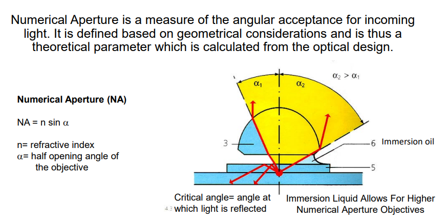
Numerical Aperture
Numerical Aperture- angular acceptance for incoming light