Histology - Reproductive
1/182
There's no tags or description
Looks like no tags are added yet.
Name | Mastery | Learn | Test | Matching | Spaced |
|---|
No study sessions yet.
183 Terms
Dense irregular connective tissue
The structure pointed in #1 is made up of what type of connective tissue?

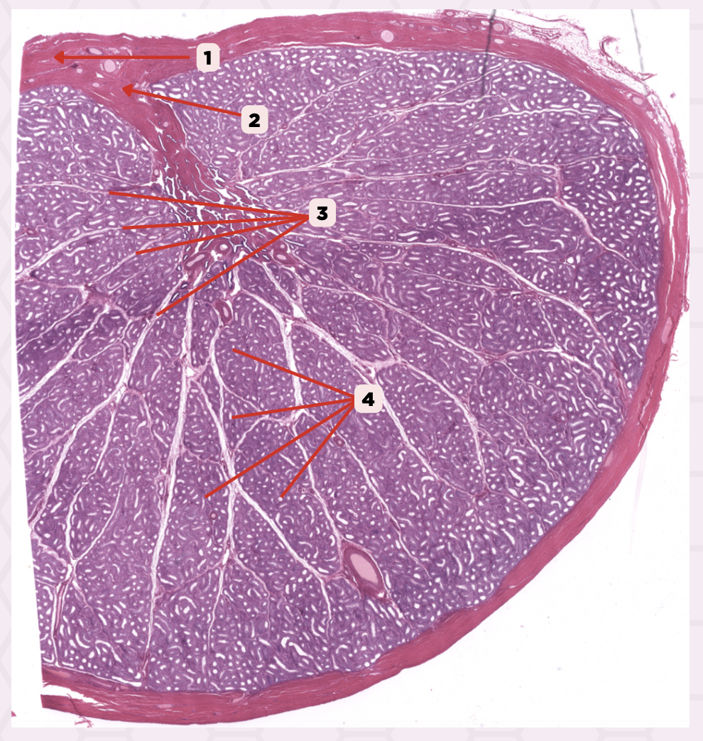
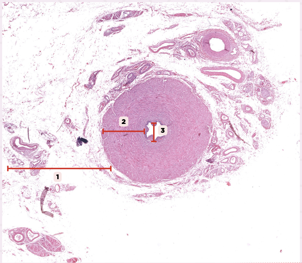
Testis
Identify the specimen.

Tunica Albuginea
Identify the structure labeled as 1.

Mediastinum Testis
Identify the structure labeled as 2.

Septulae Testis
Identify the structure labeled as 3.

Lobuli Testis
Identify the structure labeled as 4.
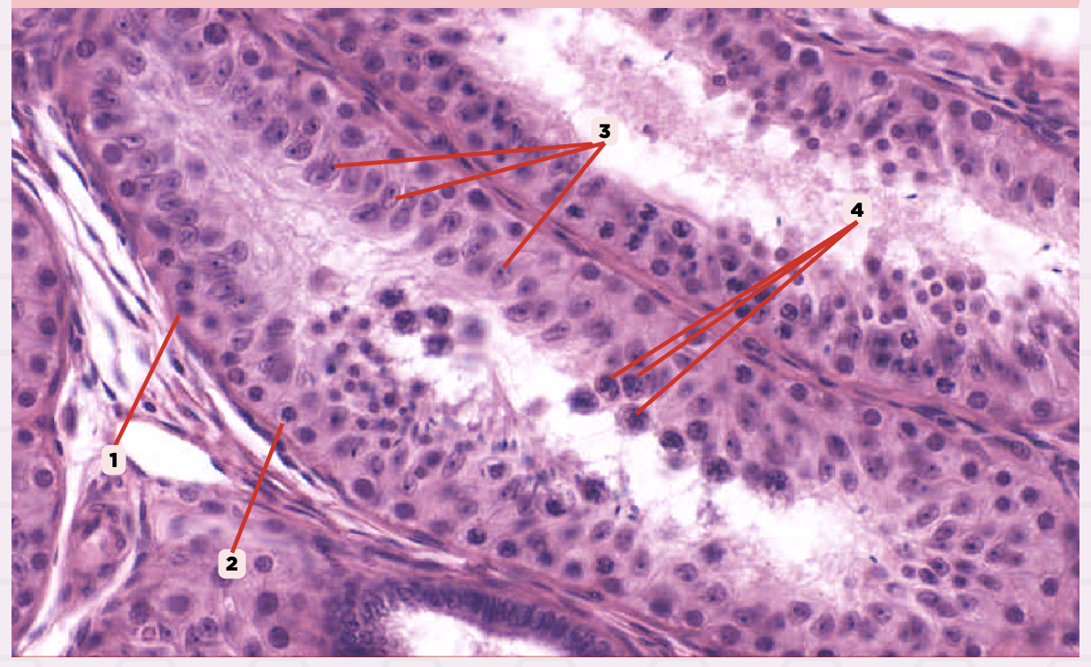
Dark type A spermatogonium
Identify the structure labeled as 1.

Pale type A spermatogonium
Identify the structure labeled as 2.

Sertoli cells
Identify the structure labeled as 3.

Primary spermatocytes
Identify the structure labeled as 4.
Testosterone
What hormone does #1 produces?
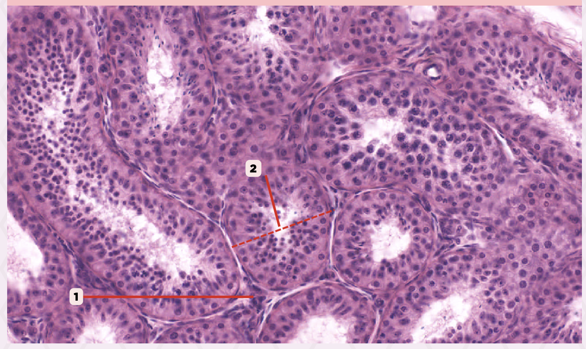
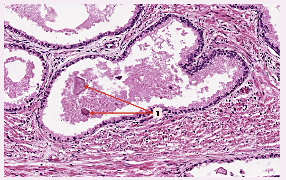
Interstitial cells of Leydig
Identify the structure labeled as 1.

Seminiferous tubule
Identify the structure labeled as 2.

Simple squamous or cuboidal
What epithelium lines #1?

Proximal
What part of the lining epithelium of #2 consists of Sertoli cells then becomes cuboidal or columnar cells with numerous microvilli?
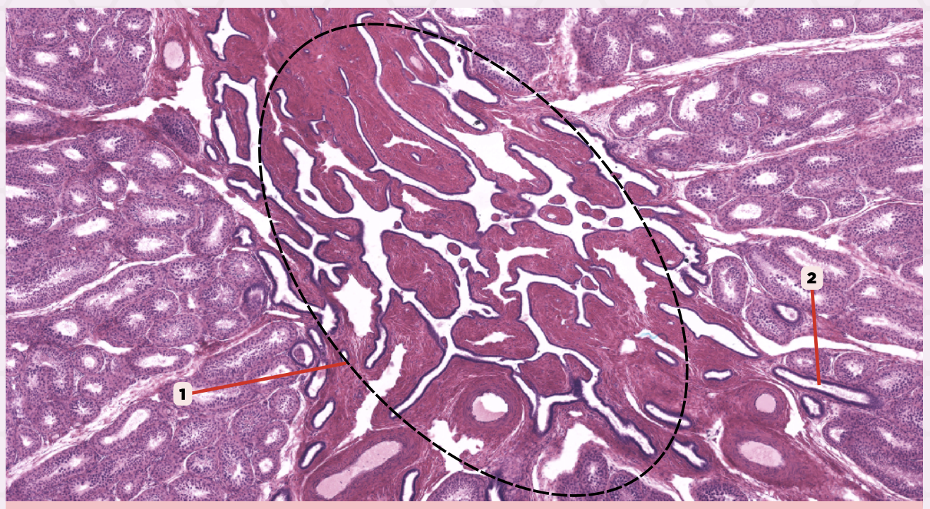
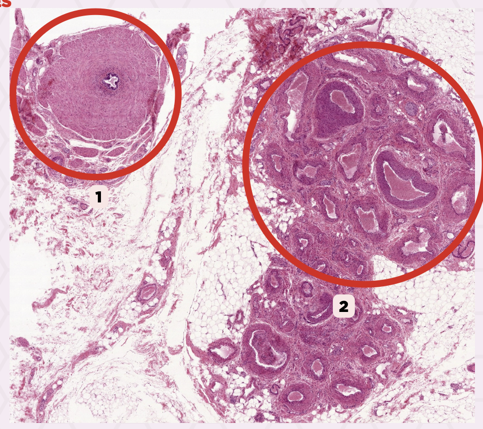
Rete Testis
Identify the structure labeled as 1.

Tubuli recti
Identify the structure labeled as 2.

Head
Which of the following structures contains an elongated nucleus and an acrosomal cap?

Middle piece
This structure is thick due to its mitochondrial sheath.

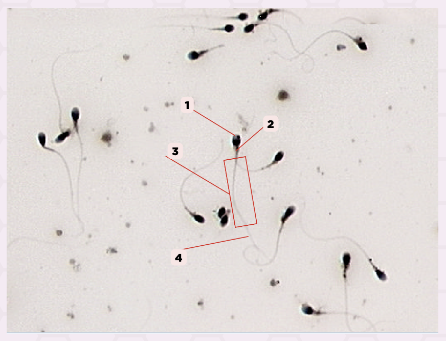
Spermatozoa
Identify the specimen.

Head
Identify the structure labeled as 1.

Middle piece
Identify the structure labeled as 2.

Principal piece
Identify the structure labeled as 3.

End piece
Identify the structure labeled as 4.
Pseudostratified columnar epithelium
What is the epithelium of this ductus?
Epididymis
Identify the specimen.

Coils of Ductus Epididymis
Identify the structure labeled as 1.

Ductuli efferentes and ductus epididymis
The epididymis consists of #2, where two types of tubular structures are embedded. Identify the embedded structures.

Scalloped Epithelium
Identify the structure labeled as 1.

Dense Connective Tissue
Identify the structure labeled as 2.
Smooth Muscle Layer
Identify the structure labeled as 3.

Sperm cells
Identify the structure labeled as 1.

Pseudostratified Columnar Epithelium
Identify the structure labeled as 2.

Smooth Muscle Cell
Identify the structure labeled as 3.
Basal Cells
Identify the structure labeled as 4.

Principal Cells
Identify the structure labeled as 5.

Stereocilia
Identify the structure labeled as 6.

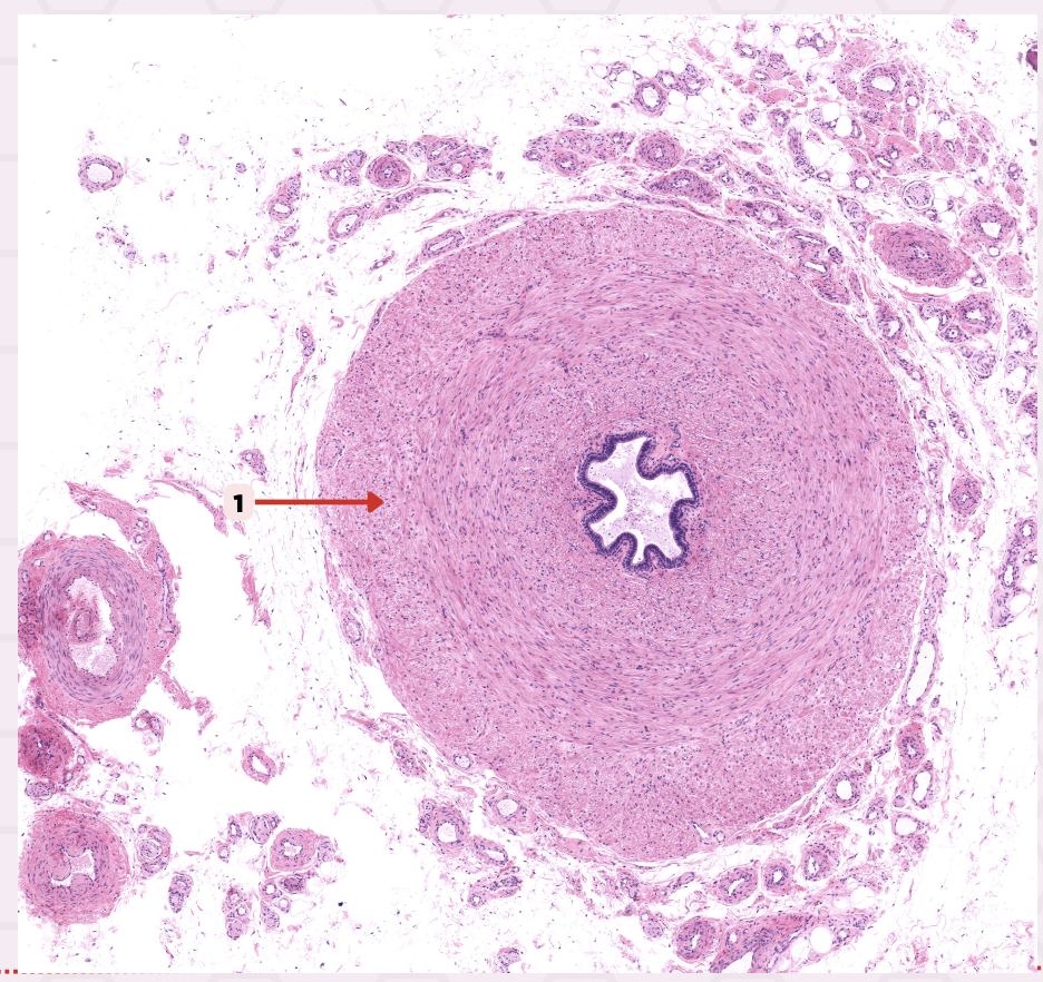
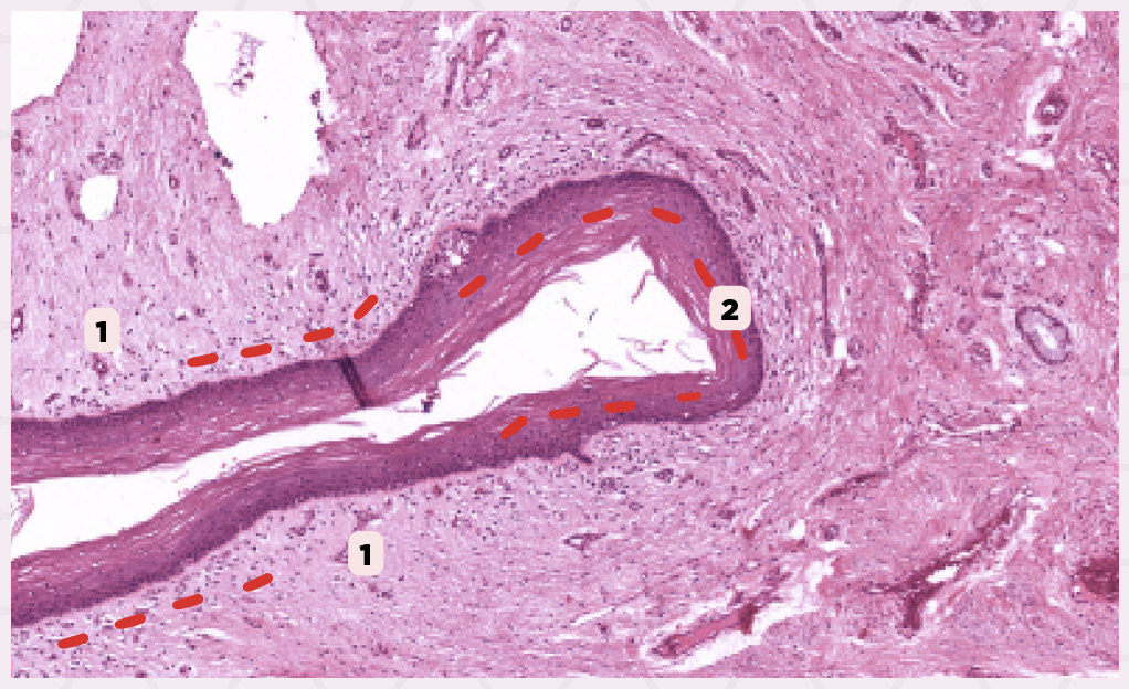
Vas Deferens
Identify the specimen.
Pseudostratified columnar epithelium
Identify the structure labeled as 1.

Lamina propria
Identify the structure labeled as 2.
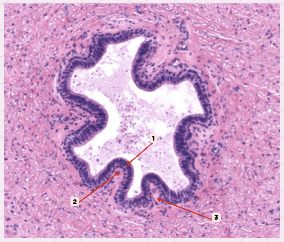
Loose Connective Tissue
Identify the structure labeled as 3.

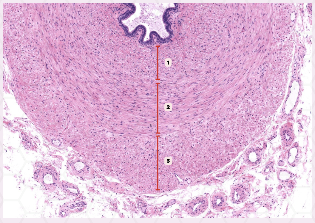
Inner Longitudinally-Oriented Layer
Identify the structure labeled as 1.

Middle Circularly-Oriented Layer
Identify the structure labeled as 2.

Outer Longitudinally Oriented
Identify the structure labeled as 3.
Adventitia
Shown is a magnified section from vas deferens. Noting the high vascularity and rich nerve supply, what layer should this be?

Loose connective tissue
What type of connective tissue are structures #1 and #2 embedded in?

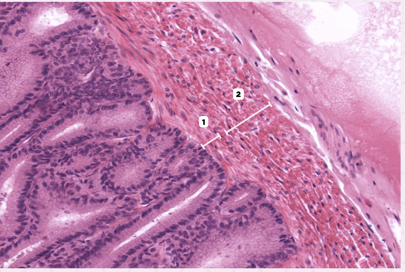
Blood Vessels (Arteries)
Identify the structure labeled as 1.
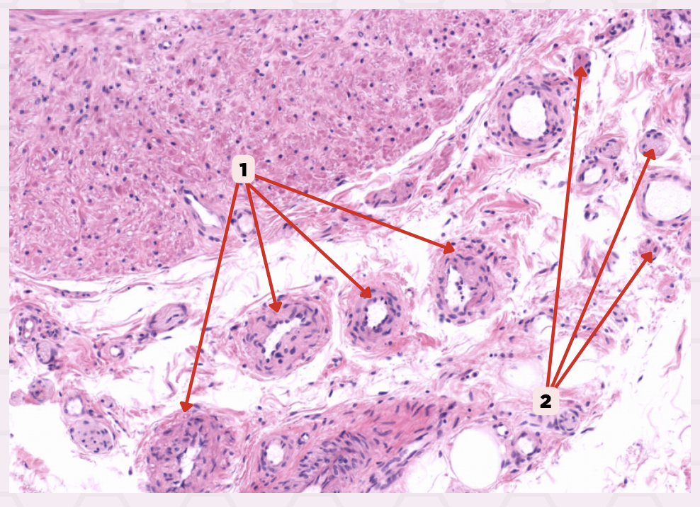
Nerves
Identify the structure labeled as 2.

Adventitia
Identify the structure labeled as 1.

Muscularis
Identify the structure labeled as 2.

Mucosa
Identify the structure labeled as 3.
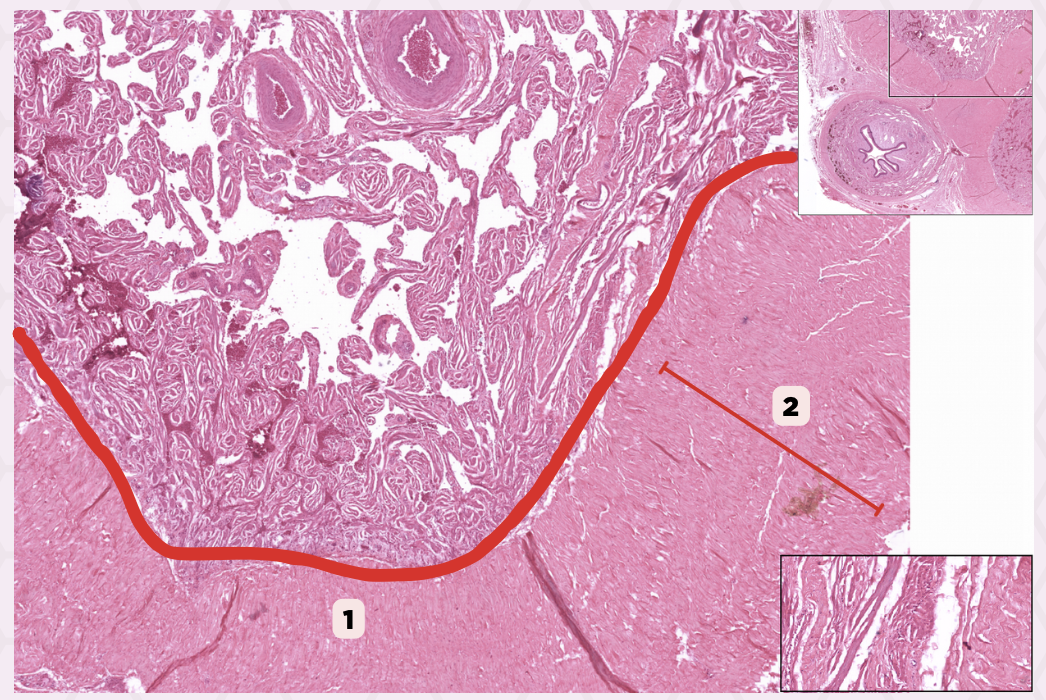
Initial Segment
The encircled structure (#1) forms part of the spermatic cord from its until it _______ enters the abdominal cavity.

Testicular Vein
The #2 drains into 2-3 larger veins, which later fuse to form the what vein?

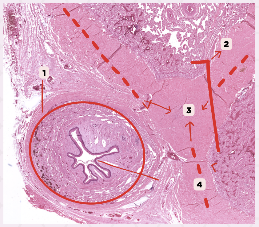
Vas Deferens
Identify the structure labeled as 1.
Spermatic Cord
Identify the structure labeled as 2.

Spermatic Cord
Identify the structure labeled as 1.

Penile erection
What process occurs due to the increased blood supply from the arteries to the vascular channels in structure 1?

Dense connective tissue
What type of connective tissue makes up structure 2?

Corpora Cavernosa
Identify the structure labeled as 1.
Tunica Albuginea
Identify the structure labeled as 2.

Penis
Identify the specimen.

Corpus Spongiosum
Identify the structure labeled as 1.

Corpus Cavernosa
Identify the structure labeled as 2.

Tunica Albuginea
Identify the structure labeled as 3.

Spongy (Penile, Cavernous) Urethra
Identify the structure labeled as 4.
Lamina Propria
Identify the structure labeled as 1.

Epithelium
Identify the structure labeled as 2.
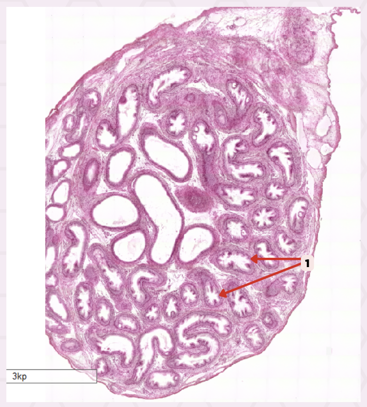
Prostate Gland
Identify the specimen.

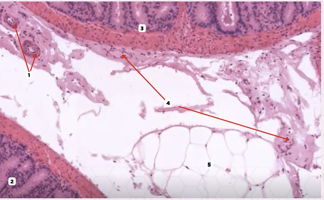
Capsule
Identify the structure labeled as 1.

Septum
Identify the structure labeled as 2.
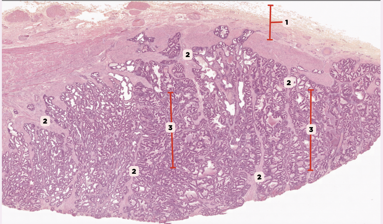
Lobule
Identify the structure labeled as 3.

Nerves
Identify the structure labeled as 1.

Blood Vessels
Identify the structure labeled as 2.

Smooth Muscles
Identify the structure labeled as 3.
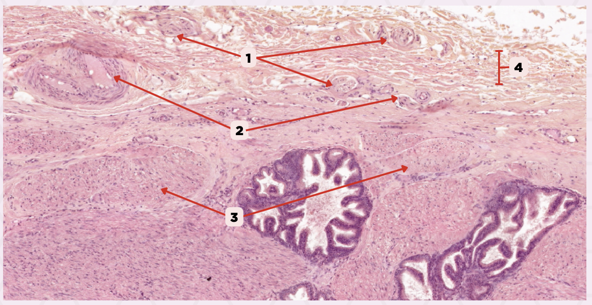
Connective tissue
Identify the structure labeled as 4.

Septum
Identify the structure labeled as 2.

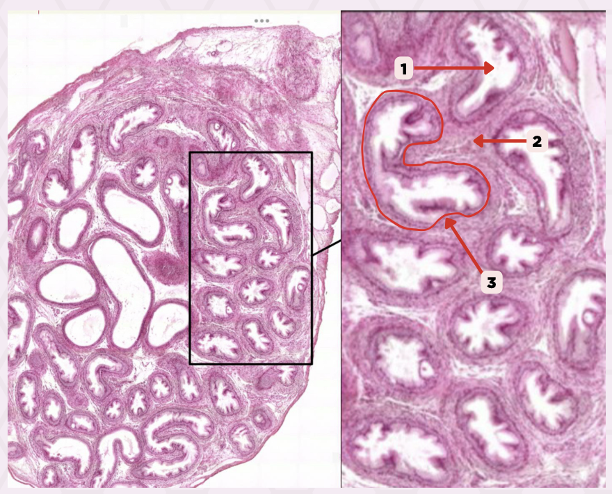
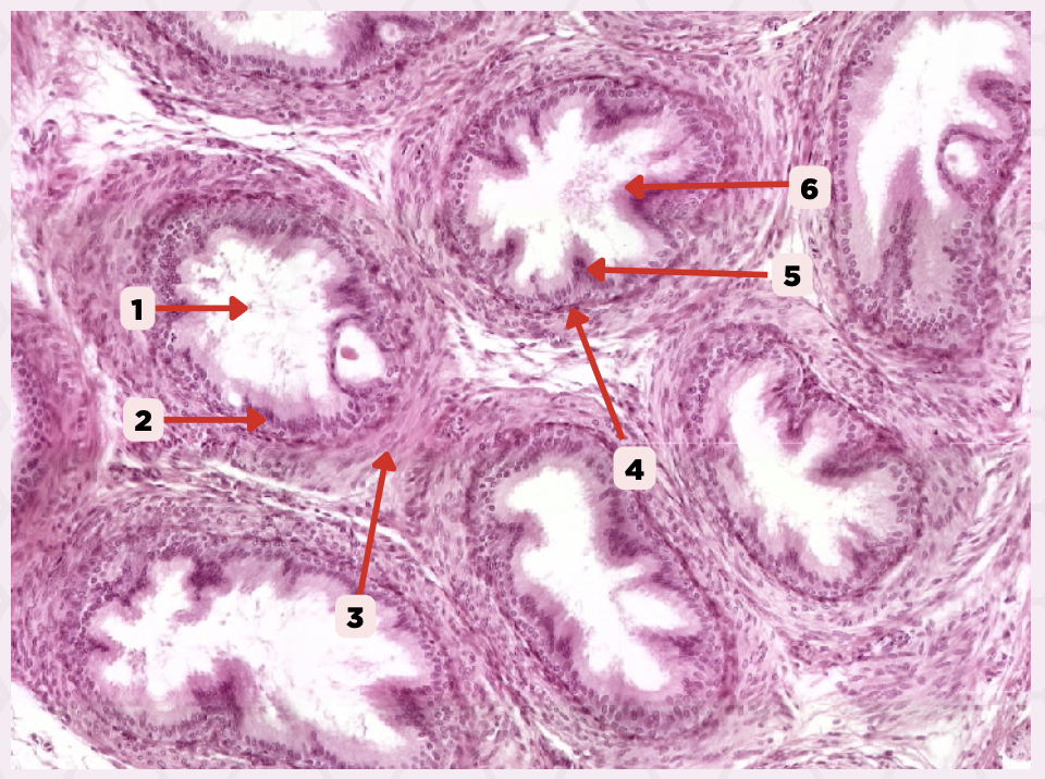
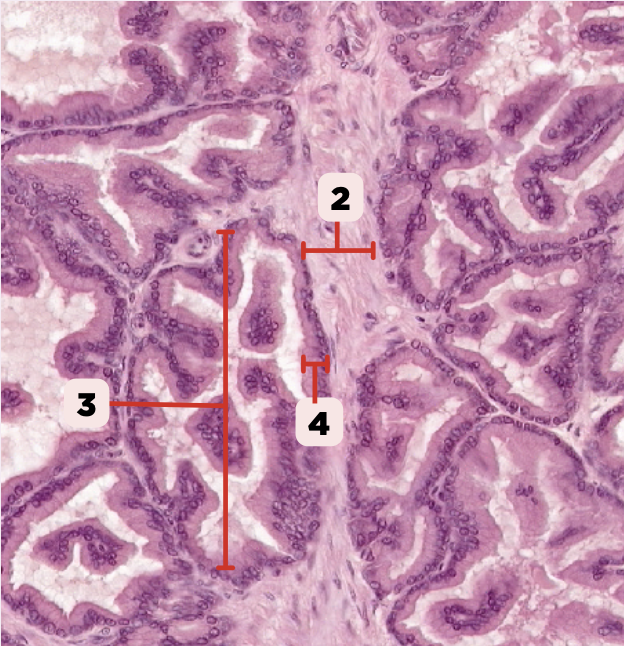
Alveoli
Identify the structure labeled as 3.
Corpora amylacea
Identify the structure labeled as 1.
Seminal Vesicle
Identify the specimen.
D. Adventitia
This slide shows what histological layer of the seminal vesicle?
A. Luminal
B. Mucosa
C. Muscularis
D. Adventitia

C. Loose Connective Tissue
What kind of connective tissue present in this layer binds the highly coiled seminal vesicle?
A. Dense Regular CT
B. Dense Irregular CT
C. Loose Connective Tissue
D. Reticular fibers
D. Pseudostratified Columnar Epithelium
The epithelial lining of the structure labelled #2 is _____.
A. Simple Squamous Epithelium
B. Stratified Squamous Epithelium
C. Simple Columnar Epithelium
D. Pseudostratified Columnar Epithelium
B. Ovarian follicles
What are the numerous round structures found in region #1 of an infant’s ovary?
A. Blood vessels
B. Ovarian follicles
C. Connective tissue fibers
D. A and B
D. Stromal tissue
What type of tissue embeds the main components found in regions #1 and #2?
A. Epithelial tissue
B. Muscular tissue
C. Adipose tissue
D. Stromal tissue

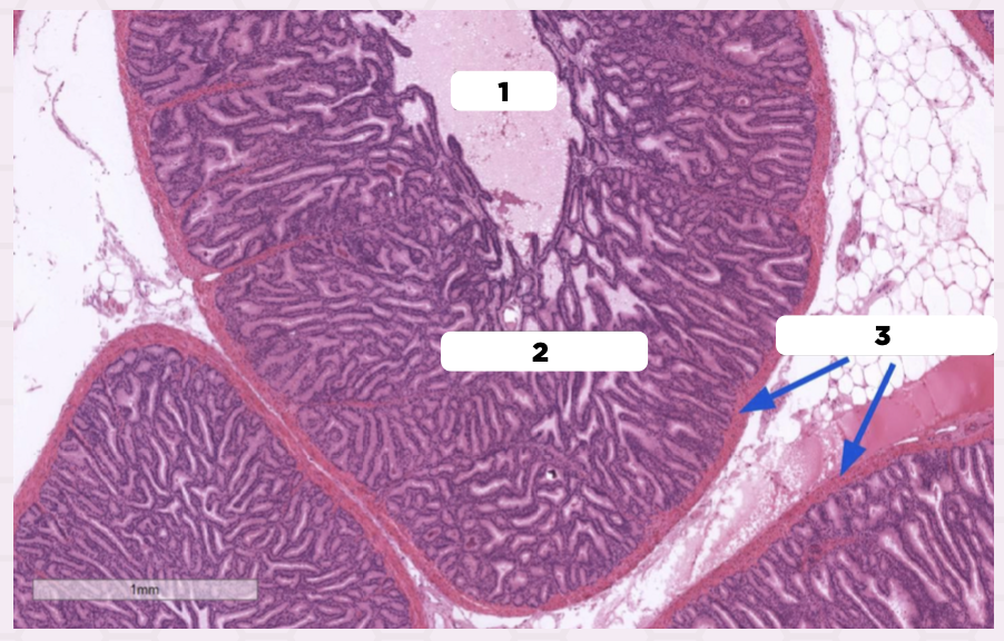
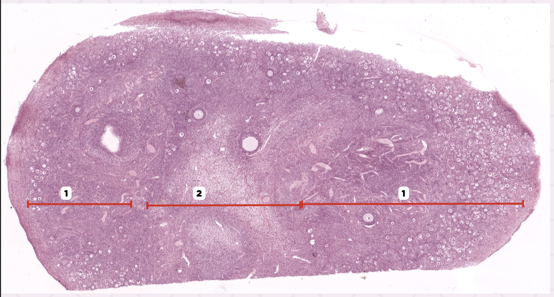
Cortex
Identify the structure labeled as 1.

Medulla
Identify the structure labeled as 2.

Ovary
Identify the specimen.

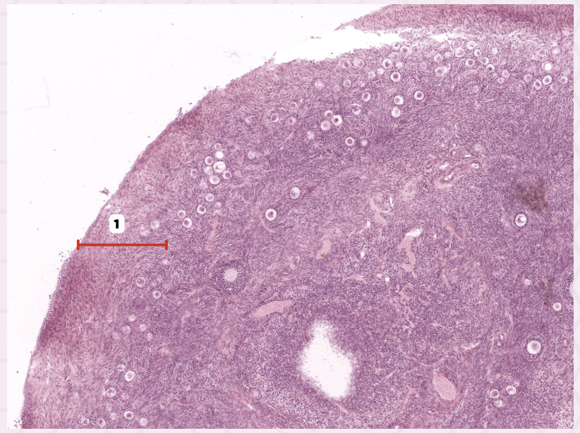
Tunica Albuginea
Identify the structure labeled as 1.
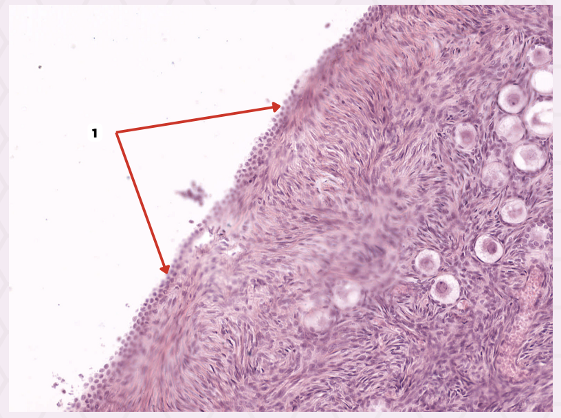
Germinal epithelium
Identify the structure labeled as 1.
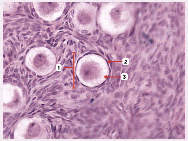
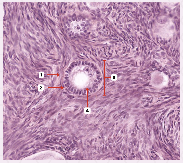
Primordial ovarian follicle
Identify the structure labeled as 1.

Granulosa cells
Identify the structure labeled as 2.

Primary oocyte
Identify the structure labeled as 3.
Theca externa
Identify the structure labeled as 1.
Theca interna
Identify the structure labeled as 2.

Primary ovarian follicle
Identify the structure labeled as 3.

Zona pellucida
Identify the structure labeled as 4.

B. Ovary
What is the roundish structure that contains several secondary ovarian follicles and lies adjacent to the Fallopian tube?
A. Uterus
B. Ovary
C. Fallopian tube
D. Cervix

C. Fallopian tube
Which structure, indicated in the image, is part of the female reproductive system and serves as a conduit for eggs from the ovary to the uterus?
A. Cortex
B. Medulla
C. Fallopian tube
D. Ovary

Ovary
Identify the specimen.
Cortex
Identify the structure labeled as 1.
