Intro to DENTAL CARIES
 Call Kai
 Learn
Learn Practice Test
 Spaced Repetition
 Match
Match Flashcards
 Knowt Play
1/88

Earn XP
Description and Tags
Operative Dentistry Exam 1
Name | Mastery | Learn | Test | Matching | Spaced | Call with Kai | Chat |
|---|
No analytics yet
Send a link to your students to track their progress
89 Terms
Dental caries
"biofilm-mediated, sugar-driven, multifactorial,
dynamic disease that results in the phasic demineralization and
remineralization of dental hard tissues
Is the localized destruction of susceptible dental hard tissue by
acidic by-products from bacterial fermentation of dietary
carbohydrates.
▪ It is a bacterial driven, generally chronic, site-specific, multifactorial,
dynamic disease process that results from the imbalance in the
physiologic equilibrium between the tooth mineral and the plaque
fluid; that is, when the pH drop results in net mineral loss over time
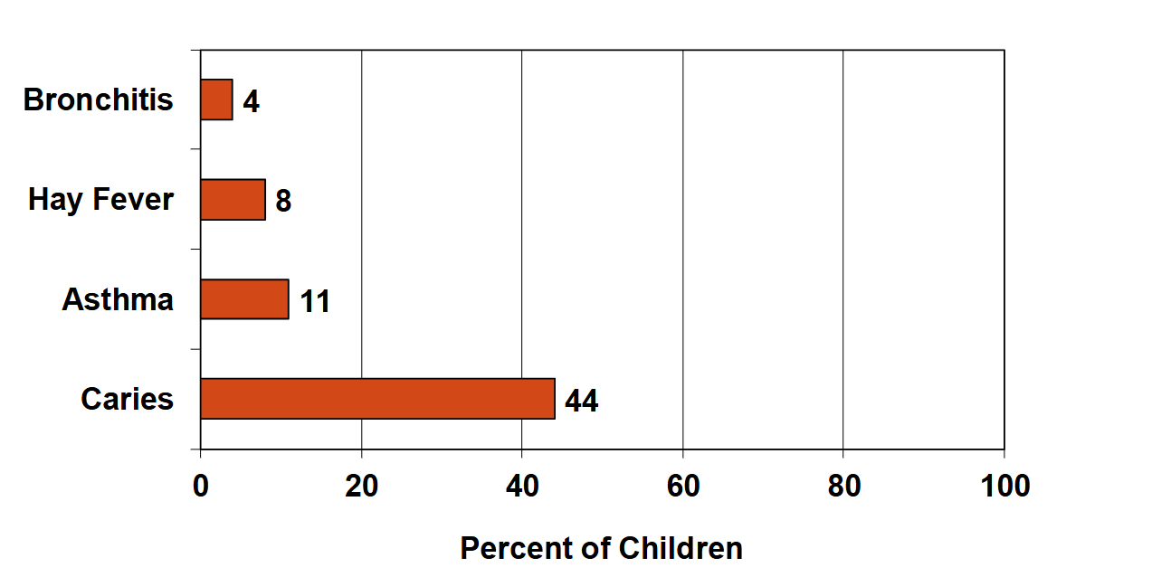
COMMON CHILDHOOD DISEASES (U.S.)
Carries is Number 1
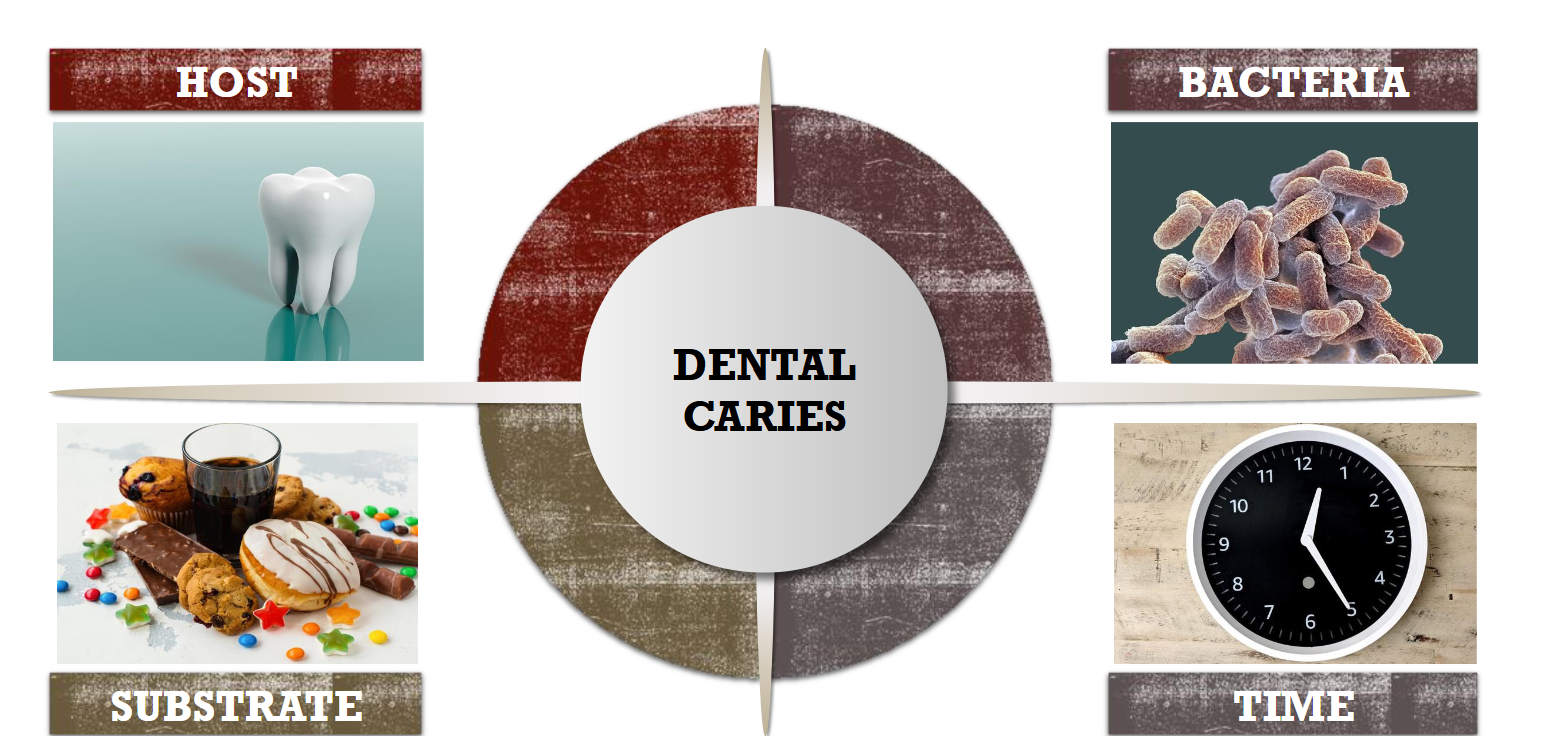
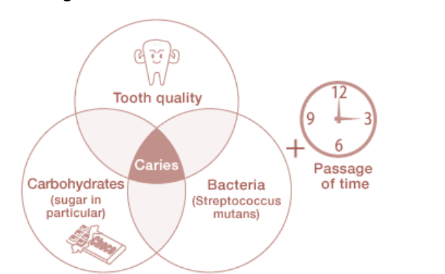
CAUSING FACTORS of Dental Caries (4)
Host Types (5)
Tooth
▪ Saliva
▪ Muscular activity
▪ Habits
▪ Environment
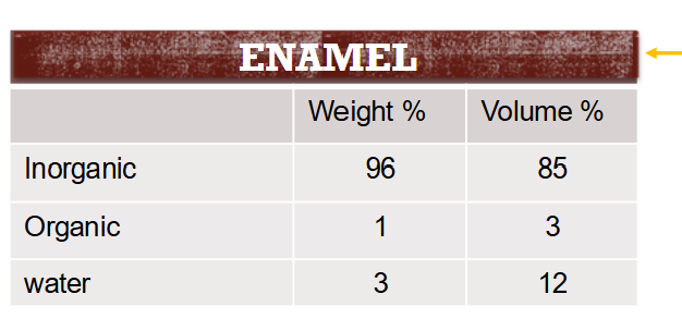
Hard Tissue Composition Enamel
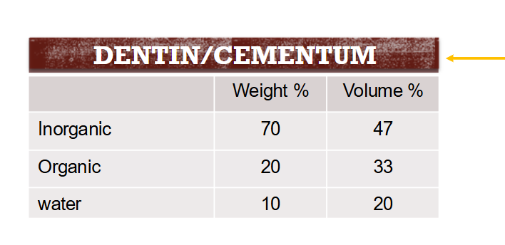
Hard Tissue Composition Dentin
SURFACE ENAMEL
Enamel is microscopically porous which makes it permeable
enamel rod
An enamel rod is the basic unit of tooth enamel. It is a tightly packed, highly organized mass of
hydroxyapatite crystals, which are hexagonal in shape and provide rigidity and strengthen the enamel.
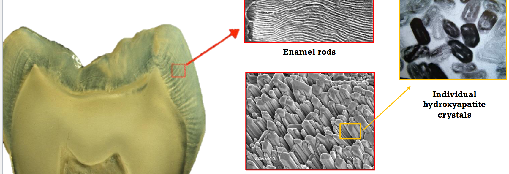
Enamel Parts (3)
Enamel rods
Interrod enamel:
Rod sheath:
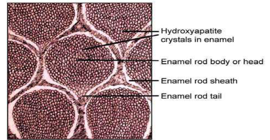
Interrod enamel
Located around the rod enamel
- Same composition as enamel rod
- Different direction
Rod sheath:
where enamel rods meet interrod
enamel
- More organic content
- Reduces stress on the rods
individual enamel crystal
About 20% of the crystal is carbonated hydroxyapatite. It is more easily dissolved
by acids
Dentin
Hydroxyapatite crystals are 30 times smaller, making
dentin somewhat softer than enamel.
Peritubular dentin
a relatively dense mineralized tissue that surrounds the tubules of
coronal tooth dentin. It is composed mainly of crystals of carbonated apatite together
with a small amount of collagen
intertubular dentin
The intertubular dentin, which lies between the tubules, is a less-calcified matrix that
consists of some apatite crystals embedded within a collagen matrix
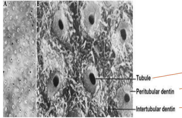
WHY IS DENTIN MORE SUSCEPTIBLE TO CARIES THAN ENAMEL?
Less inorganic content
▪ Hydroxyapatite crystals are smaller
▪ More carbonated apatite
▪ Greater numbers of impurities in the crystals
▪ Critical pH is higher than enamel
▪ Tubules offer channels for diffusion of the
bacterial acids
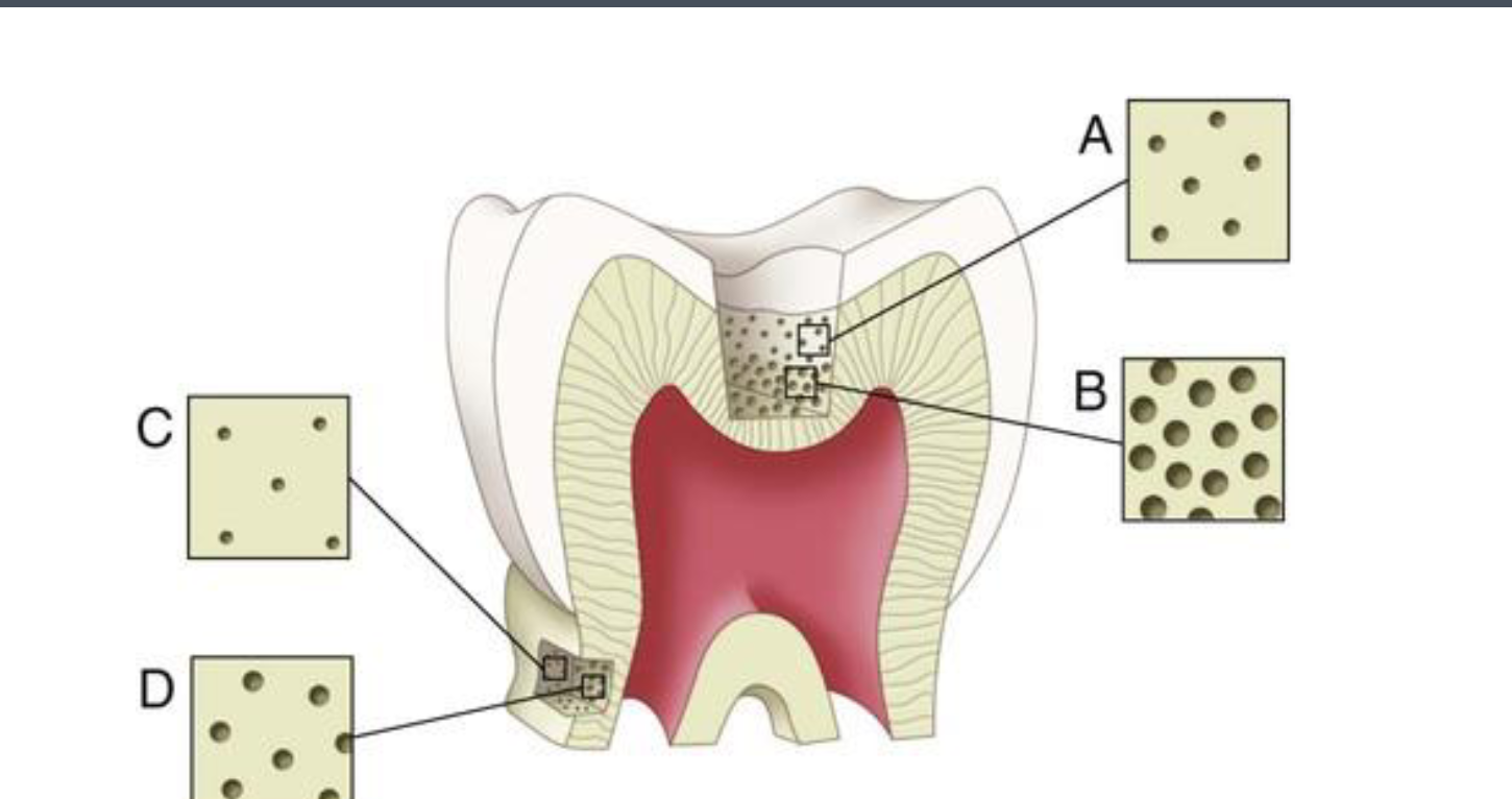
The number and diameter of the tubules increase from DEJ to the pulp
DEMINERALIZATION
Loss of minerals
When pH drops
REMINERALIZATION
Reprecipitation of minerals
When pH rises
Remineralization vs Demineralization
Remineralization is a slower process than demineralization. When remineralization is given enough time, it can eliminate the
damage done during demineralization, but in the absence of this, the caries process will progress and a lesion will develop.
STAGES OF CARIES DEVELOPMENT
It is an ongoing process, characterized by alternating periods of
demineralization and remineralization
▪ Demineralization: is the dissolving of the calcium and phosphate
from the hydroxyapatite crystals
▪ Remineralization: is the calcium and phosphate being redeposited in
previously demineralized areas
▪ It is possible to have the processes of demineralization and
remineralization occur without any loss of tooth structure
Critical pH
The critical pH is the pH at which saliva and plaque fluid cease to be saturated with calcium
and phosphate, thereby permitting the hydroxyapatite in dental enamel to dissolve.
• It is the highest pH at which there is a net loss of from dental hard tissues, which is generally
accepted to be about 5.5 for enamel.
Critical pH values (4)
Enamel → 5.5
• Dentin → 6.0
• Cementum →6.7
• Fluorapetite →4.5
PROGRESSION OF DENTAL CARIES (5)
Normal enamel surface →first stages of demineralization →Enamel surface breaking down →
dentin is carious→ cavitation
Caries Progression in Enamel
Subsurface demineralization opposite plaque surface
• Some of the dissolved mineral gets re-deposited at the surface as
it exits the tooth
• Lesion can reach dentin without cavitation
• Remineralization is still possible (if not cavitated)
TOOTH FACTOR
Certain surfaces of a tooth are more prone to decay. Eg : In mandibular 1st molar
the most prone areas in order are occlusal, buccal, mesial, distal and lingual.
▪ Position of tooth on the arch: malaligned, rotation
▪ Surface enamel is more resistant to caries than sub-surface enamel because it is
highly mineralized
▪ Dentin is more vulnerable than enamel because of structural differences and
greater numbers of impurities in the crystal lattice
COMPOSITION OF SALIVA:
Water
▪ Inorganic
▪ Organic
▪ mucus
▪ Lipids
▪ Enzymes
INORGANIC COMPONENTS IN SALIVA
Calcium and phosphate
▪ remineralization and preventing demineralization.
▪ Phosphate also acts as a buffer
▪ Potassium, sodium, chloride as in other body
fluids
▪ Bicarbonate
Bicarbonate Function in Saliva
acts as a buffer
▪ pH 6.8 - 7.3
ORGANIC COMPONENTS IN SALIVA
Neumerous proteins and lipids
Enzymes
SALIVARY FUNCTION
Clearance
2. Antibacterial
3. Lubrication
4. Mineral exchange
CLEARANCE
Saliva clears carbohydrates from the plaque
Reduced saliva flow means less of all the components, less clearance, less protective effect
Normal stimulated flow rate is about 1.0 ml/minute or more
Stimulated flow rate of less than 0.7 ml/minute is a high risk factor for dental caries
Unstimulated vs Stimulated Clearance in saliva
Unstimulated: At rest
▪ Stimulated: When secretion is promoted by different stimuli
Types Of Enzymes
She said they dont matter
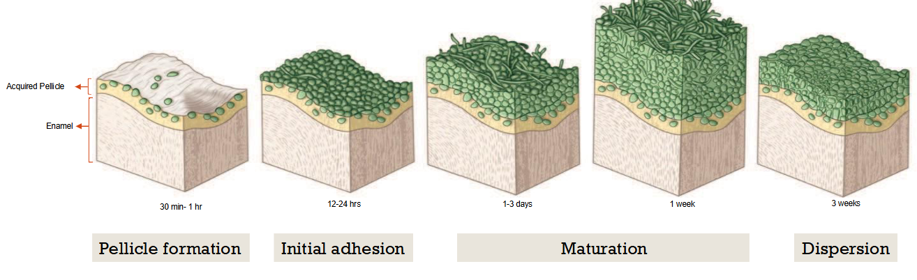
SALIVARY PELLICLE
Thin film on the surface of the enamel
▪ Comprised of strongly adsorbed specific proteins and lipids from the saliva
▪ Forms within mins/hours. Matures in days.
▪ Multiple layers
If it mature becomres bad → will start plac formation
GOOD SALIVARY EFFECTS
Salivary proteins contribute to the pellicle to
protect the outer tooth surface
▪ Salivary proteins maintain supersaturation of
calcium phosphate
▪ Salivary calcium and phosphate inhibit
demineralization and enhance
remineralization
▪ Saliva carries fluoride around the mouth
▪ Salivary components buffer plaque acids
▪ Salivary proteins have antibacterial properties
BAD SALIVARY
EFFECTS
Pellicle formed allows bacteria to colonize on the
tooth
▪ Provides a sticky surface
▪ Provides nutrients for bacteria
▪ Precursor to plaque formation
▪ With low saliva, caries activity grows
exponentially
DECREASED
SALIVARY FLOW
Slow clearance of food particles
▪ Decrease buffering of acids
▪ Encourage growth of bacteria
▪ Inhibit remineralization because calcium
and phosphate ions are reduced
Causes DRY MOUTH (XEROSTOMIA)
DRY MOUTH (XEROSTOMIA) CAN BE CAUSED BY
Many medications
▪ Radiation therapy to head and
neck
▪ Salivary gland disease
▪ Diabetes
▪ Decreased function with aging
Bacteria Criteria for Cariogenicity (In Caries) (4)
An organism must acidogenic (Produces)
An organism must be aciduric (tolerates)
An organism must exhibit tropism for teeth (sticks)
An organism must utilize refined sugar (sucrose)
Organisms responsible for dental caries (2)
Streptococcus mutans (MS): associated with the onset
of caries
▪ Lactobacilli: associated with active progression of
cavitated lesion
S.MUTANS IS THE PRIMARY CAUSATIVE AGENT OF INITIAL CARIES BECAUSE
It adheres to enamel (enzymes causes the formation of extracellular
polysaccharide which allows it to stick to smooth tooth surfaces)
▪ Acidogenic: It produces acid (it metabolizes sucrose to an end product of
lactic acid)
ACQUISITION OF MS
About 70% of children get S.Mutans from mom
- Significantly associated with maternal salivary levels
of bacteria
- Referred to as “vertical transmission”
Biofilms
Biofilms are a collective of one or more types of microorganisms that can grow on many different surfaces.
Dental plaque
Dental plaque is a structurally and functionally organized biofilm.
Dental plaque has been defined as the diverse community of micro-organisms found on a tooth surface as a
biofilm embedded in a matrix of polymers of host and bacterial origin
PLAQUE FORMATION (4 Stages)
SUBSTRATE (In Caires
Cariogenic foods
High-frequency exposure to fermentable
carbohydrates such as sucrose → produce
cariogenic biofilm
▪ Fermentable carbohydrates→ change
composition of biofilm→favors growth of
acidogenic bacteria
Moderation is very important
Cariogenic foods
Are those that contain
fermentable substrates that can be
metabolized by oral bacteria to cause a
decrease in plaque pH < 5.5 and result in
demineralization of tooth enamel and
ultimately caries
Dietary advice for caries prevention is
based on three principles
CLASSIFICATION BASED ON HARD TISSUE AFFECTED
Enamel caries
▪ Dentin caries
▪ Cementum caries
BASED ON SURFACE AFFECTED
Pit and fissure caries
▪ Smooth surface caries
▪ Root surface
BASED ON RATE OF PROGRESSION
Acute/ rampant caries
2. Chronic caries
3. Arrested caries
Acute/ rampant caries
- Rapidly invading caries involving several teeth
- Appears soft or light colored
- If unattended causes early pulp involvement
2. Chronic caries
- Slowly progressing, long standing caries
- Hard consistency and dark colored
3. Arrested caries
Sometimes chronic caries lesion can become arrested
due to change in local environment
BASED ON EXTENT OF CARIES (2)
Incipient (non-cavitated) caries
Cavitated caries
Incipient (non-cavitated) caries
First evidence of caries activity in enamel
- Demineralized enamel not extended to DEJ
- Enamel surface is hard and still intact
- Can be remineralized
Cavitated caries
- Caries has spread beyond enamel into dentin
- Enamel is broken down and remineralization is not
possible
BASED ON ONSET
Initial / primary caries
Recurrent caries
Initial / primary caries
The original carious lesion of the tooth
Recurrent caries
- Caries seen around the margins of restoration
- Occurs due to microleakage and other
favorable conditions
BASED ON WHETHER CARIES IS COMPLETELY REMOVED
OR NOT
Residual caries
- Caries that remains in the prepared cavity even after the restoration is completed
- This may be left behind either by accident, neglect or intension
CLASSIFICATION SYSTEMS (2)
▪ GV black:
- Most commonly used
- Based on affected surfaces (location)
▪ ICDAS classification:
- Based on clinical representation and progression of the
lesion (extent)
CLINICAL CHARACTERIZATION OF CARIES
The characteristic varies vary with the nature of the surface on which the lesion
develops
There are 3 different clinical sites:
Pits and fissures (occlusal surface of posterior teeth)
2. Smooth enamel surfaces (buccal, lingual, facial, palatal)
3. Root surface
PITS AND FISSURES
Most susceptible surface to development of dental caries
▪ Cross section: Inverted V with narrow entrance and progressively wide area of
involvement closer to the DEJ
SMOOTH ENAMEL SURFACES
▪ The smooth enamel surfaces of teeth are less favorable site for plaque attachment.
▪ Cross section: V shaped with a wide area of origin and the apex of the v directed
towards DEJ
ROOT SURFACE
Root surfaces are rougher than enamel and readily allows plaque formation.
▪ Cementum covering the root surface is extremely thin and provides little
resistance to caries attack.
▪ Cross section: U shaped with less well defined margins .
ZONES OF ENAMEL CARIES
a) Surface zone: appears to be intact (reprecipitation, hypermineralization).
b) Body of lesion: subsurface, the largest portion of the lesion, mineral dissolution.
c) Dark zone: surrounds body of lesion, zone where caries is in progress, many pores
d) Translucent zone: represents the advancing front of the lesion (limiting zone)
.
A sharp explorer can easily penetrate the outer enamel and cause a small cavitation on an active lesion
ZONES OF DENTIN CARIES (3)
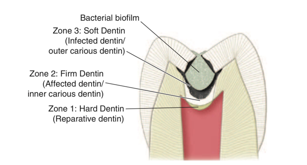
Soft Dentin
(infected dentin)
-Bacterial contamination
-Necrotic
-Irreversibly denatured collagen
-Cannot be remineralized
-Does not self repair
-Easily excavated
-Needs to be removed
Firm Dentin
(affected dentin)
- Demineralization of intertubular dentin
- Softer than hard dentin
- Intact collagen cross linking
- Leathery texture
- Can be remineralized
- Resistant to hand excavation, can only be removed by putting pressure
- Do not remove
Hard Dentin
- Cannot be easily penetrated with a blunt explorer
- Can only be removed by a bur or a sharp cutting instrument.
EXAMINATION AND DIAGNOSIS (4)
Good history
▪ Caries risk assessment
▪ Clinical evaluation
▪ Radiographic analysis
Incipient stages:
arrest and remineralize
Only when changes are irreversible: surgical treatment “drill and fill”
CARIES RISK ASSESSMENT
Clinicians treat the entire patient and not just individual teeth and caries lesions.
▪ Dental caries is a multifactorial medical disease process
▪ Individualize caries diagnosis and treatment or interventions for each patient
CAMBRA
Caries
Management
By
Risk
Assessment
.
represents a management philosophy that manages the caries disease process using a medical model.
CAMBRA Purpose
It is a process that provides an individualized evaluation of a patient’s pathologic
factors and protective factors and assesses the patient’s risk for developing future
disease.
▪ The risk assessment is then used to develop an individualized evidence- based
caries management plan that involves aspects of nonsurgical therapeutics and
dental surgical interventions
GOALS OF CAMBRA
Emphasis of the whole disease process
▪ Taking into account all factors that contribute to the
development of caries
▪ Pathological factors vs. Protective factors
▪ Shift focus from traditional restorative approach toward
the cause of the disease and the need to modify the
causes wherever possible
▪ Shift the caries balance towards the protective factors
Caries Management: The Medical Model
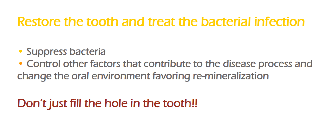
MAKE RECOMMENDATIONS BASED ON THE CARIES RISK
Increase the Protective Factors (4)
(a) Topical Fluoride delivery
(b) Saliva stimulation and supplementation
(c) Antibacterial treatment
(d) Minimally invasive restorative dentistry
General guidelines for caries management for a high- caries-risk patient
tep 1: Plaque control
• Provide prophylactic treatment followed by fluoride application
• See patient regularly to reinforce oral hygiene
Step 2: Treatment of existing caries lesions
• Treat noncavitated lesions as needed
• Restore cavitated lesions and seal surrounding pits and fissures as needed
• Salivary flow measurement to check for dry mouth and treat for xerostomia if needed
Step 3: Protection of surfaces at risk
• Seal all deep pits and fissures
• Apply fluoride varnish to exposed roots
Step 4: Maintenance care for prevention
• Review oral hygiene and dietary habits and advise patient to: Reduce the number of between-meal sweet snacks
• Substitute snacks rich in protein
• Advise patient to brush at least twice daily with high fluoride delivery toothpaste
• Advise patient to floss once daily
• Provide home treatment and/or other adjunctive therapy: Use over-the-counter fluoride rinse daily
• Chew or suck xylitol-containing gum or candies three times daily
• Recall patient every 3 months to: Reevaluate current caries risk
• Receive fluoride varnish treatment of all teeth
• Obtain bitewing radiographs every 3 to 6 months to check for lesions
eneral guidelines for caries management for a Moderate caries-risk patient
Step 1: Plaque control
• Provide prophylactic treatment followed by fluoride application
• See patient regularly to reinforce oral hygiene
Step 2: Treatment of existing caries lesions
• Treat noncavitated lesions as needed
• Restore cavitated lesions and seal surrounding pits and fissures as needed
Step 3: Protection of surfaces at risk
• Seal all deep pits and fissures
• Apply fluoride varnish to exposed roots
Step 4: Maintenance care for prevention
• Review oral hygiene and dietary habits and advise patient to: Reduce the number of between-meal sweet snacks
Substitute snacks rich in protein
• Advise patient to brush twice daily with over-thecounter fluoride toothpaste and floss once daily
• Provide home treatment and/or other adjunctive therapy: Use over-the-counter fluoride rinse daily
• Chew or suck xylitol-containing gum or candies three times daily
• Recall patient every 3 to 6 months to: Reevaluate current caries risk
• Receive fluoride varnish treatment of all teeth
• Obtain bitewing radiographs every 6-12 months to check for cavities
General guidelines for caries management for a Low caries-risk patient
Step 1: Plaque control
• Continue to reinforce oral hygiene
Step 2: Maintenance care for prevention
• Review oral hygiene habits
• Advise patient to brush twice daily with over-the-counter fluoride toothpaste
• Recall patient every 6-12 months to reevaluate current caries risk
• Obtain bitewing radiographs every 12-18 months to check for caries
OBJECTIVE OF PREVENTION
Decrease S.Mutans activity
▪ Realize that the removal of caries lesion and restoring it does not cure a caries
problem
▪ Increase protective factors
PREVENTION Antimicrobial
Chlorhexidine → Disrupts the bacterial cell wall with its positive charge
Only for 2 weeks
Fluoride
topical, varnish, dentifrices, mouthwash
Casein phosphopeptide-amorphous calcium phosphate (CPP-ACP
Provides a
reservoir of calcium and phosphate →enhance remineralization of dental hard tissues
Other Provention Le
Sucrose limitation
▪ Xylitol: stimulates saliva, enhances remineralization and has an antibacterial effect
▪ Oral hygiene
▪ Sealants
▪ Restorations