Head and Neck Anatomy: Chapter 1-3
 Call Kai
 Learn
Learn Practice Test
 Spaced Repetition
 Match
Match Flashcards
 Knowt Play
1/303

There's no tags or description
Looks like no tags are added yet.
Name | Mastery | Learn | Test | Matching | Spaced | Call with Kai | Chat |
|---|
No analytics yet
Send a link to your students to track their progress
304 Terms
Why is understanding head and neck anatomy important for dental professionals?
It is essential for performing patient examination procedures, both extraoral and intraoral.
What is anatomic nomenclature?
It is the system of names for anatomic structures, allowing for easy application during patient examinations and clinical procedures.
What is the anatomic position of the body?
The body is standing erect with arms at the sides, palms and toes directed forward, and eyes looking forward.
How does the anatomic position apply when the body is supine or prone?
The anatomic position is assumed even when the body may be lying on its back (supine) or front (prone), or when sitting in a dental chair.
What does the term 'anterior' refer to in anatomic nomenclature?
The front of an area in relationship to the entire body.
What does the term 'posterior' refer to in anatomic nomenclature?
The back of an area in relationship to the entire body.
What is the difference between ventral and dorsal parts of the body?
Ventral is directed toward the anterior (front), while dorsal is directed toward the posterior (back).
What does 'superior' mean in anatomic nomenclature?
An area that faces toward the head and away from the feet.
What does 'inferior' mean in anatomic nomenclature?
An area that faces away from the head and toward the feet.
What is the apex in anatomical terms?
The pointed end of a conical structure, such as the apex of the tongue.
What is the median plane?
An imaginary line dividing the body into equal right and left halves.
What is a sagittal plane?
A plane related to any imaginary line dividing the body that is parallel to the median plane.
What is the significance of reviewing anatomic nomenclature before studying head and neck anatomy?
It facilitates the application of anatomical terms during patient examinations and clinical documentation.
How can the body in anatomic position be divided?
By planes or flat surfaces.
What is the relationship between the terms anterior and posterior?
Anterior refers to the front, while posterior refers to the back of an area.
What is the role of the dental professional in relation to head and neck anatomy?
To apply knowledge of anatomy during patient examinations and clinical procedures.
What is the importance of the anatomic position in clinical settings?
It provides a standard reference point for describing locations and relationships of body structures.
How does anatomic nomenclature aid in patient records?
It ensures clear communication and documentation regarding anatomical structures and procedures.
What is the relationship between the apex and the conical structure?
The apex is the pointed end of the conical structure.
Why is it important to understand the terms superior and inferior?
These terms help describe the vertical relationship of body parts in relation to each other.
What is the purpose of dividing the body into planes?
To provide a framework for understanding the spatial relationships of anatomical structures.
What is a frontal plane in anatomy?
An imaginary line dividing the body into anterior (front) and posterior (back) parts.
What does a horizontal plane divide in the body?
It divides the body into superior (upper) and inferior (lower) parts.
What does it mean if a structure is described as medial?
It is closer to the median plane of the body.
What does lateral mean in anatomical terms?
It refers to a structure that is farther from the median plane.
What does peripheral refer to in anatomy?
It describes an area away from the center of the body.
What is the difference between proximal and distal?
Proximal refers to a structure closer to the median plane, while distal refers to a structure farther from it.
What does ipsilateral mean?
It refers to structures on the same side of the body.
What does contralateral mean?
It refers to structures on the opposite side of the body.
What is a superficial structure?
A structure located toward the surface of the body.
What does deep mean in anatomical terms?
A structure located inward, away from the body surface.
What is meant by internal in the context of hollow structures?
It refers to the inner side of the wall of a hollow structure.
What does external refer to in hollow structures?
It refers to the outer side of the wall of a hollow structure.
What is a midsagittal section?
A division through the median plane of the body.
What is a frontal section?
A division through any frontal (coronal) plane.
What is a transverse section?
A division through a horizontal plane.
What should dental professionals understand about anatomic variations?
They should recognize that while the number of bones and muscles is usually constant, specific details can vary among patients.
What is surface anatomy?
The study of the structural relationships of the external features of the body to the internal organs and parts.
What are the regions of the head?
The regions include frontal, parietal, occipital, temporal, auricular, orbital, nasal, infraorbital, zygomatic, buccal, oral, and mental regions.
What does the frontal region of the head include?
The forehead and the area superior to the eyes, defined by the deeper skull bone.
What is the glabella?
The smooth elevated area between the eyebrows.
What is the frontal eminence?
The prominence of the forehead.
What regions are covered by the scalp?
The parietal region and occipital region.
What defines the temporal region of the head?
The temple, which is the superficial side of the head posterior to each eye.
What is a prominent feature of the auricular region?
The external ear.
What are the components of the external ear?
The auricle (oval flap of the ear) and the external acoustic meatus (EAM).
What is the tragus?
The smaller flap of tissue of the auricle anterior to the external acoustic meatus.
What is the antitragus?
The flap of tissue opposite the tragus.
What is the function of the external ear?
To gather sound energy and focus it on the tympanic membrane (eardrum).
What separates the external acoustic meatus from the middle ear?
The tympanic membrane.
What is the primary function of the internal ear?
Sound detection and balance.
What structures are contained in the orbital region?
The eyeball and all its supporting structures in the bony socket or orbit.
What is the sclera?
The white area of the eyeball.
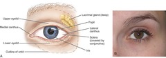
What is the iris?
The central area of coloration on the eyeball.
What is the pupil?
The opening in the center of the iris that appears black.
What are the lacrimal glands responsible for?
Producing lacrimal fluid or tears.
What is the lateral canthus?
The outer corner(s) where the upper and lower eyelids meet.
What is the medial canthus?
The inner angle(s) of the eye.
What is the main feature of the nasal region?
The external nose.
Where is the root of the nose located?
Between the eyes.
What is the landmark located inferior to the glabella in the nasal region?
The nasion, which corresponds with the junction between the underlying bones.
What forms the bridge of the nose?
The bony structure located inferior to the nasion.
What separates the nares in the nasal region?
The midline nasal septum, formed by various skull bones and nasal septal cartilage.
What are the winglike structures on each side of the nares called?
The ala (plural, alae) of the nose.
Where is the infraorbital region located?
Inferior to the orbital region and lateral to the nasal region.
What does the zygomatic region overlie?
The cheekbone, known as the zygomatic arch.
What is located inferior to the zygomatic arch and anterior to the ear?
The temporomandibular joint (TMJ), where the upper skull forms a joint with the lower jaw.
What is the composition of the buccal region?
The soft tissue of the cheek, forming the side of the face.
What muscle can be felt when a patient clenches their teeth?
The masseter muscle.
What can cause the masseter muscle to become enlarged?
Constant use, such as grinding or bruxism and clenching.
What should be examined during an extraoral examination of the facial regions?
The infraorbital, zygomatic, and buccal regions, as well as the temporomandibular joint.
What movements can be felt at the temporomandibular joint?
Opening and closing the mouth or moving the lower jaw to the right or left.
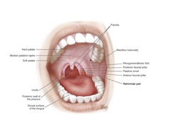
What structures are included in the oral region of the head?
The lips, oral cavity, palate, tongue, floor of the mouth, and parts of the throat or pharynx.
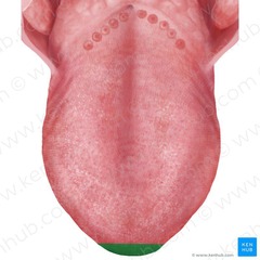
What are the three surfaces of the tongue from posterior to anterior?
Base, body, and apex.
What are the three parts of the pharynx?
Nasopharynx, oropharynx, and laryngopharynx.
Which part of the pharynx is located close to the laryngeal opening?
The laryngopharynx.
What is the opening from the oral region into the oropharynx called?
The fauces or faucial isthmus.
What forms the fauces laterally on each side?
The anterior faucial pillar and the posterior faucial pillar (also called tonsillar pillars or palatal arches).
Where is the palatine tonsils located?
Between the anterior and posterior faucial pillars.
What is the flap of cartilage located behind the base of the tongue?
The epiglottis.
What is the part of the pharynx superior to the level of the soft palate?
The nasopharynx.
What is the part of the pharynx located between the soft palate and the opening of the larynx?
The oropharynx.
What is the purpose of using a mouth mirror during a pharynx examination?
To intensify the light source and view the soft palate and throat.
How should the mouth mirror be positioned when examining the pharynx?
With the mirror side down on the middle of the dorsal surface of the tongue.
What should the patient do during the pharynx examination?
Say 'ah' to visually observe the uvula and parts of the pharynx.
What is the major feature of the mental region of the head?
The chin.
What is the mental protuberance?
The prominence of the chin.
Where is the labiomental groove located?
Between the lower lip and the chin, approximately midway between the apex of the nose and the chin.
What divides each side of the neck into anterior and posterior cervical triangles?
The sternocleidomastoid (SCM) muscle.
What major structures can be accessed through the anterior cervical triangle?
Structures that pass between the head and thorax.
What is the laryngeal prominence?
The visible part of the thyroid cartilage, also known as 'Adam's apple,' especially prominent in adult males.
What is located superior to the laryngeal prominence?
The superior thyroid notch of the thyroid cartilage.
Where are the vocal cords attached?
To the posterior surface of the thyroid cartilage.
What is the function of the hyoid bone?
Suspended in the neck, it controls the tongue and pharynx and assists in swallowing.
What forms the submandibular triangle in the anterior cervical triangle?
The main parts of the digastric muscle and the mandible.
How is the inferior part of each anterior cervical triangle subdivided?
By the omohyoid muscle into a carotid triangle and a muscular triangle.
What forms the midline submental triangle?
The two anterior bellies of the digastric muscle and the hyoid bone.
What divides the posterior cervical triangle into smaller triangular regions?
Area muscles.
What are the two subdivisions of the posterior cervical triangle created by the omohyoid muscle?
The occipital triangle and the subclavian triangle.
What structures are palpated during an extraoral examination of the neck?
The SCM muscle and the regions anterior and posterior to it.