Audiology Review Quiz #1
1/51
Earn XP
Name | Mastery | Learn | Test | Matching | Spaced | Call with Kai |
|---|
No analytics yet
Send a link to your students to track their progress
52 Terms
Eardrum (Tympanic Membrane)
Function is to transduce sound pressure into mechanic vibrations
Ossicular Chain
Three smallest bones in our body which are attached to the tympanic membrane.
Mallus, incus, and stapes
Function: Transmit sound
Function of the middle Ear
To overcome impedance mismatch (the traveling of air to fluid).
Overcome with the thumbtack and the crowbar (lever) methods
Inner Ear
Consists of 2 systems (cochlea and the vestibular system)
Cochlea: organ of hearing
Vestibular System: organ of balance
Outer Hair Cells (OHC)
Receive and Detect Sounds
Amplifiers
they exhibit motility
responsible for OAEs
connected to efferent neurons
Inner Hair Cells (IHC)
Transmits sounds to the brain
connected with afferent neurons: carry information from the cochlea to the higher auditory system.
causes the release of neurotransmitters
OHC Characteristics
3 to 5 rows
12,000 cells
50-150 Sterocilia per cell
IHC Characteristics
1 to 2 rows
3,500 Cells
50-70 Stereocilia per cell
Afferent Neurons
Carry information from the cochlea to the higher auditory system.
Efferent Neurons
Carry information from the higher auditory system (brain) to the cochlea
Functions of the Outer Ear
Collect Sounds
Localization
resonator
Protection of the middle ear
Self-mutilation
4 Different Types of output Transducers
Standard headphones (supra-aural)
Insert Earphones
Bone vibrator
Speakers in a sound field
Masking
To rule out participation of the non-test ear by using a masking noise (competing sound) in that ear
Sound can cross over based on the output transducer
Insert Earphones = 90dB
Supra-aural Headphones = 50dB
Bone Conductor= 0dB
Tympanogram
Measures the movement of the eardrum
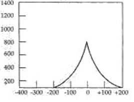
What Type of Tymp is this?
Type A
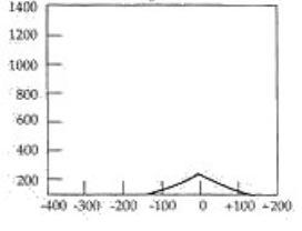
What Type of Tymp is this?
Type As
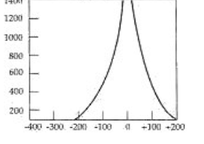
What Type of Tymp is this?
Type Ad
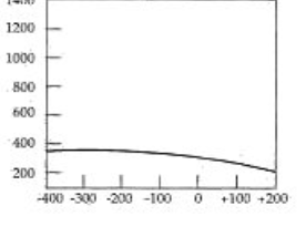
What Type of Tymp is this?
Type B
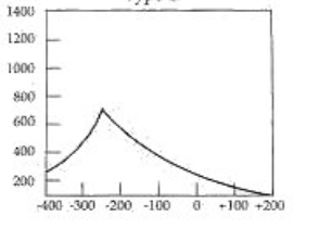
What Type of Tymp is this?
Type C
Type A Tympanogram
Normal middle ear pressure
normal eardrum movement
normal ear canal volume
Pathologies associated with this Tymp:
Otosyphillis
Presbycusis
Meniere’s Disease
Acoustic Neuroma
Microtia
Anotia
Otitis Externa
Noise Induced Hearing Loss
Ototoxicity
Auricular Tag
sometimes Tympanosclerosis
Type B Tympanogram
“Flat”
No compliance or pressure peak indicated
Middle Ear Fluid
can be normal or increase ear canal volume
Pathologies associated with this Tymp:
Perforated Eardrum
P.E Tubes
impacted Cerumen
Atresia
Chronic Otitis Media
Keratoma/ Cholesteatoma
Foreign Body in Ear Canal
Temporal Bone Fracture
Type C Tympanogram
Excessive negative middle-ear pressure
Normal or reduced compliance
Pathologies associated with this Tymp:
Eustachian Tube Dysfunction (initiation or resolution of middle ear fluid)
Type As Tympanogram
“Shallow”
the movement of the eardrum is there, but it is very slow it is hard to move
normal middle-ear pressure
reduced compliance
Pathologies related to this tymp:
Otosclerosis
Sometimes Tympanosclerosis
Fixation of the ossicles
Type Ad Tympanogram
“Flaccid”
The eardrum moves too much
increased compliance
normal middle ear pressure
Pathologies associated with Tymp:
Ossicular Disarticualtion (the bones detached from one another)
Ossicular Discontinuity (born with it)
Distortion Product Otoacoustic Emission (DPOAE)
Evoked following the presentation of two tones with frequencies that are mathematically determined to create a difference tone (the distortion product) in the cochlea.
2 tones need to go in and one need to come back out in order for an OAE to be present
When you have OAE present then it rules out any pathologies in the middle ear, outer ear or inner ear
Diagnosing Hearing Loss
Severity, configuration and Type of hearing loss
Types of Hearing Loss
Conductive Hearing Loss (temporary)
Sensorineural Hearing Loss (permanent)
Mixed Hearing Loss (both conductive and sensorineural)
Non-organic Hearing Loss (Pathological or faking)
Causes for sensorineural hearing Loss
Exposure to loud sounds
Consequences of the aging process
Genetic syndrome
Neural disorder
Vascular disorder
Infections or trauma during fetal development, at birth, in childhood, or as an adult.
true or false
The ability to hear plays a direct role in the ability to perceive and produce speech
True
4 Basic Parameters of Hearing Loss
Degree of Hearing Loss – severity of the impairment
Configuration – Shape of the hearing loss
Type of Hearing Loss – conductive, sensorineural, or mixed
Symmetry – comparison of results between ears
Air Conduction Testing
sound travels via air on a sound wave through the three parts of the ear. Traditional circumaural headphones, insert earphones, or speakers are used to transmit the sound
Bone Conducting Testing
Sound travels via bone, bypassing the outer ear and middle ear by setting the bones of the skull into vibration. The bone vibrator is placed on the mastoid bone
Conductive Hearing Loss
Involves the structures in the ear that are responsible for CONDUCTING sound to the cochlea
Can be caused by an obstruction, abnormality, or disease and is found in the outer or middle ear
This loss is typically temporary, but left untreated it can lead to a permanent condition
Sensorineural hearing Loss
Involves both the cochlear function of sensory reception and the function of the auditory nerve
Most often the problem is due to damage to the inner or outer hair cells or other structures in the cochlea
Sometimes, this problem occurs because of problems with auditory nerve fibers, in the auditory nerve, or centrally in the auditory central nervous system extending through the brain stem to the cerebral cortex
Because of its location, sensorineural hearing loss is usually permanent in nature and most often untreatable medically or surgically
Can be congenital or acquired, early onset or delayed, unilateral or bilateral
Otitis Externa
Can cause swelling, pain, and drainage
Inflammatory consequences can occlude the outer ear causing conductive hearing loss
Caused by several types of common bacteria
May be caused by fungus, which we call otomycosis
Treated with medication to fight infection and swelling
Can be extremely dangerous for diabetics if it develops into malignant otitis externa
Perforation in the Tympanic Membrane
Significant because it can create a conductive hearing loss, but also because it leaves the middle ear space open to the elements
Causes:
Spontaneous: secondary to middle ear infection or fluid buildup
Direct trauma: from an object (Q-tip, foreign object, or welding slag)
Concussive incident: (slap to the head, explosion, or waterskiing)
Barotrauma: (change in pressure frequently seen with scuba divers)
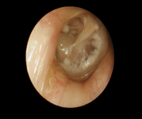
Tympanosclerosis
White plaques seen on the eardrum usually after repeated middle ear infections or after PE tubes
Plaques are caused by deposits of calcium in the tissue (collagen) of the tympanic membrane, but does not usually cause hearing loss
Type A or As Tymp
Eustachian Tube Dysfunction
Inflammatory tissue change in the nasopharynx:
Sinusitis, Adenoiditis, Tonsillitis, or growth of a mass
Treatment includes:
Antibiotics or surgical removal of the tonsils and/or adenoids
Eustachian tube dysfunction ALONE creates at most a mild conductive hearing loss, but may lead to further problems in the middle ear space
Type C Tymp
Otitis Media
General term for an inflammation or infection of the middle ear, which can invade the mastoid cavity
3 main characteristics
Fluid present in the middle ear
Fluid may or may not be infected
might be degenerative changes to the tissues of the middle ear
Otitis Media with Effusion (OME)
Common version of OM and one of the most common conditions affecting the auditory systems in children
More than 2 million episodes of OME occur annually medical costs = $4 billion dollars a year
Otitis Media with effusion is fluid build up without infection
If untreated/unresolved fluid may thicken and become “glue ear”
Conductive hearing loss varies from slight to mild depending on the thickness of the fluid
Acute Otitis Media
Fluid buildup in the middle ear is infected
this version of otitis media typically is the result of bacterial infection and may or may not present with the hallmarks of infection: (fever, pain, accumlation of whote blodd cells, etc)
conductive hearing loss is usually mild but can be moderate in some cases.
Chronic Otitis Media
Unresolved infection of the middle ear including the mastoid spaces as well as the mucosal lining with non-intact TM and discharge
Infectious, permanent, progressive, and Erosive/destructive process
Unchecked erosive infection can lead to damage to the inner ear, Facial nerve and ultimately the brain (you cause damage to the barrier it can be lethal)
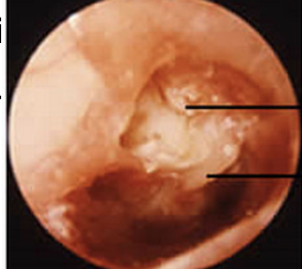
Cholesteatoma
Accumulation of dead, exfoliated skin cells from the external canal and lateral surface of the tympanic membrane also referred to as a keratoma (similar only)
Can form at any perforation site, but is most often seen on the edge of the tympanic membrane, or the upper area of the tympanic membrane when it is pulled into the middle ear space due to eustachian tube dysfunction.
Smooth, white, pearl-like growths that can erode the ME ossicles completely and may enlarge and erode into the brain cavity
Mild conductive hearing loss that is usually treated by surgical removal first and may ultimately result in the use of a hearing ai
Otosclerosis
Progressive conductive hearing loss that results in fixation of the stapes bone in the oval window
Type As Tymp
Happens because vascular and enzymatic activity degrade the old bone and new bone is structurally different – some call it spongy
Otosyphillis (Aquired Syphillis)
Symptoms of SNHL and dizziness
type A tymp
HL is usually progressive and fluctuating, bilateral but asymmetrical.
Symptoms mimic meniere’s disease and acoustic neuroma, therefore it has been called “the great imitator”
Ototoxicity
Certain medications and chemical substances cause hearing loss through sensory cell damage and interference with inner ear metabolism
Will affect the cochlea and sometimes can affect the vestibular system
Changes can be permanent and severe, or maybe revered once the appropriate medication is used
4 classes of drugs in this category
Antineoplastic Drugs
Aminoglycoside Drugs
Loop Diuretics
Analgesics and Antimalarials
Neurofibromatosis Type 2
When bilateral acoustic neuromas are found, the underlying cause is almost always NF2
Genetically/inherited condition that causes multiple neuromas, typically on sensory nerves in the head and spinal cord
Treatment is usually surgical reduction of the tumors, and the surgeon’s focus is on preserving facial nerve function and hearing
Prognosis with regard to hearing is poor with surgical reduction or without, and unfortunately hearing aids are often useless. Another option might be the Auditory Brainstem Implant (ABI)
The ABI bypasses the cochlea and auditory nerve entirely and provides direct neural stimulation to the brain stem
Treacher Collins Syndrome
Inherited
Abnormalities on the eyelids
Downward slanting eyelids
Underdevelopment of the jaw
Cleft Palate
Microtia
Atresia
Ossicular malformation or fixation
Conductive Hearing Loss
Waardenberg Syndrome
Pigmentation abnormalities of the skin, hair and eyes
Usher Syndrome
Rentinitis Pigmentosa
an inherited condition characterized by impairment of vision and hearing, resulting from the occurrence of retinitis pigmentosa and abnormal auditory neural conduction.
Type A Tymp if there is sensorineural HL
Eustachian tube Function
To equalize pressure of the middle ear
Keep the air pressure behind the eardrum the same as the pressure outside