Chater 4: Anatomy and Physiology Genes and Cellular Function
 Call Kai
 Learn
Learn Practice Test
 Spaced Repetition
 Match
Match Flashcards
 Knowt Play
1/54

There's no tags or description
Looks like no tags are added yet.
Name | Mastery | Learn | Test | Matching | Spaced | Call with Kai | Chat |
|---|
No analytics yet
Send a link to your students to track their progress
55 Terms
DNA Structure
A long, thread-like molecule with a uniform diameter but varying length. Averages about 2 inches long per chromosome.
Double helix shape.
Made up of nucleotides, which consist of three components: A sugar—deoxyribose, A phosphate group, and A nitrogenous base.
The phosphate groups and deoxyribose make up the sides of the helix facing outward.
4 DNA bases: adenine (A), guanine (G), cytosine (C), and thymine (T).
The two strands of DNA are joined via hydrogen bonds between base pairs: A pairs with T (2 H-Bonds), and G pairs with C (3 H-bonds), a stronger attachment
DNA Function
The essential function of DNA is to carry instructions (genes)
for the synthesis of proteins
Gene—segment of DNA coding for the synthesis of a specific
protein
• Humans have about 20,000 genes
• Only about 2% of total DNA are genes
• Other 98% is noncoding DNA: involved in gene regulation and chromosome structre.
What are the two base pairs that form DNA?
Adenine pairs with Thymine (2 H-Bonds).
Guanine pairs with Cytosine (3 H-bonds)—stronger attachment.
Structure of a nucleotide
A sugar—deoxyribose
A phosphate group
A nitrogenous base
DNA bases: A, T, C, G: Adenine (A), guanine (G), cytosine (C), and thymine (T).
Law of complementary base pairing
Sequence of one strand governs the sequence of the other.
Purines
Have a double-ringed structure: adenine (A), guanine (G)
Pyrimidines
Have single-ringed structure: cytosine (C), thymine (T), uracil (U)
How many chromosomes are there in most human cells?
There are 46 Chromosomes (DNA molecules in the form of chromosomes) in the nucleus of most human cells. 2 sets of 23 chromosomes one from mom and one from dad.
Chromatin structure and function
Fine filamentous DNA material compacted with proteins called histones.
Function: compacts long filamentous DNA into tightly compacted structures that can fit in a 5-micron nucleus.
Structure: DNA wraps around a cluster of 8 histones to form a core particle; those chromatin segments fold into a thicker zigzag fiber; that fiber still folds into irregular loops with increased thickening, which is where it stays in undividing cells. in diving cells, it compacts more to form chromatids, which attach to their sister chromatids to form a chromosome via centromeres.
Histones
Histones cluster in groups of eight molecules
DNA molecule winds around the cluster (like thread around spool)
Creates a granule called a core particle
Chromatin consists of thousands of repeating core particles
Chromosome territory
Each chromosome is packed into its own region of the
nucleus called a chromosome territory. Permeated with channels allowing regulatory chemicals to have access to the genes
Chromatin in nondiving cells
In a nondividing cell, the chromatin is not static
• Structure and location changes moment to moment
according to genetic activity of cell
• Genes get turned off and on.
The chromatin will be manipulated so that it is not dense around the the genes that need to be expressed, so DNA expression machinery can have acess to those genes.
Chromatin in dividing cells
When preparing to divide, cell makes copy of all nuclear DNA
• Each chromosome then consists of two parallel filaments
of identical DNA called sister chromatids
• Chromatids are joined at constricted centromere
• Kinetochores—protein plaques on each side of centromere, play a
role in cell division.
RNA structure and function
General structure:
• Contain the sugar ribose
• Bases A, U, G, C; uracil (U) replaces thymine found in DNA
• Single nucleotide chain (not a double helix) except in short regions
• Smaller than DNA, can have less than 100 or just over 10,000
bases per molecule
• Functions mainly in cytoplasm
mRNAs: encode genetic information for protein synthesis.
rRNA: ribosomol RNA forms the primary structural and catalytic backbone of ribosomes.
tRNAs: bring amino acids to ribosomes that are syntehsizing proteins from the mRNAs.
Give a working definition of the gene and explain why new
discoveries in genetics have changed our concept of what
a gene is.
Gene definition: a gene is an information-containing segment of DNA that codes for the production of a molecule of RNA that most often plays a role in synthesizing one or more proteins
The discoveries that helped us get there: (1) The body has millions of proteins and only about 20,000 genes so we know that one gene can code for one or more proteins. (2) Several genes code for non-coding RNAs like miRNAs, tRNAs, RNAi, and rRNAs.
Explain what the human genome is and what relationship
it has to the health sciences.
Genome: all the DNA in one 23-chromosome set. 3.1 billion nucleotide pairs in human genome. Individual variation comes from single-nucleotide polymorphisms.
Genomic medicine: application of knowledge of the genome to the prediction, diagnosis, and treatment of disease. Relevant to many disorders like cancer (oncogenes), Alzheimer disease, schizophrenia, obesity, AIDS, tuberculosis.
Genomics
study of the whole genome. How genes and noncoding DNA interact to affect structure and
function of the organism
Define genetic code and describe how DNA codes for
protein structure.
The genetic code is a system that enables the 4 nucleotides to code for the amino acid sequences of all proteins
• Base triplet—sequence of three DNA nucleotides that stands for one amino acid
• Codon—3-base sequence in mRNA. base triplet in DNA=codon in mRNA
• 64 possible codons available to represent 20 amino acids; 61 code for amino acids, 3 are stop codons
Stop Codons
UAG, UGA, and UAA: signal “end of message,” like a period at the end of a sentence.
Gene expression
Different genes are activated in different cells
• Any given cell uses one-third to two-thirds of its genes
• Rest remain dormant and may be functional in other types of cells
• When a gene is activated, messenger RNA (mRNA) is
made; serves as a code for a particular protein
Describe protein syntehsis and processing
DNA → RNA → protein
Transcription: DNA → mRNA; occurs in nucleus
Translation: mRNA → protein; occurs in cytoplasm
DNA Transcription
Copying genetic instructions from DNA to mRNA. Utilizes RNA polymerase, which binds to the start sequence in DNA called the TATA box and opens up the helix. It builds a complementary mRNA from the base strand of DNA. RNA polymerase rewinds the DNA helix behind it, allowing the gene to be transcribed by several polymerase molecules. Terminator: stop sequence at the end of a gene. The result is a pre-mRNA produced by transcription that then undergoes processing mRNA splicing, where enzymes remove introns and splice exons together. After mRNA splicing it is exported out of the nucleus to be translated.
C with G to the mRNA.
G with C to the mRNA.
T with A on th mRNA.
A with U on the mRNA.
what is pre-mRNA and Alternative splicing
Pre-mRNA is “immature” RNA produced by transcription; undergoes processing. Enzymes within the nucleus remove introns from the RNA and splice exons together
Alternative splicing is a process in which varitation in the way exons are spliced allow for a varity of proetisn to be produced from one gene by changing what introns are spliced.

What is DNA translation and What are the three main components?
Translation is the process where the ribosome binds to mRNA and synthesizes a protein from it using tRNAs to read the mRNA codons.
Three main participants in translation:
• Messenger RNA (mRNA): Carries code from the nucleus to the cytoplasm and has a protein cap that is the recognition site for the ribosome
• Transfer RNA (tRNA): Delivers a single amino acid to the ribosome. Contains an anticodon—series of 3 nucleotides that are complementary to the mRNA codon
• Ribosomes: Organelles that read the message and build a peptide chain. Contain a large subunit and a small subunit. Free in cytosol, on rough ER, and on nuclear envelope. E site, P site, and A site are involved in the translation process.
What are the three main steps in translation?
Three main steps in translation:
• Initiation: Ribosome assembles with mRNA in cytosol. Small subunit binds leader sequence of mRNA, large subunit joins; when AUG codon is reached, protein synthesis begins tRNA brings first amino acid, methionine
• Elongation: Next tRNA with amino acid arrives, binds to A site. Ribosome creates peptide bond between first and second amino acids. Ribosome moves down mRNA by one codon, so growing peptide is now attached to tRNA in P site. Process continues and peptide grows
• Termination: Ribosome reaches a stop codon. A site binds a protein called a release factor. Ribosome disassembles and dissociates from mRNA.
What occurs during translation of proteins that are destined for lsyosomes or secretion?
Proteins destined for lysosomes or secretion are midified by the ER. Ribsomes dock on the ER when a specific sequence of the mRNA is translated and synthesizes protein into the rough ER cisternae through a pore in the ER. The rough ER modifies and packages protein into transport vesicles for secretion or delivery to lysosomes.
Polyribosomes
multiple ribosomes can translate the smae mRNA molecule simultaneously.
Protein processing and secretion
Proteins that have been freshly synthesized must fold into precise secondary and tertiary structures (some need to be joined to other proteins in a quaternary structure). Chaperone proteins guide the folding of newly synthesized proteins prevents improper association between different proteins.
What is the pathway of a protein that is translated in the ER?
1.) Post-translational modification. After translation, enzymes in the ER modify the protein by removing some amino acid segments, facilitating folding, stabilizing it with disulfide bridges, and adding calcium.
2.) The protein pinches off from the ER in a transport vesicle via clathrin-protein-mediated exocytosis.
3.) The vesicles of the protein fuse to the membrane of the Golgi apparatus and form a new Golgi cistern, the cis cistern.
4.) The new protein from the cis cistern migrates through the Golgi to the opposite trans cistern, where the trans cistern breaks up into vesicles containing the protein.
5.) Some vesicles become lysosomes; others become secretory vesicles that fuse with membrane to release the protein into the surrounding environment.
Heat shock proetins or stress proteins
chaperone proteins that are synthesized in response to heat or stress that help facilitate the proper folding of damaged proteins into correct functions structure.
Gene regulation
Cells can turn some genes permanently off. Example: liver cells turn off hemoglobin genes.
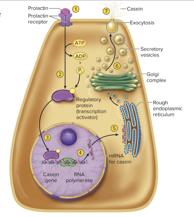
Cells can turn genes on only when needed. The level of gene expression can vary from day to day or hour to hour. This can be controlled by chemical messengers such as hormones. Example: In response to prolactin, mammary gland cells turn on gene for casein protein only when breast milk is produced.
describe the Gene regulation of Casein synthesis and secretiuon
1. Hormone prolactin binds to receptors on membrane of mammary cell
2. Receptors trigger activation of a regulatory protein (transcription
activator) in cytoplasm
3. Regulatory protein (transcription factor) moves into the nucleus and binds to the DNA near the casein gene.
4. The binding enables RNA polymerase to bind to the gene and transcribe it, producing the mRNA for casein
5. The casein mRNA moves to the cytoplasm, and ribosomes on rough ER translate it
6. The Golgi complex packages casein into secretory vesicles
7. The secretory vesicles release the casein by exocytosis, and it
becomes part of the milk.
Explain how DNA indirectly regulates the synthesis of
nonprotein molecules: Protein pathway for production of testosterone
• A cell of the testes takes in cholesterol
• Enzymatically converts it to testosterone
• Only occurs when genes for the enzyme are active
Genes may greatly affect such complex outcomes as behavior, aggression, and sex drive

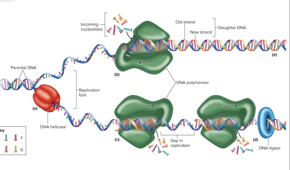
Describe how DNA is replicated.
Four steps of DNA replication:
1. Unwinding the helix from the histones
2. The DNA helicase enzyme unzips a small portion of the helix, separating the strands and forming a replication fork
3. DNA polymerase moves along each strand, reads the exposed bases, and synthesizes complementary strands
• DNA ligase enzyme binds/connects on the new but discontinuous strand.
• Semiconservative replication—each new DNA molecule contains old (parental) DNA strand and a newly synthesized DNA strand
4. New histones are synthesized to organize new DNA strands
into nucleosomes and they are further condensed into sister chromatids.
DNA Damage response (DDR)
Mechanisms in place to correct replication errors.
DNA polymerase double checks the new base pair and tends to
replace incorrect, biochemically unstable pairs with more stable,
correct pairs.
Result is only one error per 1 billion bases replicated
Discuss the consequences of replication errors.
Mutations
• Changes in DNA structure due to replication errors or environmental
factors (radiation, viruses, chemicals)
• Some mutations cause no ill effects, others kill the cell, turn it
cancerous, or cause genetic defects in future generations (oncogenes)
• Genetic variation within one’s body, due to mutations and
replication errors, is called genetic mosaicism
Genetic Mosaicism
A condition where a single person has two or more distinct populations of cells with different DNA makeup, caused by a mutation that happens after fertilization.
Somatic Mosaicism: Affects the body's non-reproductive cells; symptoms depend on how many and which tissues carry the variant.
Germline Mosaicism: Limited to the egg or sperm cells; the parent usually shows no signs of the condition, but can pass the genetic variant to offspring.
The cell cycle: Describe the life history of a cell, including the events of
mitosis
The cell cycle includes interphase and the mitotic phase
Interphase: includes three subphases: First gap phase (G1), Synthesis phase (S), and Second gap phase (G2).G1 phase: Growth and normal metabolic roles. The first gap phase is the interval between cell division and DNA repilication where the cell carries out normal tasks and synthesizes proteins and material for next phase.
S phase: synthesis phase is the phase in which the cell replicates all nuclear DNA and duplicates centrioles.
G2 phase: second gap phase is the interval between DNA replication and mitosis. The cell repairs DNA replication errors, grows, and synthesizes enzymes that control mitosis.
Mitosis: the cell replicates its nucleus and divides into two new daughter cells. The mitotic phase includes multiple subphases: Prophase, Metaphase, Anaphase, Telophase, and Cytokinesis.
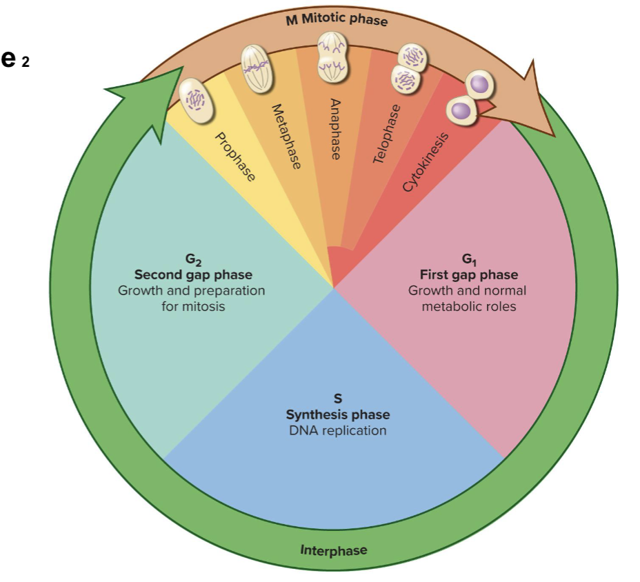
G0 or G zero phase
describes cells that have left the cycle and cease diving for a long time or permanently.
Mitosis
It is a cell division resulting in two genetically identical
daughter cells, both with 46 chromosomes. Responsible for the Development of an individual from a fertilized egg to about 50 trillion
cells and the Growth of all tissues and organs after birth, replacement of cells that die, and repair of damaged tissues.
Prophase:
Genetic material condenses into compact 46 (two chromatids per chromosome) chromosomes for easier distribution to daughter cells.
Nuclear envelope disintegrates.
Centrioles form spindle fibers (long microtubules) that push centriole pairs apart and attach to kinetochores of centromeres on chromatids.
Metaphase:
Chromosomes are aligned on the cell equator. Metaphase plate
Spindle fibers form a lemon-shaped array called the mitotic
spindle.Shorter microtubules from centrioles complete a star-like
aster that anchors itself to the inside of the two opposite sides of the cell membrane.
Anaphase:
An enzyme cleaves two sister chromatids apart at the centromere
Single-stranded daughter chromosomes migrate to each
pole of the cell as motor proteins in kinetochores crawl
along spindle fibers
Telophase and cytokinesis:
Chromosomes cluster on each side of the cell.
Rough ER makes a new nuclear envelope around each
cluster.Chromosomes uncoil to chromatin.
Mitotic spindle disintegrates.
Each nucleus forms a nucleolus.
The cell pinches in half to form two new daughter cells.
Prophase
Prophase:
Genetic material condenses into compact 46 (two chromatids per chromosome) chromosomes for easier distribution to daughter cells.
Nuclear envelope disintegrates.
Centrioles form spindle fibers (long microtubules) that push centriole pairs apart and attach to kinetochores of centromeres on chromatids.
Metaphase
Metaphase:
Chromosomes are aligned on the cell equator. Metaphase plate
Spindle fibers form a lemon-shaped array called the mitotic
spindle.Shorter microtubules from centrioles complete a star-like
aster that anchors itself to the inside of the two opposite sides of the cell membrane.
Anaphase
Anaphase:
An enzyme cleaves two sister chromatids apart at the centromere
Single-stranded daughter chromosomes migrate to each
pole of the cell as motor proteins in kinetochores crawl
along spindle fibers
Telophase and cytokinesis
Telophase and cytokinesis:
Chromosomes cluster on each side of the cell.
Rough ER makes a new nuclear envelope around each
cluster.Chromosomes uncoil to chromatin.
Mitotic spindle disintegrates.
Each nucleus forms a nucleolus.
The cell pinches in half to form two new daughter cells.
Telophase is the end of nuclear division but overlaps
cytokinesis.Achieved by the myosin protein pulling on actin in the terminal web of the cytoskeleton. Creates a crease called the cleavage furrow around the equator of the cell. Cell pinches in two.
Explain how the timing of cell division is regulated.
Cell cycle is regulated by a molecular timer and checkpoints
Proteins constitute the timer:
• Cyclins: Form during interphase; levels rise and fall through cell cycle
• Cyclin-dependent kinases (Cdks): Activated by cyclins to phosphorylate other proteins
• Events under Cyclin–Cdk complex control: (1) Replication of DNA and centrioles in the S phase, (2) Condensation of chromosomes, breakdown of the nuclear envelope, formation of the mitotic spindle, and attachment of chromosomes to the spindle in prophase, and (3) Splitting of the centromere and separation of the sister chromatids
at anaphase.
What are the three checkpoints of the cell cycle?
Start or G1 checkpoint: Either allows the cell to proceed toward the S phase or, if it doesn’t, the cell goes into the noncycling G0 phase
G2 ∕ M checkpoint: Late in the G2 phase determines whether the cell is able to proceed to mitosis.
A third checkpoint at the transition from metaphase to anaphase determines whether the cell can proceed to anaphase, leading to separation of its sister chromatids
Heredity
transmission of genetic characteristics from parent to offspring
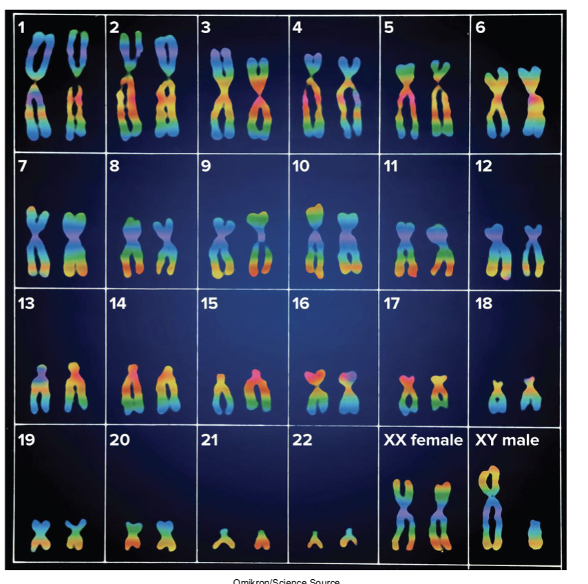
Karyotype
chart of all 46 chromosomes laid out in order by size. 23 pairs of homologous chromosomes.
Homologous chromosomes
1 chromosome from each pair inherited from each parent.
22 pairs of autosomes (look alike and carry the same genes).
1 pair of sex chromosomes where female has homologous X chromosomes and make has one X chromsome and one much smaller y chromsome.
Locus
a genes positoion on a chromsome
Alleles, dominant allele, and recessive allele
Alleles: Different forms of a paticular gene.
Alleles are found at the same locus on homologous chromosomes
• Dominant allele (represented by capital letter): If present, corresponding trait is usually seen in the individual. Masks effect of recessive allele. Often produces protein responsible for visible trait
• Recessive allele (represented by lowercase letter): Corresponding trait only seen when recessive allele present on both homologous chromosomes. Often codes for a nonfunctional variant of the protein
Gene pool
genetic make up of a whole population. multiple alleles in a population.
Codominance
both alleles equally dominant. both are phenotypically expressed.
Incomplete dominance.
intermediate phenotype between the two phenotypes like red and hwit make pink.
Polygenic inheritance
genes at two or more loci contribvute ot a single trait.