GW BGZ 2025 Case 2 - Hiking in the hills
1/43
There's no tags or description
Looks like no tags are added yet.
Name | Mastery | Learn | Test | Matching | Spaced | Call with Kai |
|---|
No analytics yet
Send a link to your students to track their progress
44 Terms
What is muscle tissue and what is its main function?
Muscle tissue is one of the four basic tissue types and is specialized for contraction and force generation. It contains actin and myosin proteins, which interact to produce movement. Muscle functions include body movement, posture, movement of substances through organs, and heat production.
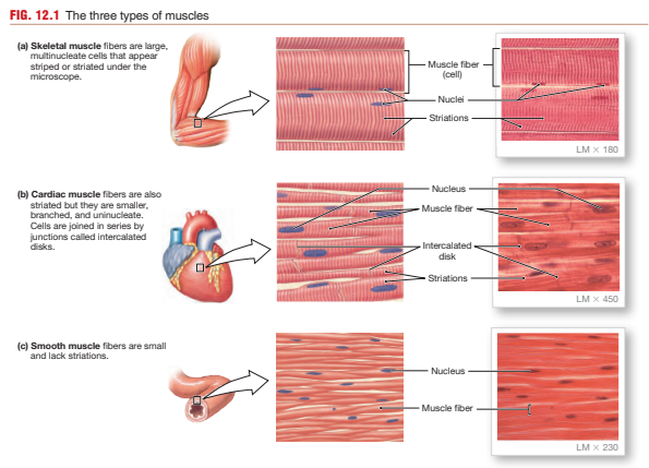
What are the three types of muscle tissue?
Skeletal muscle
Long multinucleated fibers
Striated
Voluntary control
Produces rapid, forceful contractions
Cardiac muscle
Branched striated cells
Intercalated discs
Involuntary rhythmic contraction
Found in the heart
Smooth muscle
Spindle-shaped cells
Non-striated
Involuntary contraction
Found in hollow organs and vesselsWhat are sarcoplasm, sarcolemma, and sarcoplasmic reticulum?
What are sarcoplasm, sarcolemma, and sarcoplasmic reticulum?
Sarcoplasm: cytoplasm of muscle cells
Sarcolemma: muscle cell membrane
Sarcoplasmic reticulum (SR): specialized smooth ER that stores and releases Ca²⁺ for contraction
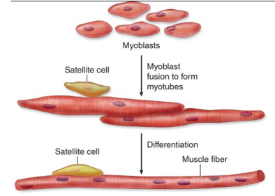
How does skeletal muscle develop embryologically?
Development sequence:
Myoblasts proliferate
Myoblasts fuse together
Multinucleated structures called myotubes form
Contractile proteins accumulate
Mature muscle fibers develop
Important features:
Mature skeletal muscle fibers are multinucleated because they formed by fusion of many myoblasts
Nuclei migrate to the periphery beneath the sarcolemma
Satellite cells
A population of stem-like cells remains between:
Sarcolemma
External lamina
Functions:
Muscle repair
Regeneration after injury
Hypertrophy support
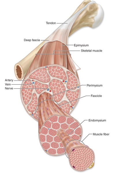
What are the connective tissue layers of skeletal muscle?
Epimysium
Dense irregular connective tissue
Surrounds the entire muscle
Continuous with tendons and fascia
Perimysium
Surrounds fascicles (bundles of fibers)
Contains:
Blood vessels
Nerves
Lymphatics
Endomysium
Delicate connective tissue around each individual muscle fiber
Contains capillaries and reticular fibers
Functional importance
These layers:
Support muscle structure
Carry vessels and nerves
Transmit force from fibers to tendons and bone
What structures are found inside a skeletal muscle fiber?
Myofibrils
Long cylindrical contractile structures
Occupy most of the sarcoplasm
Composed of repeating sarcomeres
Sarcoplasmic reticulum
Surrounds myofibrils
Stores calcium ions
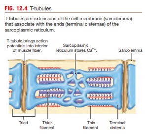
T-tubules
Invaginations of the sarcolemma
Conduct action potentials deep into the cell
Triads
A triad consists of:
One T-tubule
Two terminal cisternae of the SR
Function:
Rapid excitation-contraction coupling
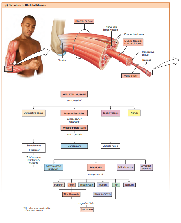
What is a sarcomere?
The sarcomere is the:
Basic functional contractile unit of skeletal muscle
Repeating unit between two Z discs
Sarcomeres are arranged end-to-end within myofibrils.
During contraction:
Sarcomeres shorten
Muscle fibers shorten
Force is generated
The highly organized arrangement of sarcomeres creates the striated appearance of skeletal muscle.
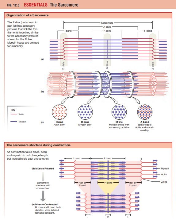
What are the bands and zones of a sarcomere?
Z disc
Boundary of a sarcomere
Anchors thin filaments
I band
Contains only thin filaments
Light band
A band
Contains entire thick filament length
Includes overlap with thin filaments
Dark band
H zone
Central region of A band
Contains only thick filaments
M line
Middle of sarcomere
Anchors thick filaments
During contraction:
I band shrinks
H zone shrinks
A band remains constant
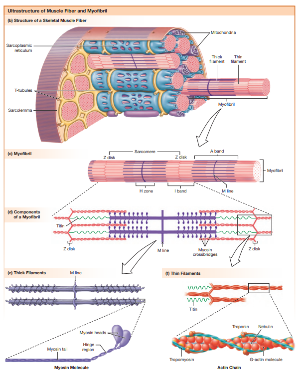
What are thick filaments composed of?
Thick filaments are primarily composed of myosin molecules.
Each myosin molecule contains:
Two heavy chains
Tail region
Two globular heads
Myosin heads:
Bind actin
Hydrolyze ATP
Generate force
The heads form cross-bridges with actin during contraction.
Myosin acts as an ATPase enzyme and converts chemical energy into mechanical movement.
What are thin filaments composed of?
Actin
Exists as F-actin (filamentous actin)
Double helical structure
Contains myosin-binding sites
Tropomyosin
Long regulatory protein
Lies along actin filament
Covers myosin-binding sites during rest
Troponin complex
Function
When calcium binds troponin:
Tropomyosin shifts away
Myosin-binding sites exposed
Contraction becomes possible
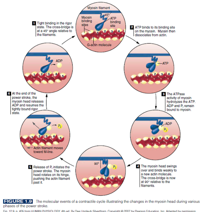
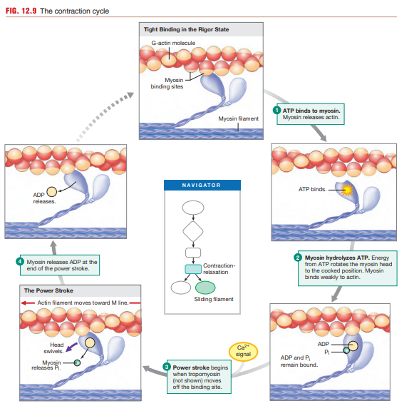
What is a cross-bridge?
A cross-bridge is the connection formed when:
A myosin head binds to actin
Sequence:
Calcium binds troponin
Tropomyosin moves
Binding sites exposed
Myosin binds actin
Cross-bridge forms
Importance
Cross-bridge cycling:
Produces force
Causes sarcomere shortening
Drives muscle contraction
What are titin and nebulin?
Titin
Giant elastic protein
Extends from Z disc to thick filament
Functions:
Stabilizes sarcomere
Maintains thick filament alignment
Contributes to elastic recoil after stretching
Nebulin
Runs alongside thin filaments
Functions:
Stabilizes actin
Maintains thin filament length
Helps organize sarcomeres
Both proteins are essential accessory structural proteins.
What are the major skeletal muscle fiber types?
Type I (slow oxidative)
Slow contraction
Aerobic metabolism
High fatigue resistance
Type IIA (fast oxidative-glycolytic)
Fast contraction
Mixed metabolism
Intermediate fatigue resistance
Type IIX/IIB (fast glycolytic)
Very fast contraction
Anaerobic glycolysis
Fatigue quickly
Muscles contain mixtures of fiber types depending on function and training.
Females more type 1 and males more type 2
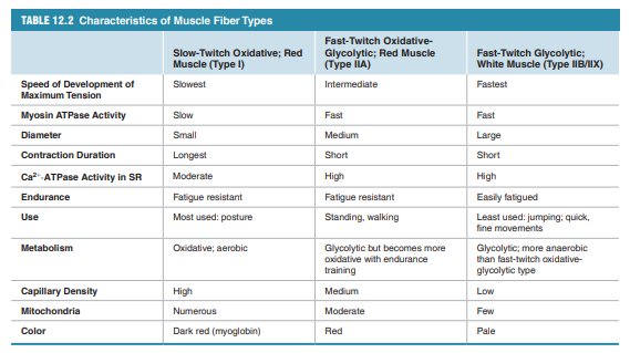
Why do Type II fibers contract faster than Type I fibers?
The contraction speed depends largely on:
Myosin ATPase activity
Type II fibers
High ATPase activity
Faster ATP hydrolysis
Faster cross-bridge cycling
Rapid force generation
Type I fibers
Lower ATPase activity
Slower cross-bridge cycling
Slower force production
Functional consequence:
Type II fibers specialized for explosive movements
Type I fibers specialized for endurance
How does calcium handling differ between slow and fast fibers?
Fast-twitch fibers
Rapid Ca²⁺ reuptake into SR
Short twitch duration
Quick relaxation
Slow-twitch fibers
Slower calcium removal
Longer contraction duration
Sustained force generation
This difference contributes to:
Explosive movements in Type II fibers
Endurance function in Type I fibers
What are the major stages of muscle contraction?
1. Neuromuscular signaling
Motor neuron stimulates muscle fiber.
2. Excitation-contraction coupling
Electrical signal triggers calcium release.
3. Sliding filament mechanism
Actin and myosin interact to generate force.
4. Relaxation
Calcium removed and contraction stops.
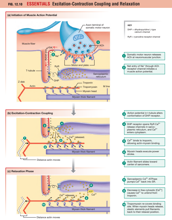
What happens at the neuromuscular junction?
Sequence:
Action potential reaches axon terminal
Voltage-gated Ca²⁺ channels open
Calcium enters neuron terminal
Acetylcholine released
ACh binds nicotinic receptors
Na⁺ enters muscle cell
End-plate potential forms
Muscle action potential generated
Acetylcholine is rapidly degraded by:
Acetylcholinesterase
What is excitation-contraction coupling?
Excitation-contraction coupling links:
Electrical excitation
toMuscle contraction
Process:
Action potential travels along sarcolemma
Signal enters T-tubules
DHP receptors detect voltage change
Ryanodine receptors open
SR releases Ca²⁺
Cytosolic calcium rises
Contraction begins
Calcium is the critical intracellular signal.
What changes occur in the sarcomere during contraction?
Structural changes:
Z discs move closer together
Sarcomere shortens
I band narrows
H zone narrows/disappears
A band unchanged
Important principle:
Filaments do NOT shorten.
They slide past each other.
This is the basis of the sliding filament theory.
What roles does ATP play in muscle contraction?
ATP is essential for multiple processes:
1. Cross-bridge detachment
ATP binding releases myosin from actin.
2. Re-cocking myosin
ATP hydrolysis energizes myosin head.
3. Calcium reuptake
Ca²⁺-ATPase pumps calcium back into SR.
Without ATP:
Cross-bridges remain attached
Calcium remains elevated
Muscle becomes rigid
This causes:
Rigor mortis after death
How does muscle relaxation occur?
Relaxation process:
Neural stimulation stops
ACh release ceases
Calcium pumped back into SR
Cytosolic calcium falls
Calcium detaches from troponin
Tropomyosin re-covers binding sites
Cross-bridge cycling stops
Muscle returns to resting length
Relaxation requires ATP because calcium pumps are ATP-dependent.
What is osteogenesis?
Osteogenesis is the process of bone formation during embryonic development, growth, remodeling, and fracture repair. Bone is formed by specialized cells called osteoblasts, which produce bone matrix and mineralize it.
There are two major mechanisms of bone formation:
Intramembranous ossification
Endochondral ossification
Both initially produce woven bone, which is later remodeled into stronger lamellar bone.
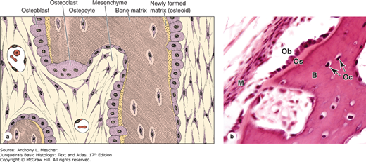
What are the major bone cell types and their functions?
Osteoblasts
Bone-forming cells
Derived from mesenchymal stem cells
Secrete osteoid
Osteocytes
Mature bone cells
Located in lacunae
Maintain bone matrix
Sense mechanical stress
Osteoclasts
Bone-resorbing cells
Derived from monocyte lineage
Break down bone using acid and enzymes
What is intramembranous ossification?
Bone formation directly from mesenchyme without cartilage.
Process:
Mesenchymal cells condense
Osteoblasts differentiate
Osteoid secreted
Matrix mineralizes
Woven bone forms
Lamellar bone replaces woven bone
Forms:
Flat bones of skull
Mandible
Clavicle
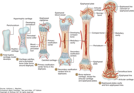
What is endochondral ossification?
Bone formation by replacing a hyaline cartilage model.
Sequence:
Cartilage model forms
Bone collar develops
Cartilage calcifies
Chondrocytes hypertrophy and die
Blood vessels invade
Osteoblasts deposit bone
Primary ossification center forms
Secondary ossification centers form later
Responsible for:
Long bones
Most bones of body
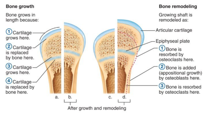
How do bones grow in length?
Length growth occurs at the:
Epiphyseal plate (growth plate)
Zones:
Reserve cartilage
Proliferation zone
Hypertrophic zone
Calcification zone
Ossification zone
Mechanism:
Chondrocytes divide
Cartilage enlarges
Cartilage replaced by bone
After puberty:
Epiphyseal plates close
Become epiphyseal lines
Longitudinal growth stops
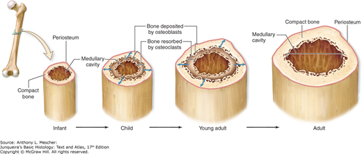
How do bones grow in width?
Width growth occurs by:
Appositional growth
Mechanism:
Osteoblasts add bone beneath periosteum
Osteoclasts remove bone internally
Result:
Increased diameter
Enlarged marrow cavity
Stronger bone without excessive weight
How do PTH and calcitonin regulate bone remodeling?
Parathyroid hormone (PTH)
Raises blood calcium by:
Stimulating osteoclast activity
Increasing bone resorption
Result:
Calcium released into blood
Calcitonin
Lowers blood calcium by:
Inhibiting osteoclasts
Promoting calcium deposition in bone
PTH and calcitonin help maintain calcium homeostasis.
Why is bone constantly remodeled?
Bone remodeling serves several essential functions:
1. Repair microdamage
Daily stress causes tiny fractures that must be repaired.
2. Adapt to mechanical stress
Bones strengthen where loads increase (Wolff’s law).
3. Calcium homeostasis
Bone acts as a calcium reservoir.
4. Replace old bone
Old bone is continuously renewed.
Remodeling cycle:
Osteoclasts resorb bone
Osteoblasts lay down new bone
Bone mineralizes
Balanced remodeling maintains skeletal strength and metabolic function.
Which types of bone are there?
Compact bone
Compact bone is dense and strong.
It forms the outer shell of bones.
It is organized into osteons.
Osteons have concentric lamellae around blood vessels.
Cancellous bone
Cancellous bone is spongy and lightweight.
It forms trabeculae inside bones.
It reduces weight but keeps strength.
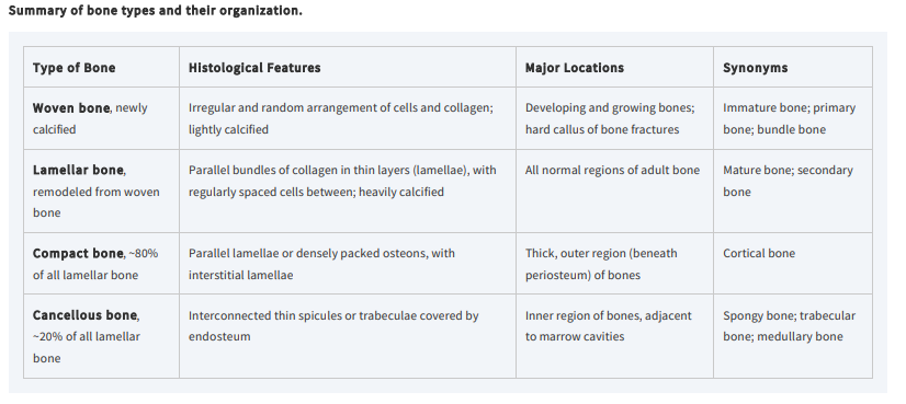
What is the difference between woven bone and lamellar bone?
Woven bone
Forms quickly
Collagen arranged randomly
Weak and disorganized
Seen in early development and fracture repair
Lamellar bone
Organized collagen layers
Strong and mature
Replaces woven bone during remodeling
What are the zones of the epiphyseal plate?
1. Reserve zone
Resting hyaline cartilage
2. Proliferation zone
Rapid chondrocyte division
Cells arranged in columns
3. Hypertrophic zone
Enlarged chondrocytes
4. Calcification zone
Matrix calcifies
Chondrocytes die
5. Ossification zone
Blood vessels invade
Osteoblasts replace cartilage with bone
Bone growth occurs mainly below the growth plate toward the shaft.
What is Wolff’s law?
Wolff’s law states that bone adapts to the mechanical stresses placed upon it.
Main principle:
Increased stress → increased bone formation
Reduced stress → bone loss
Bone structure changes to optimize strength while minimizing unnecessary weight.
Why do eccentric contractions cause more fatigue and soreness?
During eccentric contraction:
Muscle attempts to shorten
External force stretches it simultaneously
This creates:
Greater mechanical stress
More microdamage to fibers
Increased delayed-onset muscle soreness (DOMS)
Eccentric contractions are mechanically demanding even though they use less ATP than concentric contractions.
What is the role of calcium in muscle contraction?
Calcium binds to troponin, causing tropomyosin to move away from actin binding sites.
This allows:
Myosin to bind actin
Cross-bridge cycling
Force generation
Without calcium, contraction cannot occur.
What is a motor unit?
A motor unit consists of:
One motor neuron
All muscle fibers it innervates
Small motor units:
Precise control
Example: eye muscles
Large motor units:
More force
Example: hamstrings
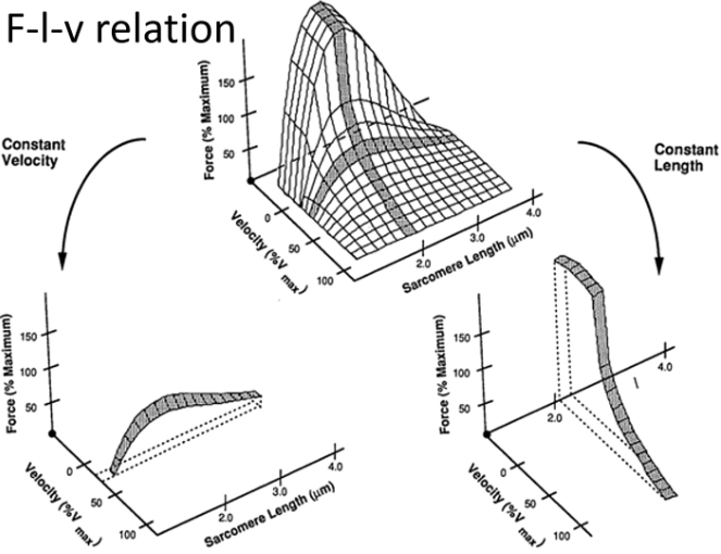
What is the force-length relationship?
Muscle force depends on sarcomere length.
Optimal overlap between actin and myosin → maximal force
Too stretched or too shortened → lower force
Force depends on how many cross-bridges can form.
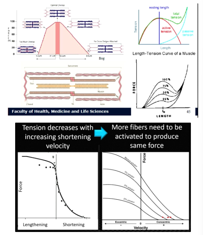
Why does overlap between actin and myosin affect force?
Force is produced by cross-bridges.
More overlap → more cross-bridges → higher force
Too little overlap → fewer cross-bridges
Too much overlap → filaments interfere
Optimal overlap produces maximal force.
What is the force-velocity relationship?
The faster a muscle shortens, the less force it can produce.
Reason:
At high velocity, fewer myosin heads are attached to actin at the same time
Slow shortening allows more cross-bridge attachment and higher force.
Why do eccentric contractions produce high force?
During eccentric contraction:
Muscle lengthens while active
Cross-bridges are stretched under tension
This creates:
High force
More muscle damage
More soreness
Example:
Walking downstairs
What is the force-frequency relationship?
Higher stimulation frequency produces greater muscle force.
Reason:
Calcium remains elevated
More cross-bridge formation occurs
Very high frequency can produce tetanic contraction (maximum force).
How does muscle cross-sectional area affect force?
Larger cross-sectional area means:
More fibers in parallel
More cross-bridges
Result:
Greater force production
Thicker muscles are generally stronger.
How does muscle fiber length affect velocity?
Longer fibers contain more sarcomeres in series.
This allows:
Greater shortening distance
Faster contraction velocity
Long fibers are specialized for speed and range of motion.
How does muscle architecture affect function?
Pennate muscles
More fibers packed together
Greater force production
Parallel muscles
Longer fibers
Greater speed and range of motion
Architecture determines muscle function.