N3 Exam 1 (copy)
 Call Kai
 Learn
Learn Practice Test
 Spaced Repetition
 Match
Match Flashcards
 Knowt Play
1/589

There's no tags or description
Looks like no tags are added yet.
Name | Mastery | Learn | Test | Matching | Spaced | Call with Kai | Chat |
|---|
No analytics yet
Send a link to your students to track their progress
590 Terms
Hypovolemic shock
Life-threatening condition in which tissue perfusion is inadequate to deliver oxygen and nutrients to support cellular function
Affects all body systems
May develop rapidly or slowly
Who is most at risk for hypovolemic shock
Any patient with any disease state may be at risk for developing shock
Regardless of the initial cause of shock, certain physiologic responses are common to all types of shock
hypoperfusion of tissues, hypermetabolism, and activation of the inflammatory response
PATHOPHYSIOLOGY OF SHOCK: Vascular responses
Proinflammatory biochemical mediators
Anti-inflammatory biochemical mediators
PATHOPHYSIOLOGY OF SHOCK: BP regulation
Mean arterial BP
Baroreceptors
Chemoreceptors
Renin–angiostien–aldosterone
Figure 11-2 in the book
P h y s i o l o g y / P a t h o p h y s i o l o g y
initial insult leading to shock state
decreased tissue perfussion and oxygen
activation of homeostatic response
decreased sympathetic response, HR, BP, cardiac contractility, which leads to decreased cardiac output;
increased respiratory rate to oxygen saturation and delivery
increased Renin-angiotensin activation, which leads to increased reabsorption of sodium and water, which leads to increased preload and decreased urine output
increased Catecholamines and cortisol to provide increased glucose for metabolism
R e s t o r a t i o n of t i s s u e p e r f u s i o n a n d o x y g e n a t i o n
TYPES OF SHOCK
Cardiogenic shock (due to heart problems)
Hypovolemic shock
Anaphylactic shock (caused by allergic reaction)
Septic shock (due to infections)
Neurogenic shock (caused by damage to the
nervous system)
A nurse is planning the care of a client who is being treated for shock.
What statement best describes the pathophysiology of this client's
health problem?
A. Blood is shunted from vital organs to peripheral areas of the body.
B. Cells lack an adequate blood supply and are deprived of oxygen and
nutrients
C. Circulating blood volume is decreased with a resulting change in the
osmotic pressure gradient.
D. Hemorrhage occurs as a result of trauma, depriving vital organs of
adequate perfusion.
B
STAGES OF SHOCK
1. Compensatory stage
2. Progressive stage
3. The irreversible (or refractory) stage
slide 9 figure
COMPENSATORY STAGE OF SHOCK
BP within normal limits
Adequate cardiac output
“Fight-or-flight” response
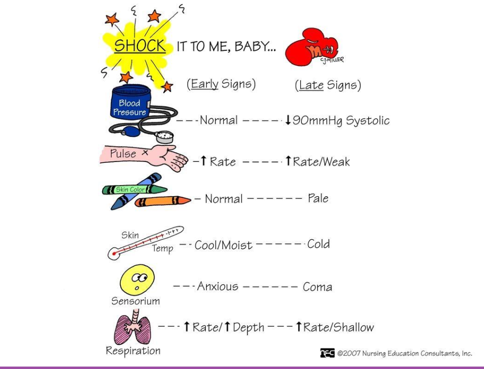
Clinical manifestations of COMPENSATORY STAGE OF SHOCK
Clinical manifestations
Inadequate perfusion
Increased respirations
Anxiety
Confusion
Table 11-1 In the book
MEDICAL AND NURSING MANAGEMENT OF
THE COMPENSATORY STAGE OF SHOCK
Treat underlying cause
Fluid replacement
Supplementation oxygen, decrease patient anxiety
Maintain BP and tissue perfusion
Frequent assessment (subtle changes)
LOC, I&O, respiratory and heart rate, BP (MAP of 65 mm Hg or less, narrowing or decreased pulse pressure)
Promote safety (advanced directives)
Gerontologic considerations, Chart 11-1
PROGRESSIVE STAGE OF SHOCK
BP no longer compensated (patient hypotensive)!!!
MAP below normal limits
Decreased mental status
PROGRESSIVE STAGE OF SHOCK Pathophysio
Heart failure
Decreased cellular perfusion
Interstitial edema
Anaerobic metabolism
Hyperactive clotting cascade
CLINICAL MANIFESTATIONS OF THEPROGRESSIVE STAGE OF SHOCK
Rapid and shallow respirations
Chest pain (increased cardia biomarkers)
Changes in mental status
Acute kidney injury (decreased GFR)
Liver failure
GI bleeding
Abnormal bruising and petechiae with prolonged clotting times
MEDICAL AND NURSING MANAGEMENT OF THE PROGRESSIVE STAGE OF SHOCK
Treat underlying cause
Restore tissue perfusion with fluid management
Respiratory support
Nutritional support for energy
Assessment (subtle changes) including ABGs, hemodynamic monitoring, ECG
monitoring, mental status changes, and serum electrolytes
Oral care if on ventilator to prevent VAP
Promote rest and comfort to reduce stress, decreased chance of postintensive
care syndrome
IRREVERSIBLE STAGE OF SHOCK
Severe organ damage
Unable to maintain BP with treatment
Unable to oxygenate with ventilatory support
Unable to maintain MAP
Multiple organ dysfunction MODS
Complete organ failure
IRREVERSIBLE STAGE OF SHOCK:Complete organ failure
Organ damage too severe to respond to treatment
Acute metabolic acidosis
Lactic acidosis
Depleted energy stores
CLINICAL MANIFESTATIONS OF THE
IRREVERSIBLE STAGE OF SHOCK
Patient unable to respond to treatment
Ventilator dependent
Severe hypotension
Complete decline in mental status
organ failure
MEDICAL AND NURSING MANAGEMENT OF THE IRREVERSIBLE STAGE OF SHOCK
Treat underlying cause
Respiratory support
Circulatory support
Nutritional support
Experimental treatments
Simple comfort measures (palliative care)
Support and education for the friends and family
Be honest regarding prognosis
Which stage of shock is characterized by a normal blood
pressure?
A. Initial
B. Compensatory.
C. Progressive
D. Irreversible
B
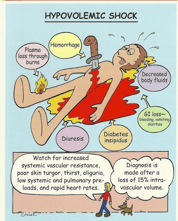
HYPOVOLEMIC SHOCK: External fluid flossess
most common type of shock
Trauma
Surgery
Diarrhea
Hypovolemic shock: Internal fluid losses
Occurs with15% to 30% reduction in intravascular volume
Hemorrhage
Burns
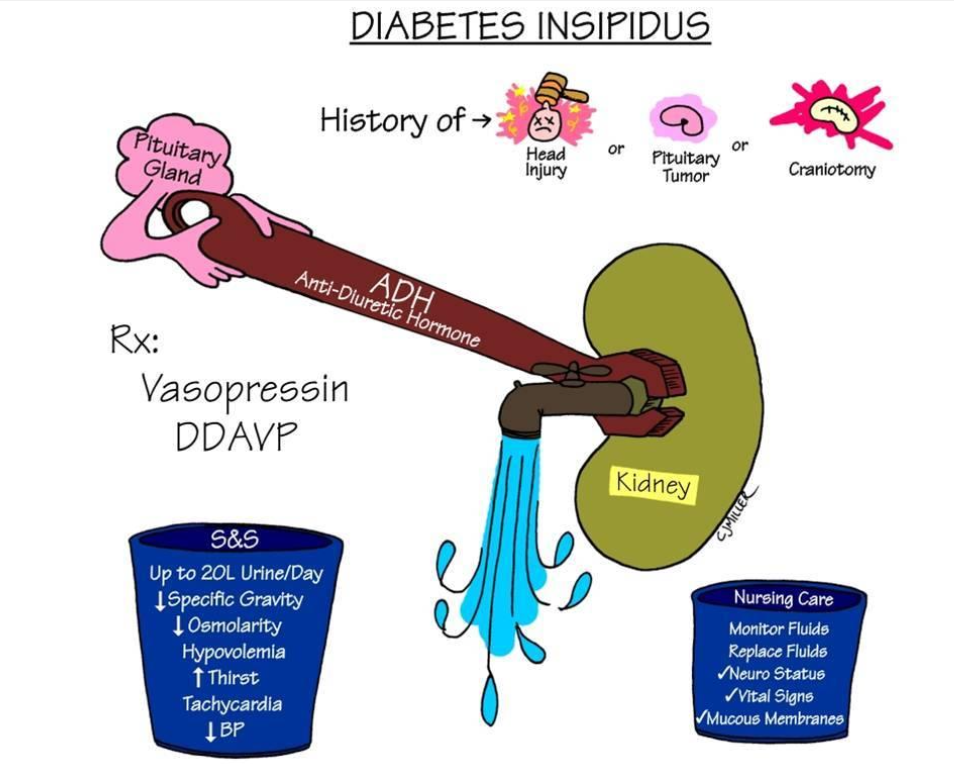
CLINICAL MANIFESTATIONS
OF HYPOVOLEMIC SHOCK
Decreased blood volume
Decreased cardiac output
Decreased tissue perfusion
MEDICAL AND NURSING MANAGEMENT OF
HYPOVOLEMIC SHOCK
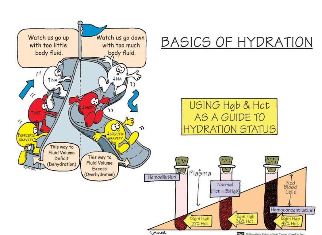
Restore and redistribute intravascular volume with fluid, blood
replacement
Treat underlying cause
Vasoactive medications
Focus on prevention
Ongoing monitoring of those at risk for hypovolemia
Safe administration of fluids and medications
Supplemental oxygen
Comfort measures
When caring for a patient in hypovolemic shock who is
receiving large volumes of IV isotonic fluids, the nurse should
monitor for symptoms of:
A. Hyperthermia
B. Pain
C. Pulmonary edema.
D. Tachycardia
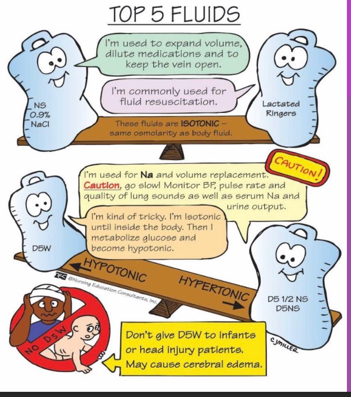
IVF ISOTONIC Solution types
D5W
Normal NS
LR
• 0.9% saline (NS)
• So/odextrose in vvater (5%DvV)
• 5% dextrose in 0.225 dosaline (5 o D/1/4NS)
Lactated ringer's solution
D5W Function
• Is an isotonic carbohydrate (sugar) solution that contains glucose (sugar) as the solute.
• When this is absorbed, the glucose is usually quickly taken up by cells and utilized for energy, leaving only water, which is then a hypotonic solution.
D5W Purpose
Provides 170 cal/L
Prevents ketosis associated with starvation
Often used for diabetic patients who are NPO
D5W Risks
avoided in patients with renal failure, cardiac compromise, or those who are at risk for increased intercranial pressure.
it can sometimes lead to fluid overload in cardiac and renal patients, and cerebral edema those with intercranial pressure issues
Normal NS function
More NaCl than ECF
Expands IV volume
What is normal Saline preferred for
Preferred fluid for immediate response
only IV that can be given with blood transfusion
fluid of choice for resuscitation efforts
one of the most common IVF: it works for most hydration needs due to vomiting, diarrhea, hemorrhage, or even shock
only fluid used in conjunction with blood administration.
NS risks
Risk for fluid overload higher
sometimes used with caution or even avoided in patients with cardiac or renal compromise because of the sodium causing fluid retention that may put extra stress on the heart or kidneys
LR Function
Similar in composition to plasma
except contains no magnesium
Expands ECF—treat burns and GI losses
No free water or calories
LR Purpose
Especially great for burn victims or those with hypovolemia due to fluid shifts.
Used in operating room frequently
LR risks and contradictions
• It is avoided in patients with liver disease because the liver may not be able to efficiently process the lactate
• Contraindicated with hyperkalemia and lactic acidosis
Hypertonic solutions
D51/2
D10W
D5 ½ NS
Common maintenance fluid
Replaces fluid loss
KCl added for maintenance or replacement
D10W
•Provides 340 kcal/L
•Provides free water but no electrolytes
•Limit of dextrose concentration may be infused peripherally
True or False: Hypovolemic shock occurs where there is low
fluid volume in the interstitial system
a. True
b. False
B.
A patient who is experiencing hypovolemic shock has
decreased cardiac output, which contributes to ineffective
tissue perfusion. The decrease in cardiac output occurs due to?
A. An increase in cardiac preload
B. An increase in stroke volume
C. A decrease in cardiac preload
D. A decrease in cardiac contractility
C
P r e l o a d
Pressure from volume of blood in ventricles at end of diastole
(end diastolic pressure)
Preload increased in
Hypervolemia
Regurgitation of cardiac valves
Heart Failure
Afterload
Resistance left ventricle must overcome to circulate blood
Afterload increased in
Increased in:
Hypertension
Vasoconstriction
1Afterload =
T Cardiac workload
Central venous access
• Who can insert these
lines?
• What are nursing considerations when providing care for patients with these access?
Intravenous (IV) Therapy
Goal of IV therapy
• Maintain fluid balance
• Prevent fluid and electrolyte imbalances
• Administer parenteral medications
• Replenish blood volume
• Assist in pain management
Thinking Nurse: What are patient conditions
that require IV therapy?
Principles for Practice
• Successful IV therapy depends on patient preparation,
site selection, catheter selection, and catheter insertion
• A____ plays key role in decision making for IV
therapy
• Follow rights of medication administration:
• WHAT ARE THE 8 PATIENTRIGHTS?
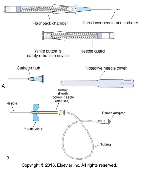
Intravenous Catheters- Angiocatheter
An Angiocatheter = venous access device (VAD) is inserted into a
vein
•Can be peripheral or central venous access devices, depending on where the final tip resides
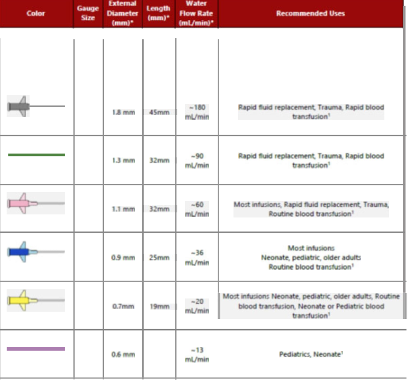
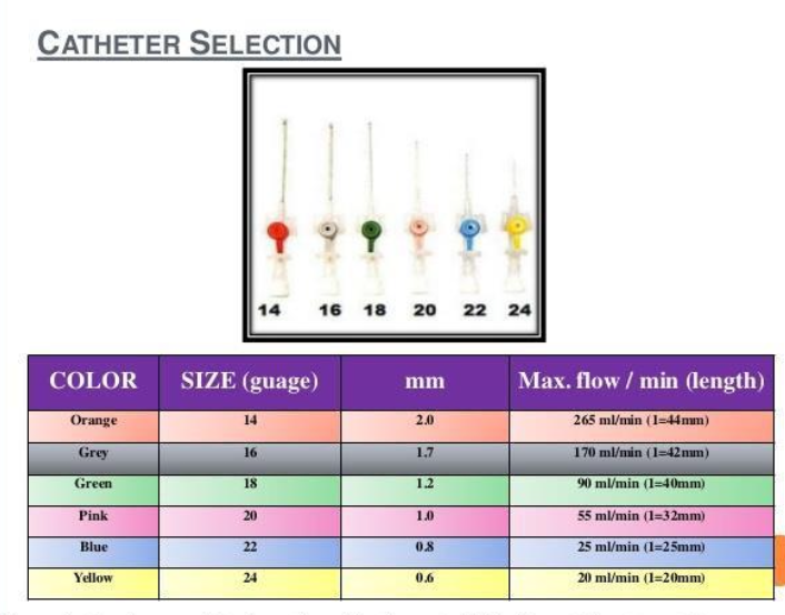
Choosing the right Catheter***When selecting the appropriate
VAD, consider:
• a patient’s prescribed therapy
• length of treatment
• duration the device remains
in place
• vascular characteristics
• patient’s age, co-morbidities,
history of infusion therapy,
preference for VAD location,
and pt’s a________
Saline Lock (S.L.)
• When continuous infusion of solutions or medications is not necessary, a short-peripheral IV can be locked with preservative-free 0.9% sodium chloride (NS).
• The “old” term is HepLock
WHY?
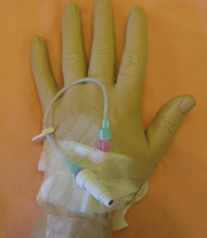
Thinking Nurse: What gauge is the PIV on the photo?
Intravenous solution categories
isotonic
hypotonic
hypertonic
• Hypotonic: can exacerbate a
hypotensive state
• H
______: irritating to the vein; can
cause increased risk of heart
failure and pulmonary edema
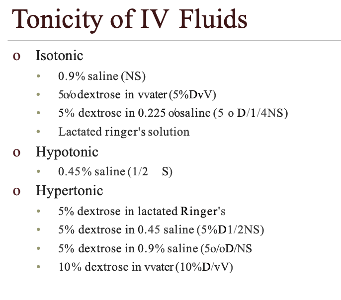
Isotonic
Have same osmolality
as body fluids; can cause
increased risk for fluid overload
Hypotonic Purpose
0.45% NS (1/2 NS)
fluid is designed to treat cellular dehydration, caused by such conditions as hypernatremia or diabetic ketoacidosis
Hypotonic avoided in
always avoided in patients with burns, trauma, or liver disease because of its tendency to deplete intravascular fluid levels.-
Infusing half normal saline, causes water to shift from the extracellular fluid compartment to the ICF compartment
Hypotonic can be harmful:
Sudden shift of fluid from blood vessels to the cells – cardiovascular collapse
Hypotonic solutions - potential to cause cellular swelling
Monitor for changes in mentation →indicate cerebral edema
Examples- Hypotonic IV solutions
D5NS.45 (5% dextrose in ½ normal saline)
5% Dextrose and water (D5W)- Provides calories and water
Hypertonic solution types
D5 ½ NS
D10W
D5 ½ NS
Common maintenance fluid
Replaces fluid loss
KCl added for maintenance or replacement
D10W
•Provides 340 kcal/L
•Provides free water but no electrolytes
•Limit of dextrose concentration may be infused peripherally
IV Solution Additives- Potassium
Potassium chloride (KCl) – common additive to IV fluids
Measured in milliequivalents (mEq)
order usually written to indicate the amount of milliequivalents per liter
IV solutions are often available with Potassium premixed in them`
Evidence-Based Practice
• Implementation of quality indicators and benchmarks to prevent CLABSIs develops a culture of accountability
• Remove VADs appropriately
• Use disinfection caps that contain a sponge saturated with 70% alcohol
• Primary and secondary continuous administration sets used to administer solutions other than lipid, blood, or blood products should be changed no more often than every 96 hours
Safety Guidelines: IV Therapy
• Prior to initiating IV therapy, assess the patency and functioning of the VAD.
• Maintain sterility at all time
• Assess S/S of IV complications
• Ensuring correct IVF as ordered
• Know which IV can be given via PIV VS Central access
• Trace the line!!!!
Thinking Nurse: How do you assess IV patency?
What are possible IV complications?
IV complications: To prevent infusion-related complications
solutions and medications with an osmolarity greater than 900 mOsm/L are infused through a C___ C____
IV complications: Short-peripheral catheters
should not be used for vesicant therapy, parenteral nutrition, or infusates with an osmolarity greater than 900mOsm/L
Solutions or medications with low or high pH have the potential to cause
infusion-related complications such as phlebitis when administered with a short-peripheral or midline catheter.
Thinking Nurse: What is the common low pH IV solution?
Phlebitis
inflammation of vein caused by mechanical, bacterial, or chemical irritation
Signs/symptoms of Phlebitis?
Pain, ↑ skin temp, redness
Treatment for Phlebitis?
D/ CIV line, apply moist, warm compress, monitor IV site q hr for redness/ tenderness to prevent
solution should be administered in another site
0-4 grading scale for Phlebitis?
A: Erythema, pain, edema, red streak along vein, palpable venous cord, purulent drainage
What is Infiltration?
Leakage of intravenous solution or medication (non-vesicant) into the extravascular tissue
Happens when the needle or catheter in which the product is entering the blood vessel (vein) is dislodged from the vein
Signs/symptoms of Infiltration?
Edema, pallor, ↓ skin temp around the site, and pain
Treatment for Infiltration?
D/C IV line, elevate extremity, warm compress to absorb fluid
Product should be administered at another site
0-4 grading scale for Infiltration?
Skin blanched, tight/leaking, bruised, swollen, edema, pitting, circulation, pain
What is Extravasation?
IV catheter becomes dislodged and medication infuses into the tissues (vesicant)
The accidental administration of IV drugs into the surrounding tissue, because the needle has punctured the vein and the infusion goes directly into the arm tissue. The leakage of high osmolarity solutions or chemotherapy agents can result in significant tissue destruction
Signs/symptoms of Extravasation?
Pain, stinging, burning, swelling, redness at site
Treatment for Extravasation?
D/C IV line, apply cool compress, admin antidote if one exists formed
0-4 grading scale for Extravasation?
Skin blanched, tight/leaking, bruised, swollen, edema, pitting, circulation, pain
VIP Score 0?
IV site appears healthy — No sign of phlebitis — Observe cannula
VIP Score 1?
One of: slight pain near IV site OR slight redness near IV site — Possible first sign of phlebitis — Observe cannula
VIP Score 2?
Two of: pale near IV site, erythema, swelling — Early stage of phlebitis — Resite cannula
VIP Score 3?
All of: pain along path of cannula, erythema, induration — Medium stage of phlebitis — Resite cannula, consider treatment
VIP Score 4?
All of (evident & extensive): pain along path of cannula, erythema, induration, palpable venous cord — Advanced stage of phlebitis/start of thrombophlebitis — Resite cannula, consider treatment
VIP Score 5?
All of (evident & extensive): pain along path of cannula, erythema, induration, palpable venous cord, pyrexia — Advanced stage of thrombophlebitis — Initiate treatment, resite cannula
IV-Site Infection
Does not produce much (if any) pus or inflammation at the IV site. This is the most common cannula-related infection, may be the most difficult to identify
Cellulites
Warm, red and often tender skin surrounding the site of cannula insertion; pus is rarely detectable
Thrombolism / thrombophlebitis
occur when a small clot becomes detached from the sheath of the cannula or the vessel wall - prevention is the greatest form of defence. Flush cannula regularly and consider re-siting the cannula if in prolonged
Bruising
commonly results from failed IV placement - particularly in the elderly and those on anticoagulant therapy
Air embolism
occurs when air enters the infusion line, although this is very rare it is best if we consider the preventive measures - Make sure all lines are well primed prior to use and connections are secure
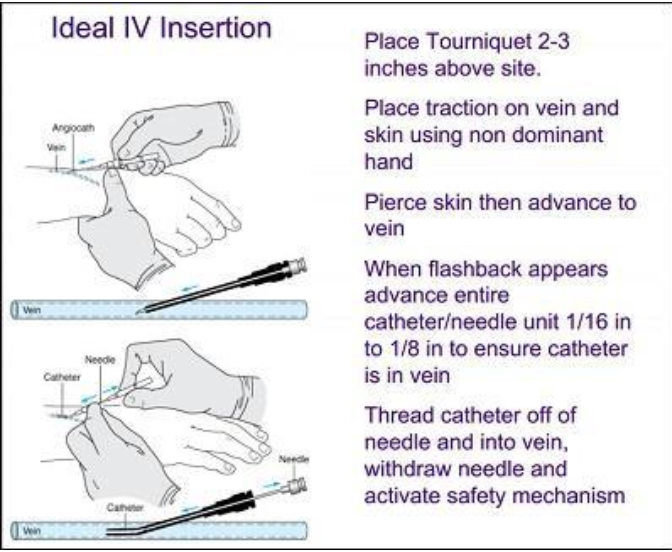
Insertion of a Short-Peripheral Intravenous Device
You will learn this
in Nursing 5
• Intravenous devices provide
access to the venous system for
fluid delivery
• Various catheters available
• BEVEL UP!!!
PIV Sites
Hands and arms
Antecubital fossa (AC space)
Cannulation
• What are the signs of a good vein ?
• Bouncy
• Soft
• Above previous sites
• Refills when depressed
• Visible
• Has a large lumen
• Well supported
• Straight
• Easily palpable
Gerontologica: Agerelated considerations
• Veins of the older population are very fragile; they have less subcutaneous support tissue, and their skin is thinning. Avoid sites that are easily moved or bumped.
• If possible, avoid the back of the older adult’s hand or the dominant arm for venipuncture because use of these sites interferes with the individual’s independence.
• As older adults lose subcutaneous tissue, the veins lose stability and roll away from
the needle. To stabilize the vein, pull the skin taut and toward you with your nondominant hand and anchor the vein with your thumb.
• Reduce the angle of insertion (e.g., 5 to 15 degrees on insertion) to accommodate more superficial veins.
Regulating Intravenous Flow Rate
• Proper regulation reduces
complications
• Regulate infusion rates
• Manual flow-control
devices
Regulation of IV drip rate manually
1. Tubing for these sets contain a roller camp
2. Can open or close & regulate rate manually
3. Use a watch with a second hand to count number of drops per minute in the chamber
Thinking Nurse: When would you need to give IV without a pump?
Changing Intravenous Solutions
• Change a container when
• An order for a new solution is received
• It is time to add a sequential container to avoid
exceeding
hang time
• The patient’s condition indicates the need
• The IVF is expired