oncol mt 1 pt 2
 Call Kai
 Learn
Learn Practice Test
 Spaced Repetition
 Match
Match Flashcards
 Knowt Play
1/119

There's no tags or description
Looks like no tags are added yet.
Name | Mastery | Learn | Test | Matching | Spaced | Call with Kai | Chat |
|---|
No analytics yet
Send a link to your students to track their progress
120 Terms
tumor suppresor genes role
inhibit cell proliferation and survival
what happens when TSG is completely or partially lost
increase likelihood of cancer development
is activation of oncogenes adequate to cause cancer? if not, what else is involved
NOOOOO, TSG are also dysregulated in cancer
loss of TSG happens more frequently that activation of proto-oncogenes
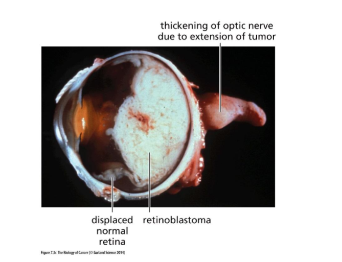
retinoblastoma what and origin
tumor of retina from photoreceptor cells (stem cell precursor of cones)
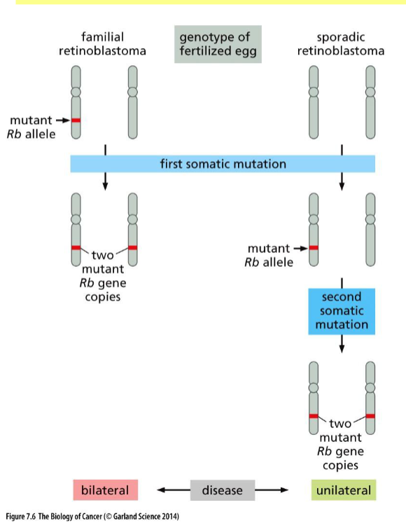
sporadic retinoblastoma
non heritable
single eye tumor (unilateral)
after treatment, no futher risk of other tumors
familial form retinoblastoma
heritable bilateral tumor in retina
after treatment, increase risk of bone and other cancers
Rb
TSG gene involved in retinoblastoma
mutations are recessive and cause cancer
show with DNA the difference between sproadic and familial retinoblastoma
two mutations is much more rare than one
deletions in retinoblastoma
Rb is deleted

what happens when one TSG allele is mutated
it becomes inactive, and the other one compensates
if they both become mutated they transform cells
why are sequence mutations not likely and what is more plausible
one mutation is 1/10^6 two mutation would mean 1/10^12,,,,
translocations and deletions (meh)
epigenetics (gene silencing)
one copy mutated second copy altered to same mutation
examples of where one copy of a gene is mutated so the second one alters
mitotic recombination (more common) + gene conversion (most common)
mis-segregation at mitosis or deletion (less common)
mitotic recombination
2 homologous chromosomes exchange DNA during mitosis in somatic cells

LOH
loss of heterozygosity
when a heterozygous gene becomes homozygous dominant or recessive
gene conversion
homologous chromosomes

chromosomal mis-segregation
during mitosis

DNA methylation
add methyl group to cytosine at CpG site by DNA methyltransferase
heritable epigenetics
effect of methylated CpGs in DNA
recruit protein complexes that functions as histone deacetylase
removal of acetyl groups from histone in chromatin
more densley packed chromatin (inactive)
transcription stop
what are the 2 mechanisms that inactivate both copies in promoter methylatio
both copies are methylated independently (less common)
one copy is methlylated, second copy lost through duplication of methylated copy
2 types of methylation in cancer
global hypo: dec DNA methylation of highly repeated sequences = instability
localized hypermethylation gene silences
how are oncogenes identified
transformation assay
why are biological identification of TSG more complicated
TSG prevent transformation, so its harder
how can we ID TSG
genomics
ID region where LOH happen → find TSG here

SNP in TSG finding
SNP can help us distinguish alleles, distinguished can help us map sites

gatekeepers
½ proteins encoded by TSG
direct control by hindering cell proliferation (cell division)
increasing cell differentiation and/ or death
caretakers
½ protein encoded by TSG
maintain integrity of genome
two types of bad miRNA
no miRNA = proliferation of oncoprotein… bruh (loss of miRNA suppresor)
miRNA inhibits TSG from producing cancer killers …. bruh (oncogenic miRNA)

what do gatekeepr proteins respond to
growth inhibitory signals
DNA damage
unkown :o
familal adenomatous polyposis
heritable susceptibility to develop adenomatous polyps in colon → colon carcinoma
linked to loss of tumor suppressor APC
APC
tumor suppressor gene commonly found inactivated in sporadic colon carcinomas
why is APC an important tumor suppressor

wild type vs mutated intestinal crypt

what is the most important molecular consequence of APC inactivation
beta-catenin accumulation…
percentage of sporadic colon carinomas
90% = APC inactivation
10% = wild type APC is inactivated by promoter methylation
beta-catenin mutations that cannot be phosphorylated for GSK degredation
cell cycle clock what and where
what: network of interacting proteins
where: nucleus
cell cycle basic
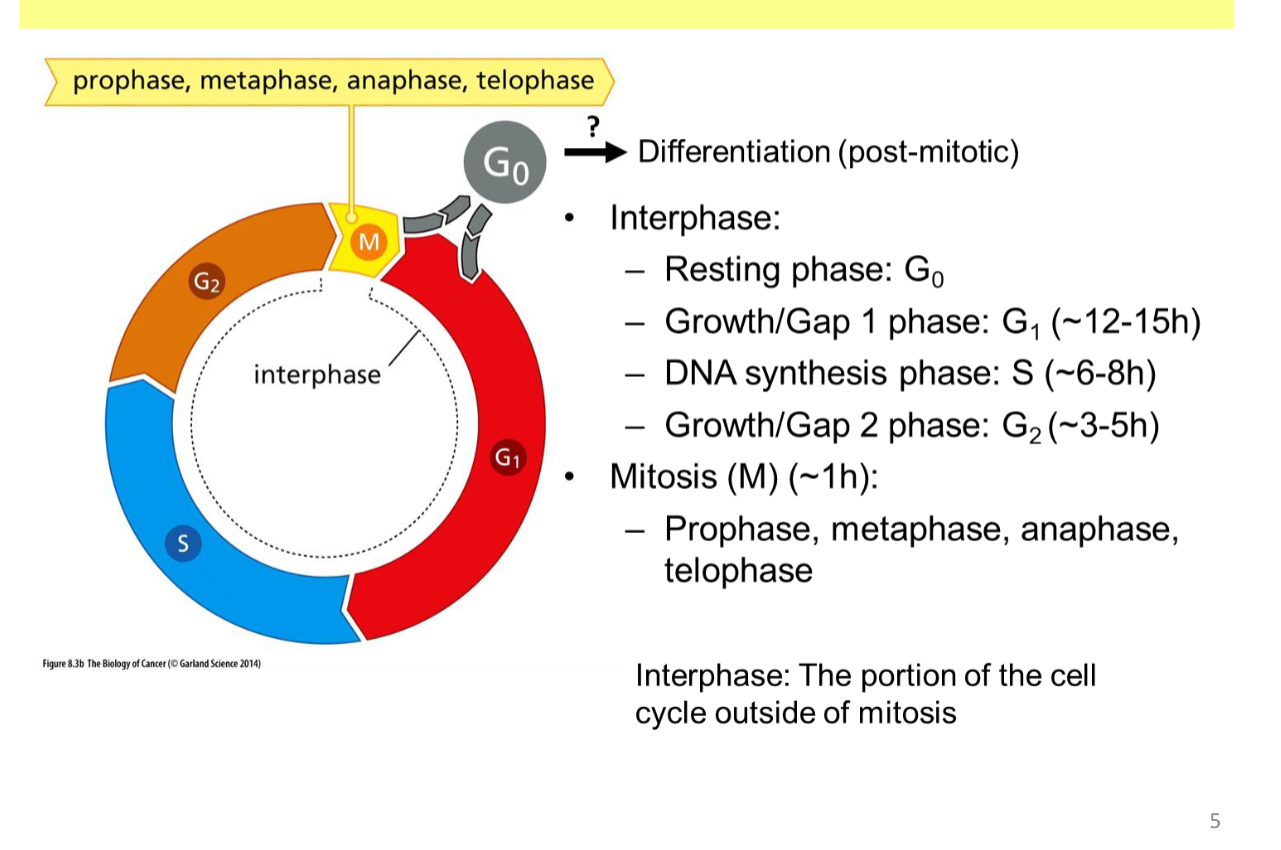
prophase
chromosmes condense
centrosomes assemble
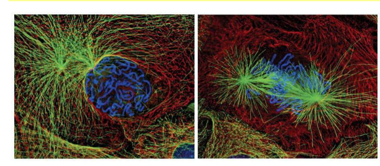
metaphas
align on metaphase plate
mitotic spindle

anaphase
chromatids pulled apart by mitotic spindles

telophase
chromosomes decondense, nuclear membrane forms, cytokinesis begins
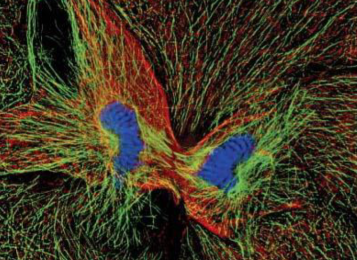
general checkpoints of mitotsis

which period are cells responsive to mitogenic GF and TGF-beta


green and yellow arrow
green: where cells are responding to mitogenic GF and TGF-beta
yellow: R point
when does R point occur
late G1
several hours before G1/S transition
what happens at R point
→ divide (continue) or no (g0)?
CDK
cyclin dependent kinase (serine/threonin kinase)
always present
cyclins
regulate CDK → activation + substrate specificity
cyclin + CDK combo in each part of cell cycle
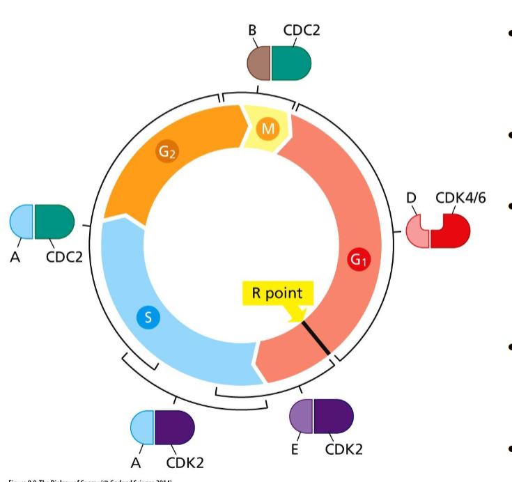
cyclin amount graph
cyclins are degraded within minutes

how is each cyclin-CDK activated and how is it shut down
activated: D + 4/6 = mitogens (only extracellular part), the rest by the subsequent phase
shuts down: by predecessor
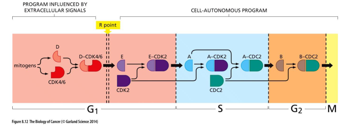
how is cyclin D activated
extracellular signals: ECM (Fak), GF, Wnt, others
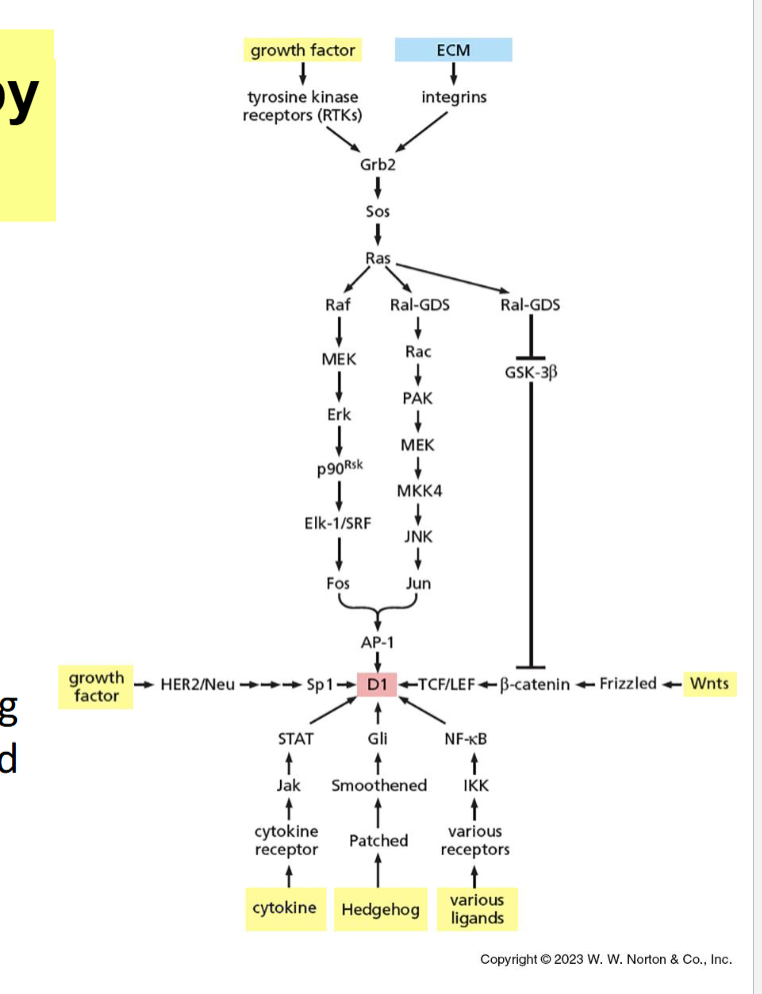
how are other cyclin families (EAB) activated
regulated by cell cycle
intracellularly
CDK inhibitor names and what do they inhibit

where must CDK inhibitors be to inhibit cyclin/CDK
nucleus
p15 activation and what stage
early and mid G1
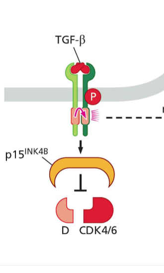
p21 activation and what stage
almost every stage

how are CDK inhibitors inactivated
CDK can only be active in nucleus

explain this image

pRb origin and main goal
encoded by RB TSG
guardian of R-point gate
phosphorylation cycle of pRb

after mitosis, pRb is (hypophosphorylated/ dephosphorylated/ hyperphosphorylated) by _______
dephosphorylated; protein phosphatase 1

in early and mid G1, pRb is (hypophosphorylated/ dephosphorylated/ hyperphosphorylated) by _____
hypophosphorylated; cyclin D- CDK 4/6
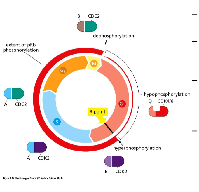
after R point pRb is (hypophosphorylated/ dephosphorylated/ hyperphosphorylated) by
hyperphosphorylated by EAB CDK complexes
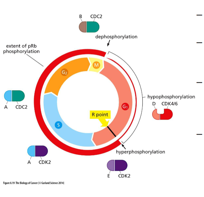
in S and G2 phase phosphorylation of pRb can be ______ by ______
transiently reversed by unknown phosphatases
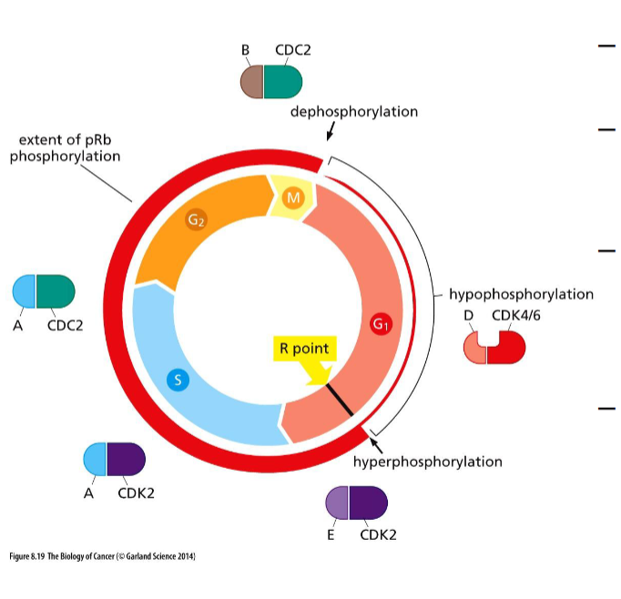
diagram of pRb being hyperphosphorylated

pRb and E2F interaction
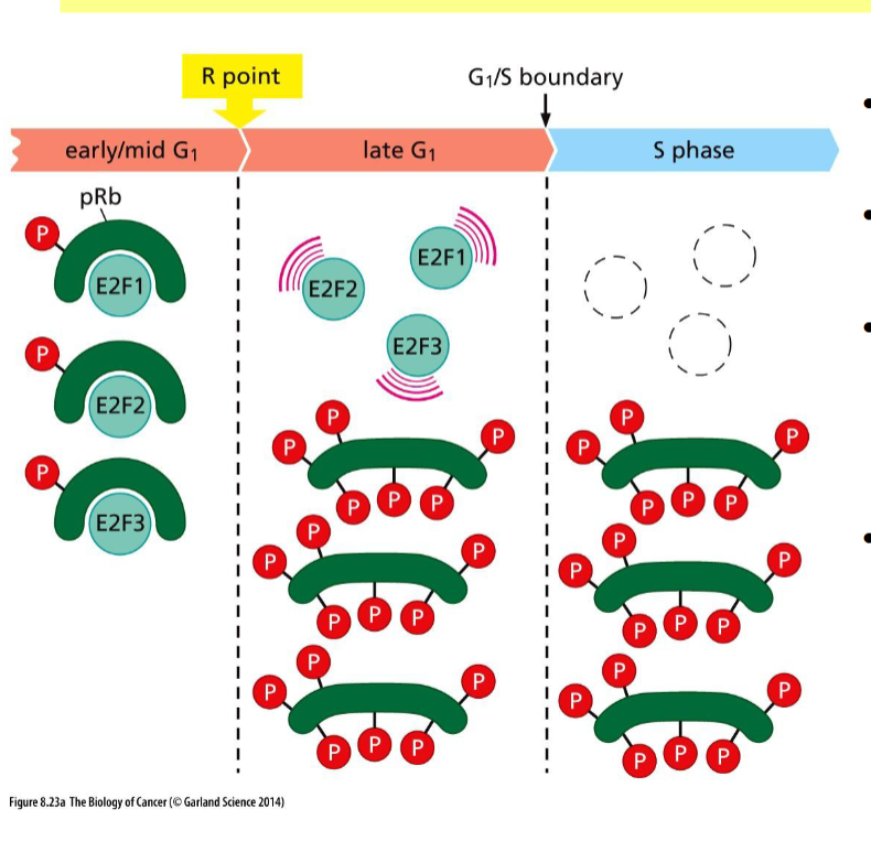
what is E2F and what are its target genes
E2F= transcriptional activators
targets: DNA precursor nucleotide synthesis, and DNA replication
positive feed back loop of cell cycle

3 ways pRb can be inactivated
mutation of RB gene followed by LOH
methylation of RB gene promoter = silencing
inactivation of pRb by HPV E7 proteins (human cervical caner)
how does inactivation of pRb by HPV E7 work
E7 proteins bind strongly to pRb, prevents E2Fs from binding, E2F constant activity
cyclin D1 _____ in cancer by
expression is elevated; Ras/Raf and PI3K and AKt signalling paths
(increased transcription and reduced degradation)
correlation between cyclin levels and deaths

myc reulgation of cell cycle
also promote degradation of CDK inhib (Miz)

5 molecular changes leading to deregulation of cell cycle clock
inactivation of pRb
overexpression of cyclin
increase activity of CDK (activation mutation)
improper localization of underexpression of CDK inhibitors
alterations induced by Myc

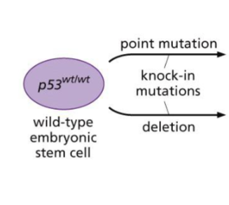
explain this picture
initiallt, p53 was thought to be an ongogene
however, cDNA had point mutation
most mutations carried by p53 gene are______ that lead to ______
missence point mutations; amino acid substitutions
dominant negative
p53
a mutant allele of a gene interferes with wild type when co-expressed (becomes dominant)
majority of p53 mutations affect where
DNA binding domain
not tetramization
in most mutations, how is wild type p53 discarded
through LOH
finish

half life of p53
20 minutes
signals that increase p53 protein level

radiation ______ p53 levels which _____ p21
increases; induces → cell cycle arrest at almost every stage
cytostatic function of p53
pro-apoptotic function of p53
p53-/- were resistant to irradiation induced apoptosis

Mdm2 mechanism
induce p53 ubiq. + degradation
inhibit p53 transcriptional activity

what happens when there is cell damage vs cell survival signals

where does Mdm2 and p53 complex go to get degraded
nucleus
high E2F induces what protein
p14ARF (human) and p19ARF (mice) binds to Mdm2 (TSG)
sequester in nucleus → cannot bind p53 → increase p53 → apoptosis

p 53 targets + mechanism

p53
TF and regulates gene expression
how is p53 anti-angiogenesis
induces expression of thrombosponding (TSP1) antiangiogenic protein
familial cancer symdrome
mother and father have one mutated copy of p53 (point mutation) → leads to very common family cancers

apoptosis plays important role in _____ that do not involve ______
physiological processes; p53
mitrochondrial path way
aka stress-activated pathway
apoptosis that is dependent on mitochondrial release of cytochrome C
death receptor pathway
apoptosis that does not require cytochrome c release (but it can accompant activation)
what is on mitochondria in relation to intracellular signals
balance between pro-survival and pro-apoptotic proteins on surface
what happens if apoptotic signals predominate on mitochondrial surface
channel in outer membrane opens
cytochrome c released in cytosol
activation of cascade proteases → caspases
cleavage of “death substrates”
apoptotic phenotype
pro-survival gene family

bax family
pro-apoptosis family #1

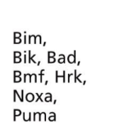
BH3 only family
pro-apoptosis family #2