Echo Board Pt. 2
 Call Kai
 Learn
Learn Practice Test
 Spaced Repetition
 Match
Match Flashcards
 Knowt Play
1/368

There's no tags or description
Looks like no tags are added yet.
Name | Mastery | Learn | Test | Matching | Spaced | Call with Kai | Chat |
|---|
No analytics yet
Send a link to your students to track their progress
369 Terms
What is bacterial endocarditis?
Infection of epithelial cells that causes vegetation formation on valves
What is the most common cause of bacterial endocarditis?
Streptococcus viridans or staphylococcus aureus
What are the symptoms of bacterial endocarditis?
Fever of unknown origin
New murmur
Positive bacterial cultures
What is the most common valve affected by bacterial endocarditis?
MV
Where are the vegetations located in those with bacterial endocarditis?
Atrioventricular valves (TV/MV): Atrial side of valve
Semilunar valves (AV/PV): Ventricular side of valve
What is Lambl excrescence?
Normal protrusion from aortic leaflets that can be mistaken for aortic valve endocarditis
What is the sonographic appearance of bacterial endocarditis?
TEE preferred for visualization
Thickened leaflets
Mobile mass formation on leaflets
Pericardial effusion
Complications: Valve ring abscess, fistula formation
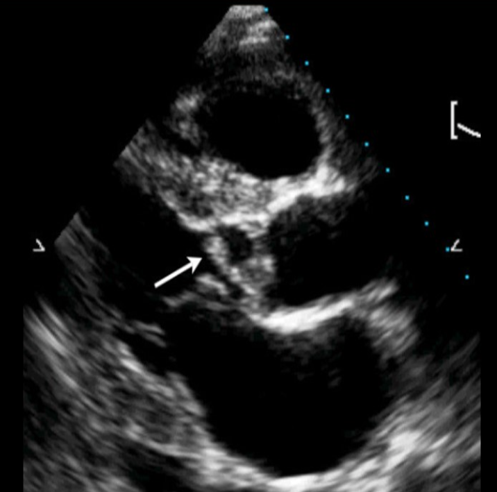
Identify this image.
Endocarditis
What is myocarditis?
Inflammation of myocardium
What is pericarditis?
Inflammation of outer later of tissues surrounding heart
What is constrictive pericarditis?
Chronic inflammatory process of pericardium that may cause calcification, adhesion, and fibrosis of pericardial layers
What are the symptoms of constrictive pericarditis?
Dyspnea
Ascites
Jugular vein distension
Pericardial “knocking” murmur due to restricted diastolic filling
What is the sonographic appearance of constrictive pericarditis?
Thickened pericardium with increased reflectivity
LAE
Paradoxical motion or early diastolic notching of IVS
IVS position varies with respiration
MV E/A ratio > 1.5
Septal E’ > Lateral E’ or annulus reversus
MV velocity increases > 25% with EXPIRATION
TV velocity increases > 40% with INSPIRATION
Deceleration time < 160 ms
No MR or TR
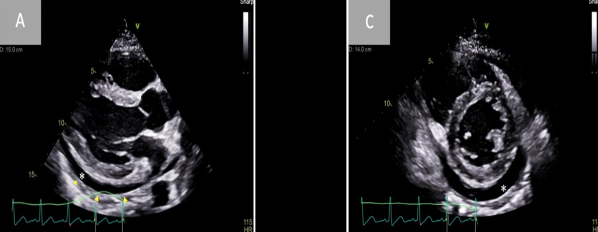
Identify this image.
Thickened pericardium and pericardial effusion seen in those with constrictive pericarditis
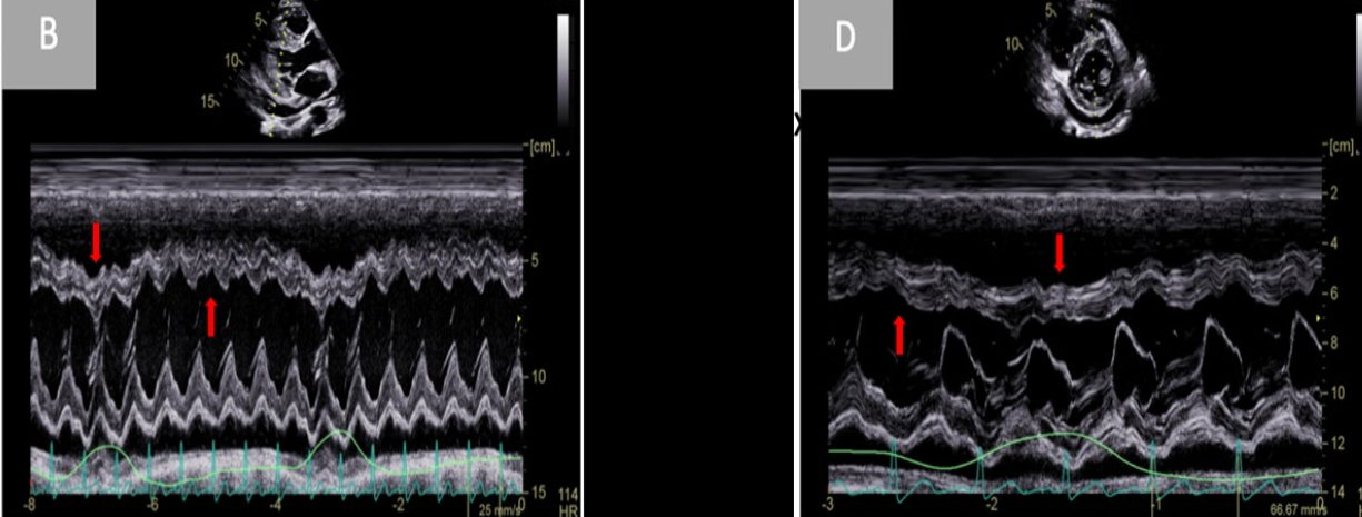
Identify this image.
IVS position varies with respiration seen in those with constrictive pericarditis
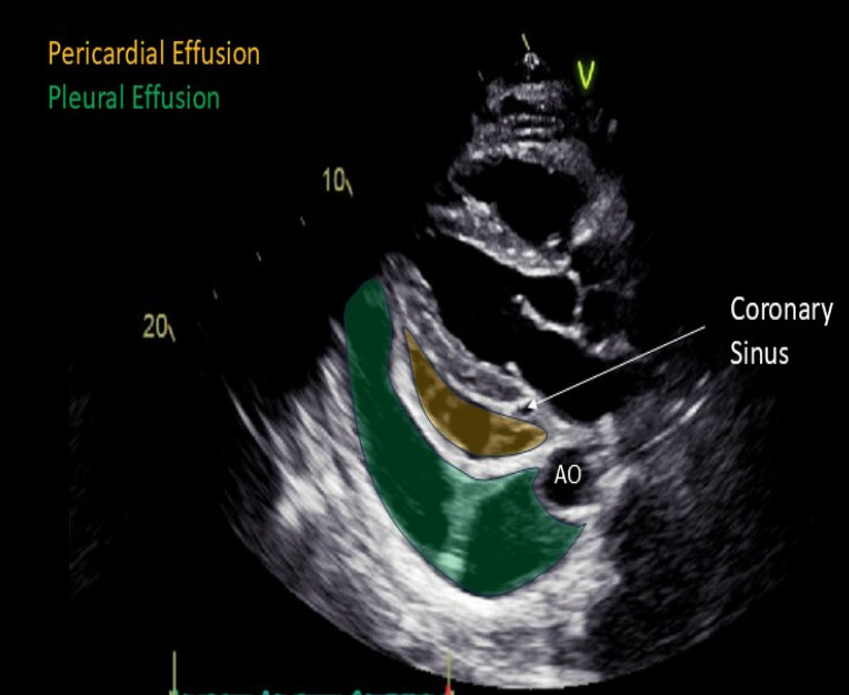
What is pericardial effusion?
Fluid located anterior to descending aorta
What is the sonographic appearance of pericardial effusion?
Small < 1 cm fluid
Large > 2 cm fluid
What is pleural effusion?
Fluid located posterior to descending aorta
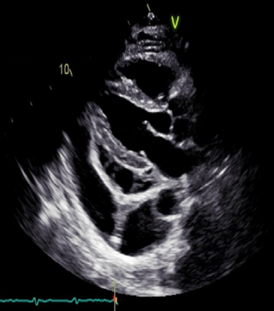
Identify this image.
Pericardial and pleural effusion
What is cardiac tamponade?
Rapid collection of pericardial fluid that causes pressure build up outside of heart and restricted diastolic filling
What are the symptoms of cardiac tamponade?
Beck’s Triad
IJV distension
Hypotension
Muffled heart sounds on auscultation
What is the sonographic appearance of cardiac tamponade?
Diastolic right heart collapse
MV velocity decreases with inspiration
TV velocity increases with inspiration
Pulsus alternans or paradoxus
Swinging motion of herat in fluid
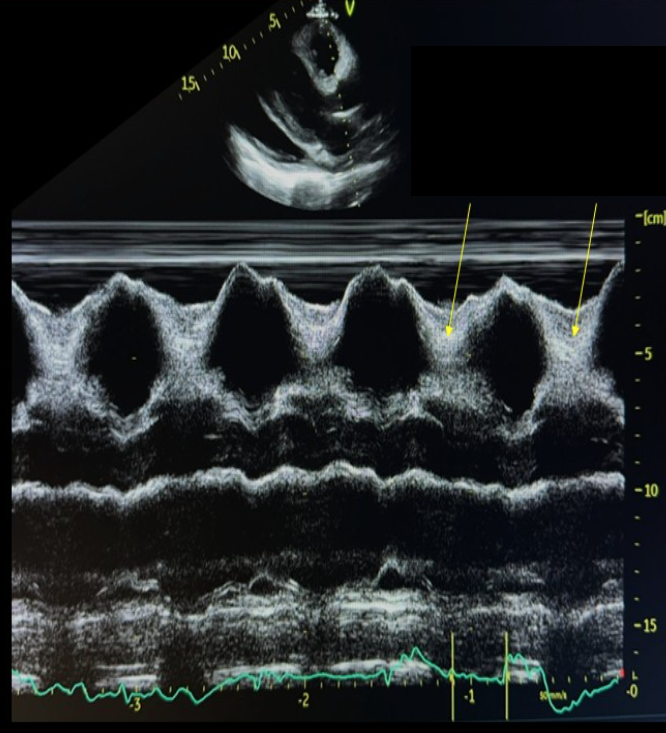
Identify this image.
Diastolic RV collapse seen in those with cardiac tamponade
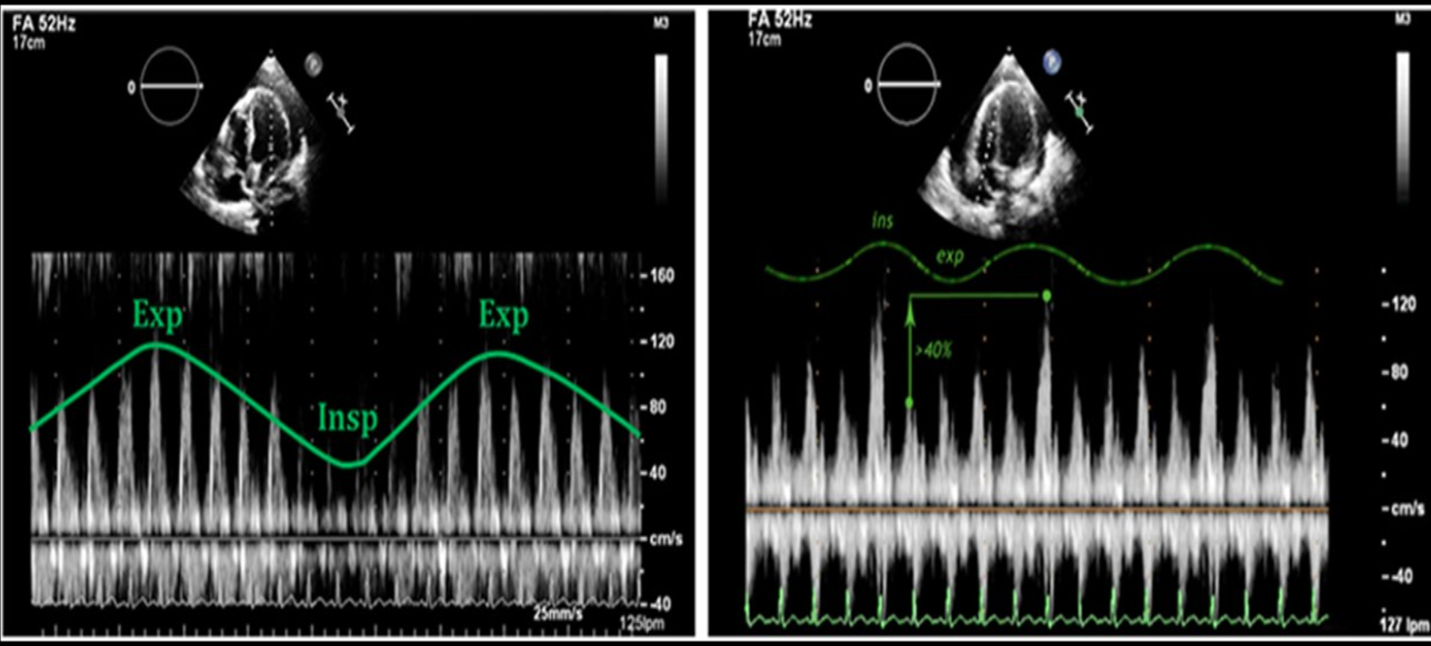
Identify this image.
Cardiac tamponade
MV velocity decreases with inspiration
TV velocity increases with inspiration
What percentage of cardiac tumor are benign?
80%
What is the most common benign heart tumor in adults?
Myxoma
What is a myxoma?
Mobile mass in LA with “grape cluster” appearance
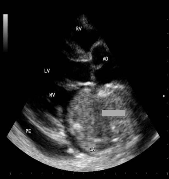
Identify this image.
Myxoma
What is the most common benign heart tumor in children?
Rhabdomyoma
What is a rhabdomyoma?
Mass in RV
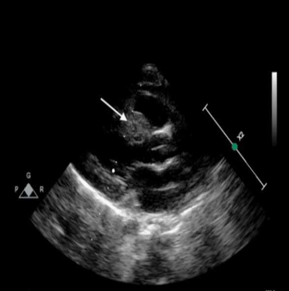
Identify this image.
Rhabdomyoma
What is the most common valvular tumor?
Papillary fibroelastoma
How is a papillary fibroelastoma differentiated from a valvular vegetation?
Papillary fibroelastomas are located on opposite sides of valvular vegetations
Atrioventricular valves (TV/MV): Ventricular side
Semilunar valves (AV/PV): Atrial side
What is the most common primary heart malignancy?
Angiosarcoma
What is an angiosarcoma?
Mass located in RA
What is the most common form of heart malignancy?
Metastasis to heart from another organ
What are the sources of metastasis to the heart?
Melanoma
Lung
Kidney
What is a hiatal hernia?
When stomach protrudes into chest cavity and causes cardiac complications such as LA compression
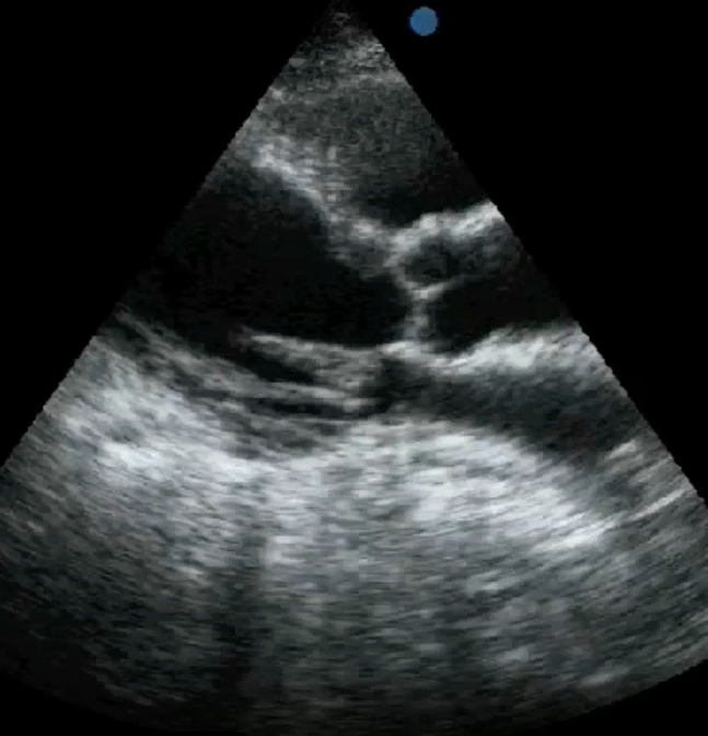
Identify this image.
Hiatal hernia
What is a mechanical valve?
Man-made valve used to replace damaged structures
What are examples of mechanical valves?
Ball-Cage such as Starr-Edwards
Floating disk
Monocuspid tilting disk such as Bjork-Schiley
Bicuspid tilting disk such as St Jude
What is a bioprosthetic valve?
Natural and nonviable tissue used to replace damaged structures
Bioprosthetic valves contain…
3 leaflets
What is a homograft or allograft?
Bioprosthetic valve that comes from human donor OTHER than patient themself
What is an autograft?
Bioprosthetic valve or portion of pericardium that comes from patient themself
What is a heterograft?
Bioprosthetic valve that comes from an animal donor
What is the most common heterograft?
AV from pig
What is a transcatheter bioprosthetic valve?
Valve is inserted via catheter through femoral artery and into heart in order to replace open heart surgery
What are the differences between mechanical and bioprosthetic valves?
Mechanical
Durable and lasts forever
Requires anticoagulant therapy
Produces valve masking or shadowing from metallic elements
Thrombus formation is most common cause of failure
Increased risk of vegetations or endocarditis
Bioprosthetic
Lasts up to 10 years
Does NOT require anticoagulant therapy
Produces less valve masking or shadowing
Tissue degeneration or calcification is most common cause of failure
Vegetations or endocarditis appear similar to native infection
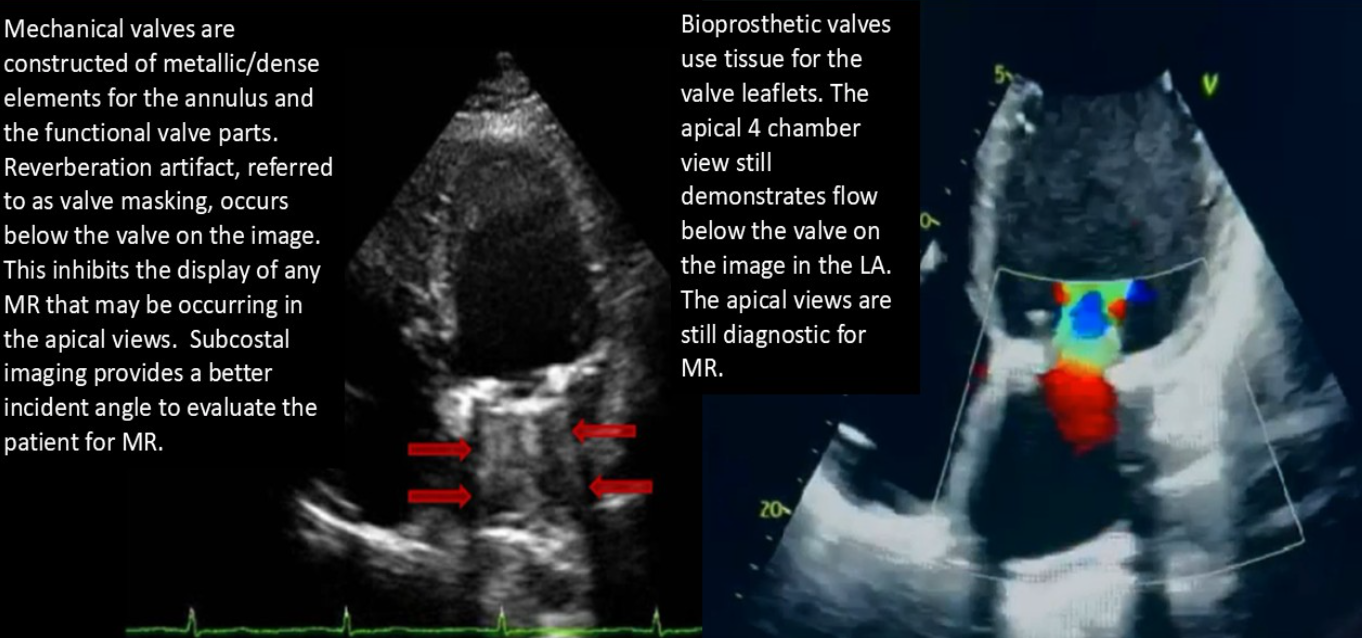
What is the preferred method for evaluating a prosthetic aortic valve?
Velocity ratio or dimensionless index in apical windows
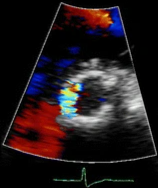
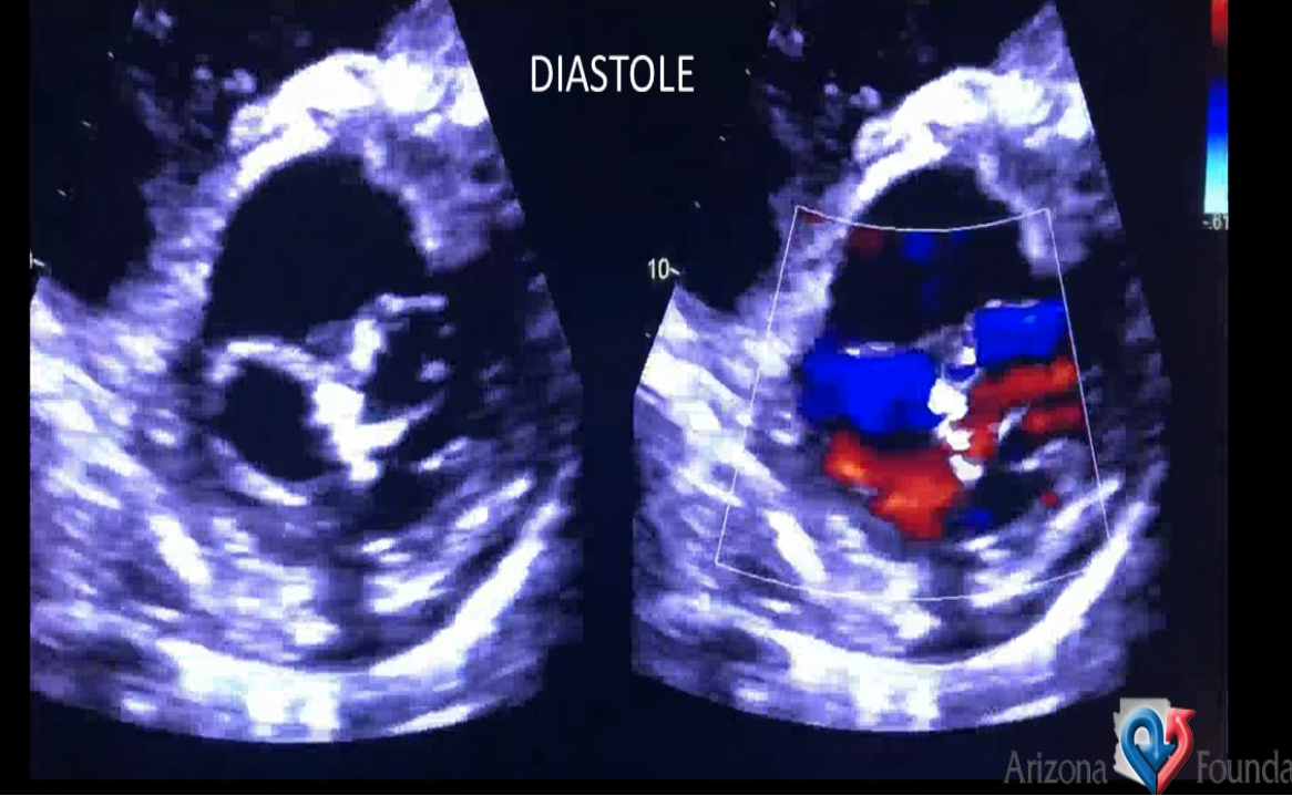
Identify this image.
Paravalvular TAVI/TAVR leak associated with valve failure
What is a transcatheter edge-to-edge repair?
Procedure used to treat MR in patients with EF of 20-50%
Identify this image.
Mitra-clip or transcatheter edge-to-edge repair
What is pannus formation?
Prosthetic valve complication that refers to excessive overgrowth of tissue around valve
What is dehiscence?
Prosthetic valve complication that refers to strut detachment from heart
What is angina?
Chest pain caused by myocardial ischemia
What is stable angina?
Chest pain controlled by medication or rest
What is unstable angina?
Chest pain at rest
What is shortness of breath (SOB)?
Difficulty breathing when at rest
What is dyspnea of exertion (DOE)?
Difficulty breathing with exercise
What is orthopnea?
Difficulty breathing when lying down associated with CHF
What is paroxysmal nocturnal dyspnea?
Difficulty breathing that interrupts sleep associated with pulmonary edema
What is apnea?
Cessation of breathing
What is hemoptysis?
Bloody sputum associated with pulmonary edema, hemorrhage, or MV disease
What is cyanosis?
Blue discoloration of skin associated with pulmonary disease, Eisenmenger syndrome, and Tetralogy of Fallot
What is edema?
Accumulation of fluids associated with heart failure or systemic HTN
What is jugular vein distension?
Increased right side pressure associated with TV disease, constrictive pericarditis, and tamponade
What is pulsus paradoxus?
> 10 mmHg drop in systolic BP with inspiration associated with constrictive pericarditis, tamponade, pulmonary embolism, or COPD
What is syncope?
Fainting associated with hypotension, arrhythmia, MI, or AV disease
What is a fever?
Increase in body temperature associated with endocarditis
What is cachexia?
Malnurtition and wasting caused by chronic heart disease
What lab values indicate the occurrence of an MI?
Elevated levels of
Troponin
Creatine kinase-MB
Lactic dehydrogenase (LDH)
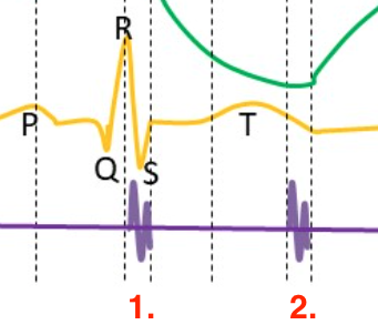
Identify this image.
Normal heart sounds
S1 “lub”: Closure of AV valves after atrial contraction
S2 “dub”: Closure of semilunar valves after ventricular contraction
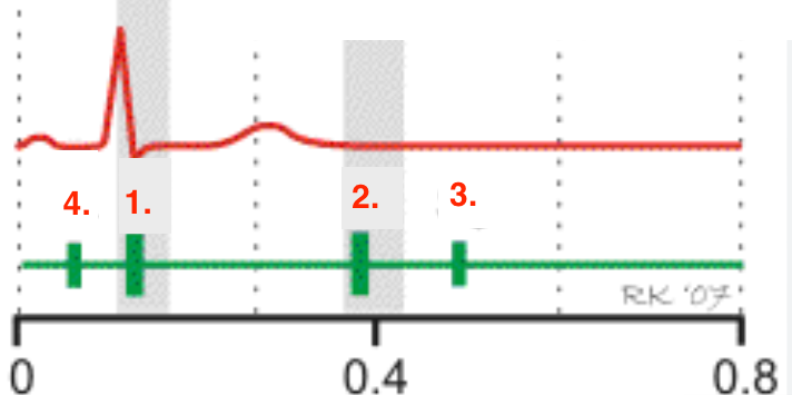
Identify this image.
Abnormal heart sounds
Normal S1 “lub”: Closure of AV valves after atrial contraction
Normal S2 “dub”: Closure of semilunar valves after ventricular contraction
Abnormal S3: Ventricular gallop
Abnormal S4: Presystolic or atrial gallop during atrial kick
What are the causes of a third heart sound or S3?
Normal heart sound in child or adults < 40
Indicates rapid ventricular filling due to
Pregnancy
Anemia
CHF
Dilated CM
Severe valvular regurgitation
Left to right shunting
What are the causes of a fourth heart sound or S4?
HTN
Severe AS
Severe PS
CAD
CM
What is a pericardial rub?
Heart sound due to inflammation associated with acute pericarditis
What is a pericardial knock?
Heart sound heard in early diastole associated with constrictive pericarditis
What is a murmur?
Heart sound caused by turbulence of blood as it crosses a narrow path
What is a functional murmur?
Heart sound caused by rapid flow across normal cardiac valve
What is a pathologic murmur?
Heart sound caused by turbulent flow across diseased cardiac valves or cardiac defects
What is a systolic murmur?
Murmur that occurs during S1 and S2
What are the causes of a SYSTOLIC EJECTION murmur or EJECTION CLICK?
Congenital AS (BAV)
Congenital PS
HOCM
What are the causes of a MID SYSTOLIC murmur?
AS or dilation of aortic root
PS or dilation of pulmonary trunk
Increased flow through normal semilunar vales
What is a midsystolic click?
Heart sound heard in mid to late systole associated with MVP
What are the causes of a LATE SYSTOLIC murmur?
MVP
TVP
Papillary muscle dysfunciton
What are the causes of a HOLOSYSTOLIC murmur?
MR
TR
VSD
What is a diastolic murmur?
Murmur that occurs between S2 of one cardiac cycle and S1 of another
What are the causes of EARLY DIASTOLIC murmurs?
AR
PR
What are the causes of DIASTOLIC EJECTION murmurs or an OPENING SNAP?
MS
TS
What are the causes of LATE DIASTOLIC murmurs?
Complete heart block
Large left to right shunts (PDA, ASD)
What is an Austin Flint murmur?
Functional diastolic murmur caused by severe AR
What is a Dock murmur?
Early diastolic murmur associated with LAD stenosis
What is a continuous murmur?
Murmur caused by continuous flow throughout systole and diastole
What are the causes of a continuous murmur?
PDA
AVF
Anomalous coronary origin
How can the location of a murmur identify the cause?
2nd RIGHT parasternal space: AS
2nd LEFT parasternal space: PS
3rd parasternal space: AR
4th parasternal space: TV disease
5th midclavicular: MV disease
What is the effect of the valsalva maneuver on murmurs?
Increased murmur in those with HOCM and MVP
What is the effect of amyl nitrate on murmurs?
Increased murmur in those with LVOT stenosis and MVP
Decreased murmur in those with AR or MR
What are the effects of alcohol abuse on the heart?
MI
Reduced systolic function
Dilated CM
What are the effects of diabetes on the heart?
CAD
LVH
Diastolic dysfunction