📸 Wiley Plus Chapter 20 Part 1
 Call Kai
 Learn
Learn Practice Test
 Spaced Repetition
 Match
Match Flashcards
 Knowt Play
1/35

There's no tags or description
Looks like no tags are added yet.
Name | Mastery | Learn | Test | Matching | Spaced | Call with Kai | Chat |
|---|
No analytics yet
Send a link to your students to track their progress
36 Terms
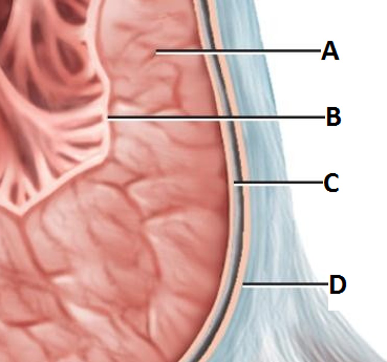
The visceral layer of the serous pericardium:
is the outer layer of the heart
The thickest wall in the heart due to its increased work load is the:
left ventricle
What is the correct pathway of blood starting at the right atrium?
right atrium, right ventricle, pulmonary trunk, pulmonary veins, left atrium, left ventricle, aorta
A skeletal muscle may undergo tetanus but cardiac muscle cannot.
True
Comparatively, which phase is the longest during an action potential in a ventricular contractile fiber?
Refractory
What is occurring during isovolumetric contraction?
All four valves are closed
The walls of the atria are thicker because the atria receive blood under pressure.
False
The walls of the ventricles are thicker because the ventricles pump blood under higher pressure over greater distances.
True
The wall of the right ventricle is thinner than that of the left ventricle because the right side of the heart pumps blood a shorter distance at lower pressure.
True
Although the right side of the heart has a smaller workload, the right and left ventricle simultaneously eject the same amount of blood.
True
Prolapse of the atrioventricular valves is prevented by
contraction of the papillary muscles
The semilunar valves open when pressure in the right and left ventricles exceeds that in the pulmonary trunk and aorta, respectively.
True
The intercalated discs seen in cardiac myocytes
1. have desmosomes which help to hold the muscle fibers together.
2. have gap junctions which allow action potentials to conduct from one muscle fiber to the next
3. are the result of actin/myosin overlap.
1 and 2 are correct
Which represents the correct flow of excitation through the cardiac conduction system?
SA node, AV node, bundle of His, right and left bundle branches, Purkinje fibers
Which of the following places the phases of the cardiac cycle in correct order?
atrial contraction, isovolumetric contraction, ventricular ejection, isovolumetric relaxation, ventricular filling, atrial contraction
The plateau seen in action potentials recorded from ventricular fibers is due to inflow of Ca+2.
True
Isovolumetric contraction and isovolumetric relaxation both occur when all four valves are closed.
True
Purpose of the structure indicated is to: (right auricle?)
slightly increase the capacity of the atrium (see diagram)
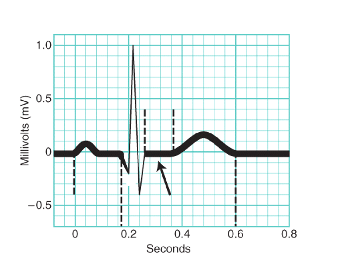
What occurs after the QRS phase of the ECG?
the ventricles contract
The pericardium consists of a fibrous and serous layer. The visceral layer of the serous pericardium may be referred to as the epicardium.
True
The right atrium receives blood from the superior and inferior vena cava and the coronary sinus while the left atrium receives blood from the 4 pulmonary veins.
True
Autorhythmic fibers are self-excitable
True
Autorhythmic fibers: are pacemakers
True
Autorhythmic Fibers: Form the conduction system of the heart
True
The left ventricular pressure must be greater than the aortic pressure during ventricular ejection so that blood will move out through the pulmonary arteries.
False
The membrane that surrounds and protects the heart is called the
pericardium
Which of the following is used to reduce friction between the layers of membranes surrounding the heart?
pericardial fluid
Which layer of the heart wall consists of cardiac muscle tissue?
myocardium
Contraction of the ventricles of the heart leads to blood moving directly
into arteries
Which of the following chambers of the heart contains deoxygenated blood?
right atrium and right ventricle
Cardiac muscle fibers are electrically connected to neighboring fibers by
gap junctions
Which network of specialized cardiac muscle fibers provides a path for each cycle of cardiac excitation to progress through the heart?
cardiac conduction system
Which term refers to the period of time during a cardiac cycle when contraction of a chamber occurs and pressure within the chamber rises?
systole
Which wave in an electrocardiogram represents repolarization of the ventricles?
T wave
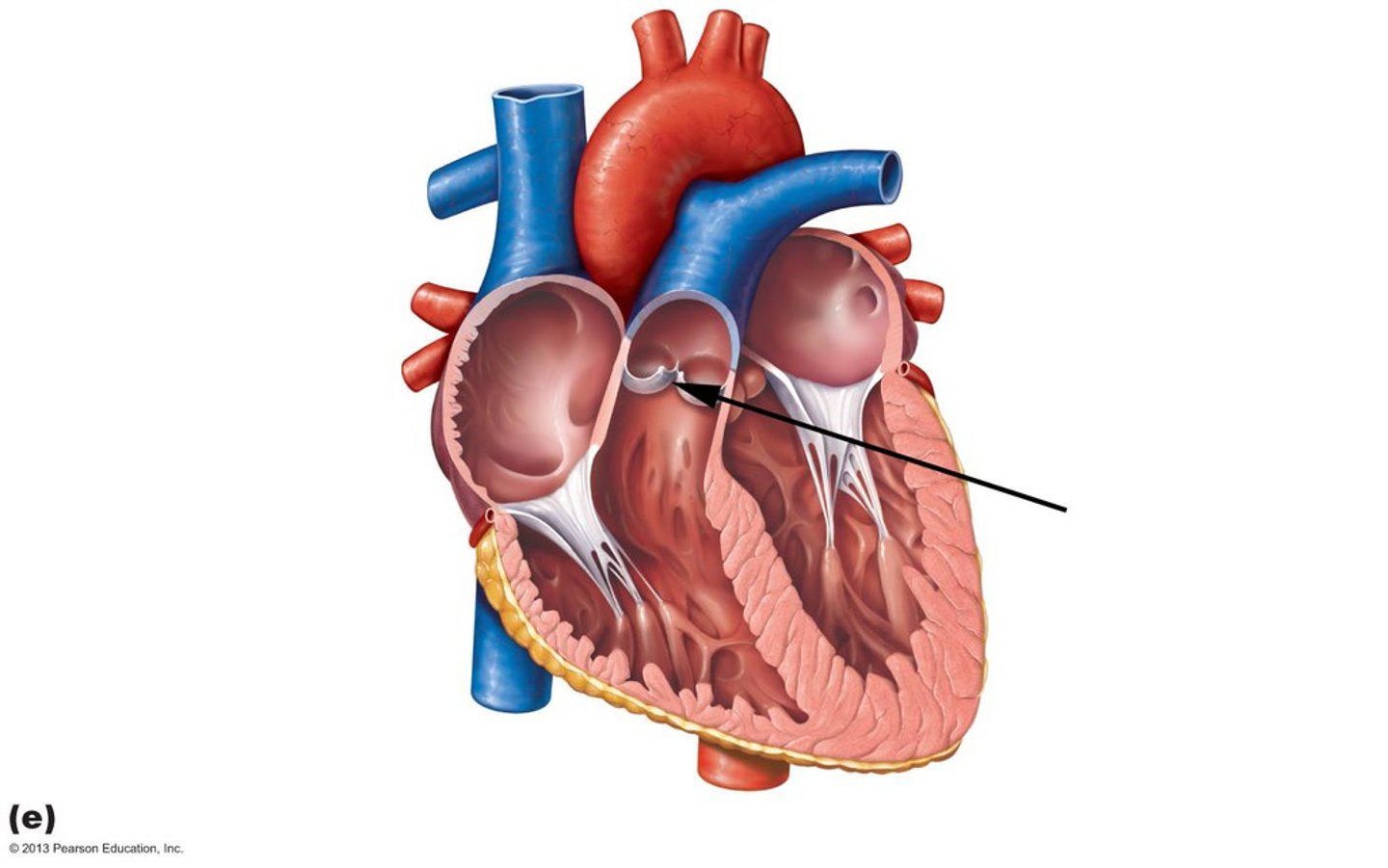
Identify the structure indicated in this diagram
pulmonary valve
The structure indicated is the:
endocardium (C)