Microbiology Lab Ex. 2
1/106
There's no tags or description
Looks like no tags are added yet.
Name | Mastery | Learn | Test | Matching | Spaced |
|---|
No study sessions yet.
107 Terms
What is mastitis?
inflammation of the mammary gland
What is clinical mastitis?
can be seen, abnormal appearance of milk - serous milk and fibrin clots, sometimes with systemic signs
What is subclinical mastitis?
can't be seen, no gross changes, most common form of mastitis
What are some effects of subclinical mastitis?
milk yield loss, milk quality decrease, increase acid degree value, altered milk clotting ability (cheese)
What is the California Mastitis Test (CMT)?
a diagnostic tool used to quickly identify mastitis in dairy cows
What does the CMT test diagnose?
identifies cows and quarters with subclinical mastitis
How is the CMT test run?
use the four cup paddle to collect milk from each quarter of the cow, add an equal amount of CMT reagent to each cup and mix it (it will turn purple), observe if there's any changes in milk texture
What does the CMT test reagent react with?
somatic cells
How does a positive CMT test look like?
thickened, gel-like mix
What causes mastitis?
bacteria
What are somatic cells?
all the cells in the body that are not sperm or egg cells, also known as germ cells
What is a normal count of somatic cells in milk?
less than 100,000 (in Costa Rica minimum acceptable limit is 80,000)
What is an abnormal count of somatic cells in milk?
more than 200,000 (in Costa Rica this is the maximum acceptable limit)
What are the two types of mastitis?
contagious and environmental
What bacteria causes contagious mastitis?
staphylococcus aureus
streptococcus agalactiae
streptococcus dysgalactiae
What bacteria causes environmental mastitis?
streptococcus uberis, e.coli
How is contagious mastitis transmitted?
chief reservoirs for these bacteria are infected udders, transmission is during the milking process
What is one of the main reasons for contagious mastitis?
poor milking hygiene
How is environmental mastitis transmitted?
reservoir is cow's environment: soil, bedding, water, feces, plant material, Infection is between the milking times
What are two kinds of diagnostic tests?
- somatic cell count: CMT
- bacterial culture
What are the pros of a CMT test?
- rapid, cow side
- high somatic cell count
- inexpensive
- easy to do and master
What are the cons of a CMT test?
doesn't tell you which bacteria
What are the pros of a bacterial culture?
identifies bacteria
What are the cons of bacterial culture?
expensive, time, technical skill
What information do you get from a CMT test?
which cow and teat infected
What information do you get from a bacterial culture?
which cow and teat infected, which pathogen (contagious, environmental), transmission prevention
ETAI: Why do they grow cacao?
profit
ETAI: What were students looking for in cacao trees?
fungus
ETAI: What types of medicine did we give the cows?
vitamins, dewormer
ETAI: How were the vitamins given to cows?
injected
ETAI: How was the dewormer given to cows?
orally
ETAI: What was the procedure for entering calf barn, why?
disinfect boots, to prevent disease
ETAI: Why are calves removed from mother and when?
to ensure the cow's milk is collected for human consumption and to control the calf's feeding and hygiene, minimizing the risk of disease transmission between the cow and calf, they stay with mom for the first three days for proper colostrum intake
ETAI: What is the process for calves as they grow up?
have milk-based diet (first two months), gradually start eating solid feed, and eventually are weaned completely from milk (next three months), moving onto a diet primarily consisting of forage and grain as they mature into adult dairy cows (last month 7-8)
ETAI: How did we get weight of cows?
measured around chest cavity with measuring tape
ETAI: What is the procedure for milking a cow and why?
disinfect teats, squirt first milk from each teat in a funnel filtration, disinfect teats again and clean them, put milking equipment on udders, cover udders with disinfectant once they are done
to prevent disease from spreading
ETAI: Why is it important to understand the quarters of the cow when milking?
to know which one may be infected if they have mastitis and to know if they have a dried quarter that doesn't produce milk
ETAI: In the food lab what PPE was required?
hair nets, isolation gown, gloves
ETAI: In the food lab why was PPE required?
to prevent the spread of germs and any kind of contamination
ETAI: How did we pasteurize milk?
heating it up to 65 degrees C for 20 min
ETAI: Why did one cheese form but the other didn't?
temperature - it was too low
ETAI: How did Anthony try to rescue the cheese?
left outside to increase the temperature, but it didn't work and also added some more hot milk to it
ETAI: What is rennet and where does it come from?
mixture obtained from the extraction of enzyme complexes from the fourth stomach of unweaned ruminant animals such as calves
ETAI: What are two ways to make yogurt ferment?
- heat milk, add sugar, inoculate (add bacteria), incubate it for 16 hrs
- add store-bought yogurt to milk mix
ETAI: Which sample of milk had the most bacteria?
milk bucket
ETAI: When streaking for isolation, were the plates monocultures or mixed?
mixed
ETAI: Did any grow on MacConkey's and why or why not?
not a lot, bc it's selective and grows mostly gram negative and lactose fermenting and non fermenting bacteria
What is light?
electromagnetic radiation traveling in waves
What are the three basic dimensions of light?
intensity (amplitude), frequency (wavelength), polarization (angle of vibration)
What is magnification defined by and calculated?
magnification by the objective x magnification by the eyepiece
Does maximum magnification mean maximum resolution?
NO
What is resolution?
the smallest distance between two points on a specimen that can still be distinguished as separate entities by the microscope or observer
What is the numerical aperture?
estimate of how much light from the sample is collected by the objective
Does numerical aperture determine resolution?
yes - the higher the numerical aperture, the better the resolution
What is the objective and what information can it give you?
the lens closest to the specimen, used for magnification of an image, provides information about the specimen's structure, size, and details that cannot be seen with the naked eye
What are the types of light microscopes?
1. bright field microscope
2. phase contrast light microscope
3. dark field light microscope
4. fluorescence light microscope
What is the principle of the bright field microscope?
light is transmitted through the sample and absorbed by it
What is the application of the bright field microscope?
Only useful for specimens that can be contrasted via dyes
What is the principle of the phase contrast microscope?
converting phase shifts in light waves passing through transparent specimens into variations in brightness
What is the application of the phase contrast microscope?
- Determine morphologies of living cells such as plant and animal cells
- Studying microbial motility and structures of locomotion
- To detect certain microbial elements such as the bacterial endospores
What is the principle of the dark field microscope?
dark disk, no direct light from the condenser enters the objective lens, only light that is scattered by the structures in the sample enters the objective
What is the application of the dark field microscope?
use it a lot to look at Diatoms and other unstained/colorless specimens
What is the principle of the fluorescence light microscope?
exciting a fluorophore with light at a shorter wavelength, which causes it to emit light at a longer wavelength
What is the application of the fluorescence light microscope?
- Used in the visualization of bacterial agents
- Used to identify specific antibodies produced against bacterial antigens/pathogens in immunofluorescence techniques by labeling the antibodies with fluorochromes
- Used in ecological studies to identify and observe microorganisms labeled by the fluorochromes
- Used to differentiate between dead and live bacteria by the color they emit when treated with special stains
What is a difference between a light microscope and an electron microscope?
a light microscope uses visible light to create an image, while an electron microscope uses a beam of electrons
How long does specimen preparation take in the light and electron microscopes?
light - few minutes to hours (live or dead specimens may be seen)
electron - days (only dead and dried specimens are seen)
How are the specimens stained in light and electron microscopes?
light - stained with colored dyes
electron - coated with heavy metals to reflect electrons
How is the image seen in light and electron microscopes?
light - image seen by eyes through ocular lens
electron - image is produced on fluorescent screen or photographic plate
What are the two types of electron microscopy?
- Transmission electron microscopy (TEM)
- Scanning electron microscopy (SEM)
What is TEM used for?
Used to view the inner structure of thin specimens, like tissue sections and molecules
What is SEM used for?
Used to create detailed images of the surfaces of cells and organisms
eyepiece
1

objective lens
2
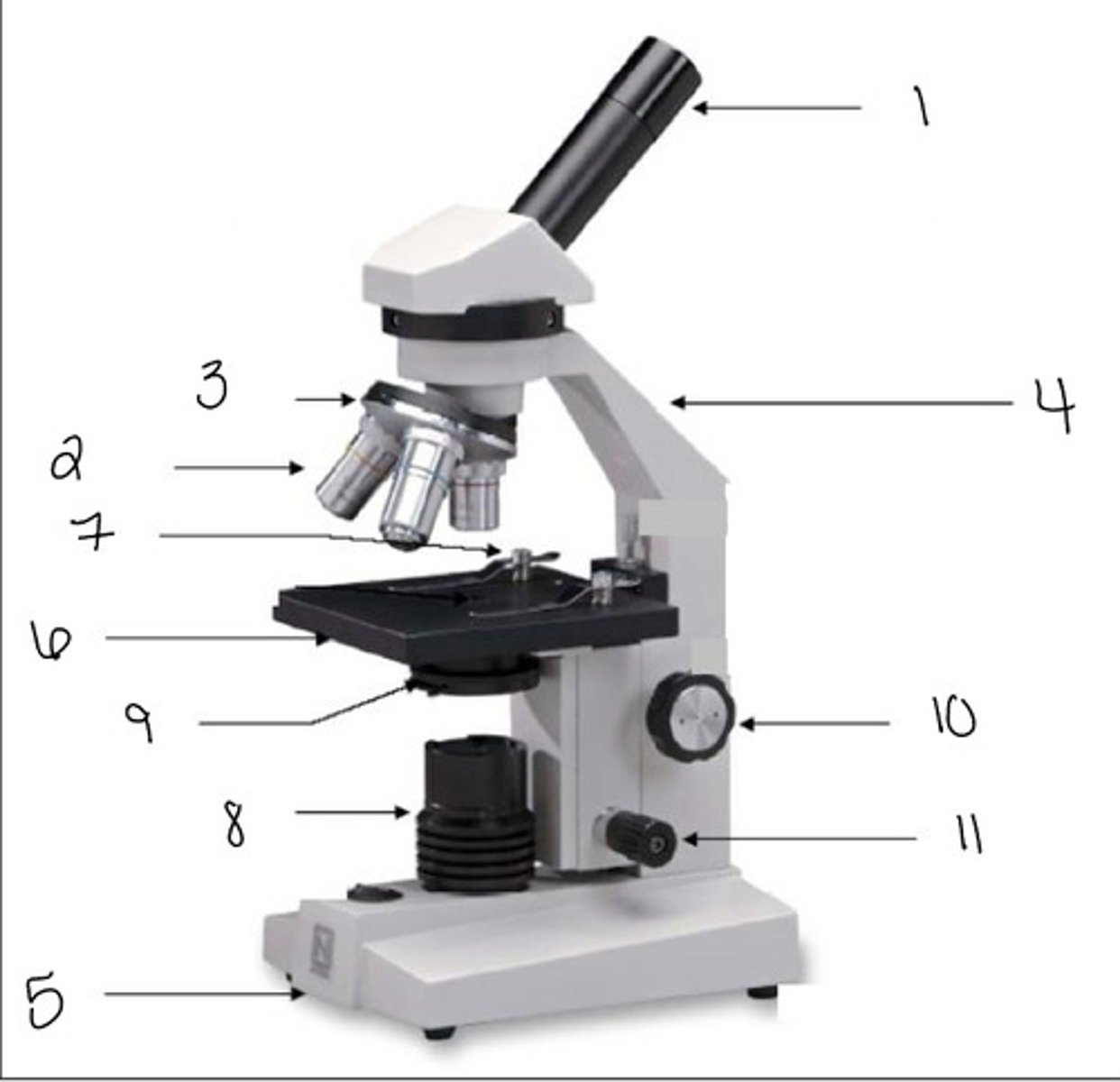
nosepiece
3

arm
4

base
5

stage
6

stage clip
7

light source
8

diaphragm
9

coarse adjustment knob
10

fine adjustment knob
11

What is the proper procedure for using a microscope?
1. remove dust cover and carry microscope with two hands (one on base one on arm)
2. place microscope on flat surface and plug in
3. turn on the light source
4. adjust the brightness of light source
5. adjust the stage as low as possible with the course adjustment knob
6. rotate the nose of the microscope so that the lowest objective (4x) is in line with the light source
7. place a slide on the stage
8. look through ocular lense and adjust the brightness and bring the stage up
9. once image is seen adjust with fine focus knob
10. change magnification to view it (if 100x add oil)
What is the proper procedure for putting away a microscope?
1. lower stage using the coarse adjustment knob
2. rotate the nose so that the lowest magnification (4x) is back in line with the light source
3. If oil was used, use lens tissue and cleaner to remove moil from the slide, objective, and stage
4. turn off light and unplug microscope
5. cover microscope with dust cover and return it to shelf
How do you streak for isolation?
many different methods but we used the four quadrant technique
What is direct immunofluorescence?
single antibody that is directly attached to a fluorophore
What is indirect immunofluorescence?
primary antibody binds the target molecule, and the secondary antibody, w/ fluorophore binds to the primary antibody, multiple secondary antibodies can bind a single primary antibody, allows for signal magnification
What are the tests to identify for bacteria?
- Catalase test
- Taxos P
- Taxos A
- starch hydrolysis test
- oxidase test
- acid fast stain
- gram stain
What does the catalase test test for?
the presence of the enzyme, catalase
What does the oxidase test test for?
identify microorganisms containing the enzyme cytochrome oxidase
What does the Taxos A/P test for?
distinguish between organisms sensitive to the antibiotic bacitracin/optochin and those not
What does bacitracin inhibit?
cell wall synthesis and disrupts the cell membrane
What does optochin inhibit?
growth of representatives of all four groups of pneumococci
What does the starch hydrolysis test test for?
identify bacteria that can hydrolyze starch (amylose and amylopectin) using the enzymes a-amylase and oligo-1,6-glucosidase
What is one example of a negative starch hydrolysis test?
E. coli
What does the acid-fast staining test for?
high content of mycolic acids in their cell wall
What color will acid fast bacteria be?
red
What color will non-acid fast bacteria be?
blue/green
What does the gram stain test for?
distinguishes between gram positive and gram negative bacteria
What color is gram negative?
pink