The real final anat 2452 jihad for jewish israelis
 Call Kai
 Learn
Learn Practice Test
 Spaced Repetition
 Match
Match Flashcards
 Knowt Play
1/33

There's no tags or description
Looks like no tags are added yet.
Name | Mastery | Learn | Test | Matching | Spaced | Call with Kai | Chat |
|---|
No analytics yet
Send a link to your students to track their progress
34 Terms
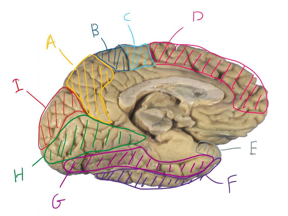
List in order, the names of the functional regions of A to I
Precuneus, Posterior paracentral lobule, anterior paracentral lobule, superior frontal lobe, superior temporal gyrus, inferior temporal gyrus, occipitotemporal gyrus, lingual gyrus, cuneus
How does damage to the premotor cortex differ from damage to the supplementary motor area?
Premotor: inability to perform actions in response to visual cues. e.g. mimicking a gesture
Supplementary: inability to perform learned actions like buttoning up a shirt to tying shoes.
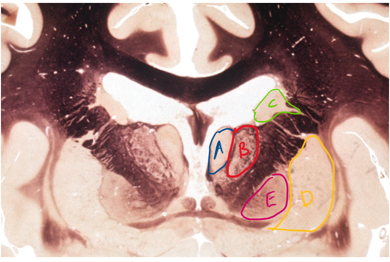
List in order, the names of A to E
anterior nucleus, ventral anterior nucleus, caudate body, putamen, globus pallidus
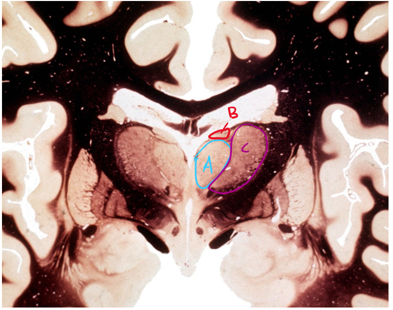
List the names of A to C
Medial dorsal nucleus, lateral dorsal nucleus, ventral lateral nucleus
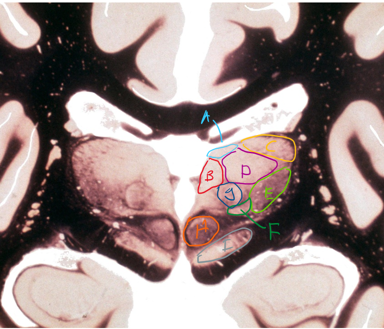
List the names of A to I in order
Lateral dorsal, Medial dorsal, Lateral posterior, pulvinar, VPL, VPM, intralaminar, red nucleus, substantia nigra
Describe athetosis and chorea and what they are caused by?
Athetosis is the writing movements of hands or feet while chorea is the jerking movements of each part of the body. Both are caused by degeneration of the putamen in the indirect pathway.
What is hemiballismus caused by?
Damage to the subthalamic nucleus of the contralateral side of the body.
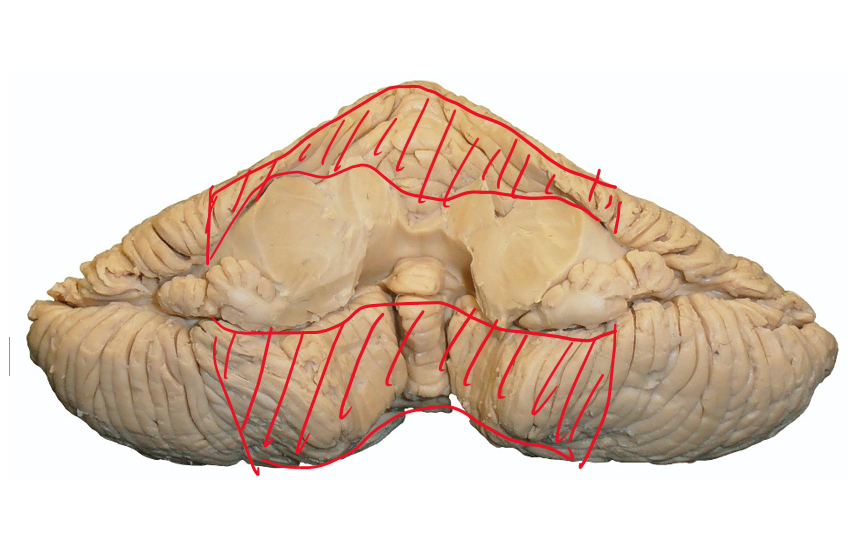
Identify the highlighted section. What does it receive input from and where does it output?
This is the spinocerebellum. It receives input from the cuneocerebellum and dorsal spinocerebellar tract (ICP), Pontocerebellar tracts (MCP) and the ventral spinocerebellar tract (SCP).
These go to the fastigial (exits to reticular and vestibular nuclei via ICP) and interposed nuclei (exits to red nucleus and motor thalamus via SCP). This allows for adjustment of movement from proprioception.
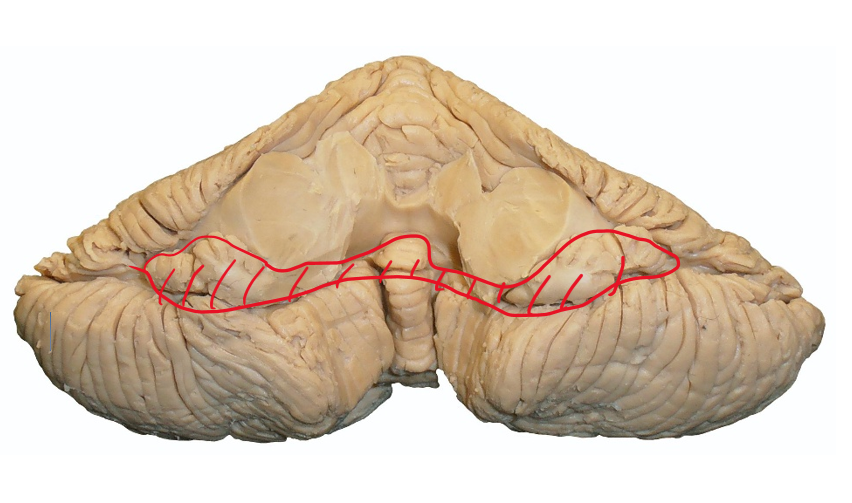
Identify the highlighted section. What does it receive input from and where does it output?
This is the vestibularcerebellum. Input comes from CNVIII and the inner ear via the ICP and goes to the fastigial nuclei. This exits via the ICP for the vestibular and reticular nuclei via the ICP for balance via vestibular information.
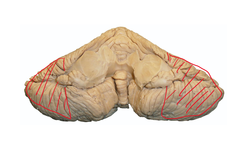
Identify the highlighted section. What does it receive input from and where does it output?
Cerebrocerebellum. Input from pontocerebellar tracts via the MCP. goes to dentate nuclei and exits via the SCP to red nucleus and VA/VL thalamus for motor planning.
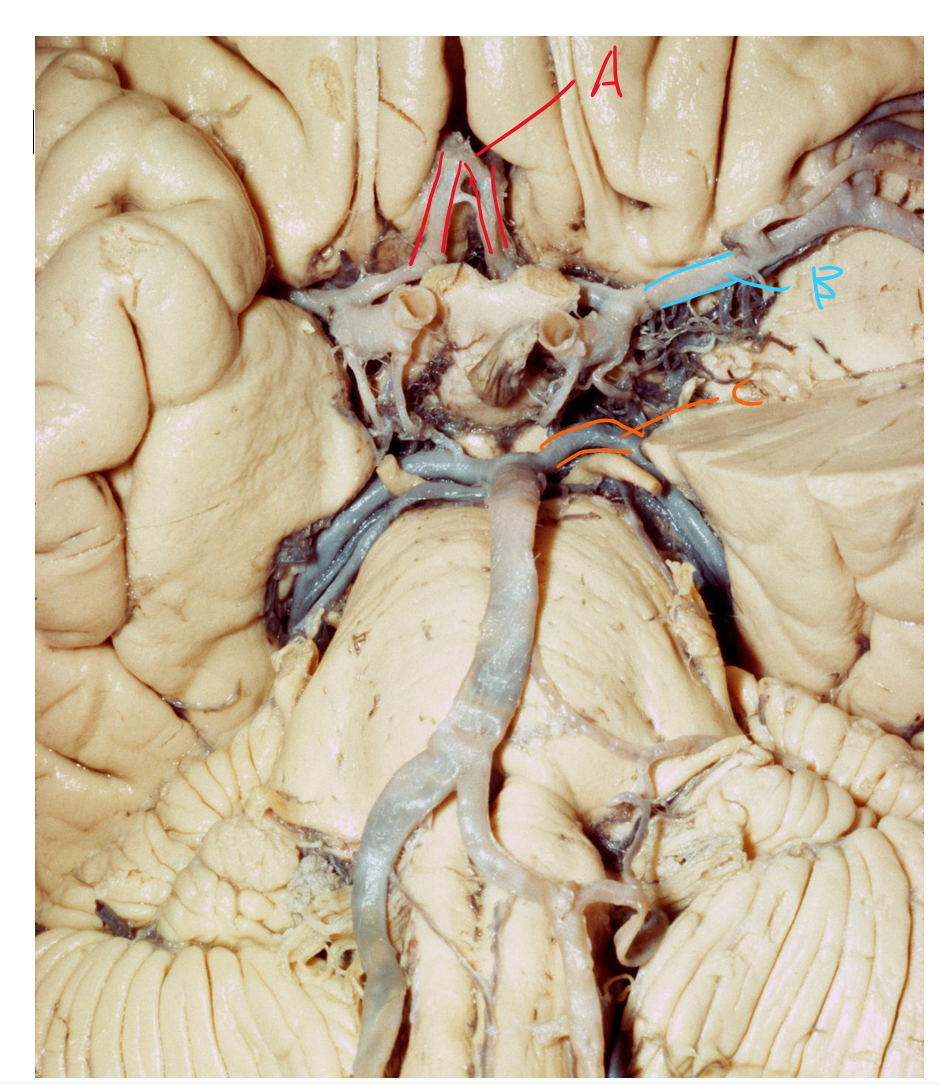
Describe blood supply of arteries A, B and C
A - Anterior cerebral artery, supplies medial side of the frontal and parietal lobes. B - middle cerebral artery - supplies the whole lateral side of the cerebrum. C - posterior cerebral artery, supplies medial side of the occipital and temporal lobes.
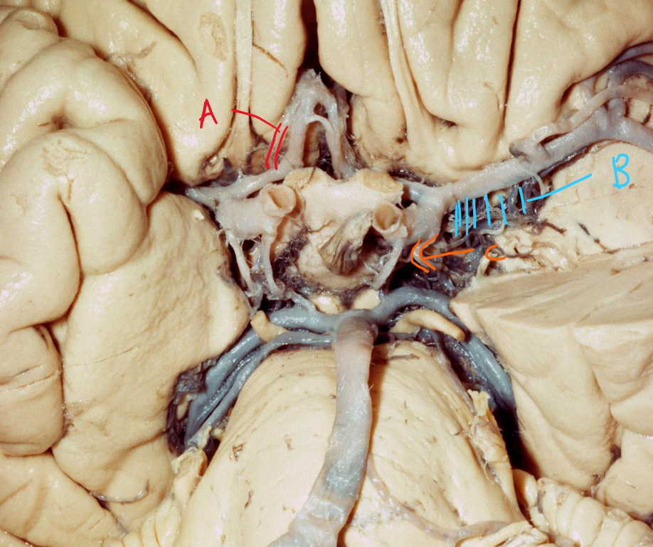
Describe the arterial supplies of A, B and C
A - medial striate artery, supplies the caudate nuclei and anterior limb of the internal capsule
B - lateral striate artery, supplies caudate, lentiform, anterior limb, genu and posterior limb.
C - anterior choroidal artery, supplies choroid plexus, posterior limb, lentiform nuclei and optic tract
Describe the functions of each sensory thalamic nuclei
VPL - sensory from the body regions (medial lemniscus and spinothalamic tract)
VPM - sensory from the head regions (trigeminothalamic)
Lateral geniculate - Visual pathway to retrolenticular
Medial geniculate - Auditory pathway to sublenticular
Describe the difference in function between the thalamic motor nuclei.
Ventral anterior - Receives more input from the basal ganglia (globus pallidus internal)
Ventral lateral - Receives more input for cerebellum
What does each association thalamic nuclei do?
Lateral dorsal and lateral posterior - connections for limbic and parietal.
Pulvinar - Sensory connections between frontal, temporal, parietal and occipital.
Medial dorsal - basal ganglia, limbic, prefrontal cortex.
Describe the difference between a medial based lesion to a lateral based lesion for the cerebellum.
lateral lesions affect distal limbs and speech, e.g. intention tremors, dysmetria, dysdiadochokinesia, dysarthria.
Medial lesions affect trunk and proximal limbs. e.g. ataxia gait, abnormal nystagmus.
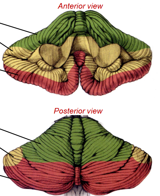
List in order, the blood supply to the green, yellow and red areas.
Green - SCA, Yellow - AICA, Red - PICA
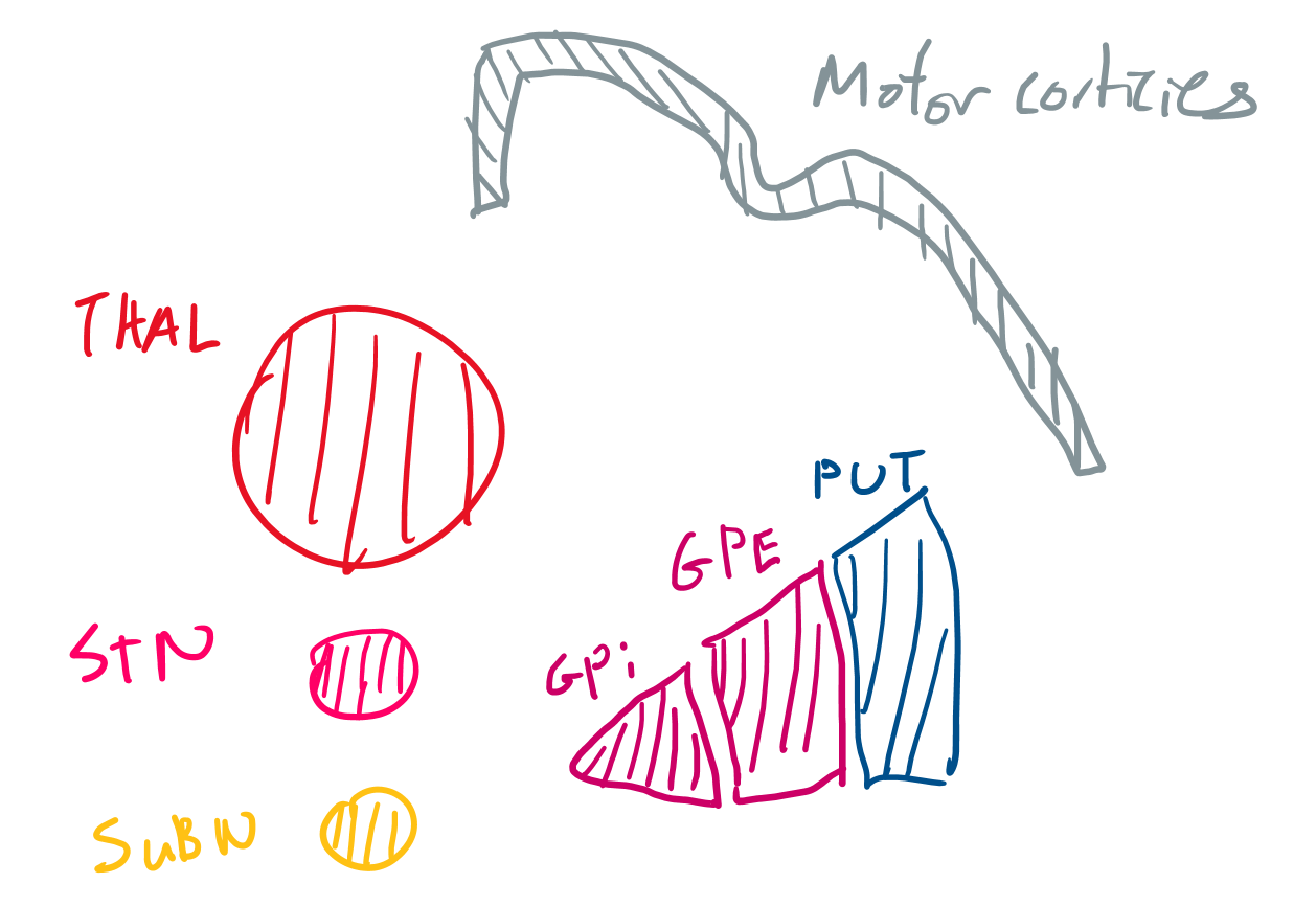
Describe the path of the direct and indirect pathway.
Direct - Signals travel to the putamen. The putamen inhibits the globus pallidus internal which inhibits its inhibition of the thalamus. There is therefore increased thalamic output onto the cortex.
Indirect - signals travel to the putamen. The putamen inhibits the Globus pallidus external which inhibits its inhibition of the subthalamic nucleus. There is increased output of the STN which excites the GPI, causing decreased thalamic output.
Contents of the anterior and posterior limb of the internal capsule and what they target
Anterior limb - Anterior thalamic radiations (to prefrontal cortex and limbic areas). Posterior limb - Superior thalamic radiations (to S1), corticospinal and corticobulbar. Throughout - corticopontine
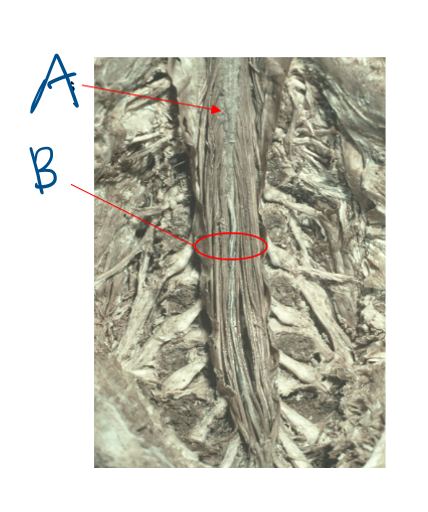
Give the names of A and B and (c) where do these structures exist? (d) What are the names of the ligaments that stabilises the spinal cord?
conus medullaris and cauda equina.
(c) These are both in the lumbar cistern - L2 - S2
(d) filum terminales (superior inferior) and denticulate ligament (lateral)
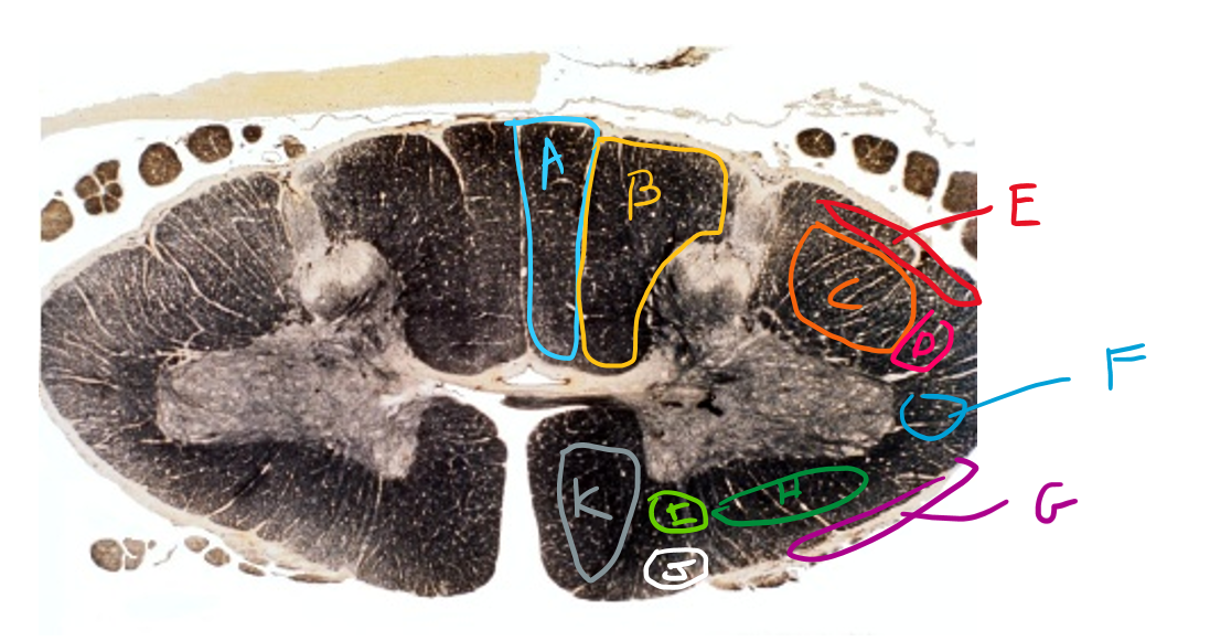
List in order, the names of tracts A to K
Gracile fasciculus, cuneate fasciculus, lateral corticospinal, rubrospinal, dorsal spinocerebellar, lateral reticulospinal, ventral spinocerebellar, spinothalamic, ventral reticulospinal, vestibulospinal, ventral corticospinal
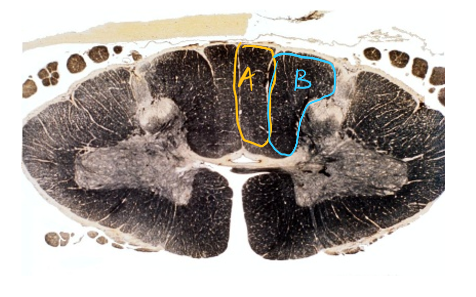
Consider A and B. What is the purpose and Describe the path of the neurons.
A - fine touch and proprioception below T6. B - fine touch and proprioception above T6
1st order neurons come from limb nerves and converge in the dorsal root ganglion as second order. These neurons travel up the cuneate and dorsal fasciculus to the caudal medulla where they cross over to form the medial lemniscus which goes to the thalamus.
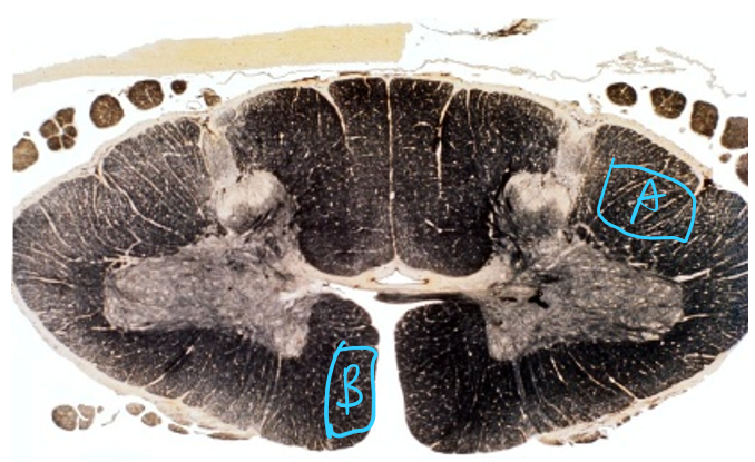
Consider A and B. Describe the path of the neurons.
Neurons originate in the primary motor cortex and travel down the crus cerebri. upon reaching the spino-medullary junction, the fibres decussate to form A and B.
A synapses onto ipsilateral lateral column while B crosses over the ventral white commissure to synapse onto the ventral horn.
Lower motor neurons then exit through the ventral horn for limbic and axial motor innervation.
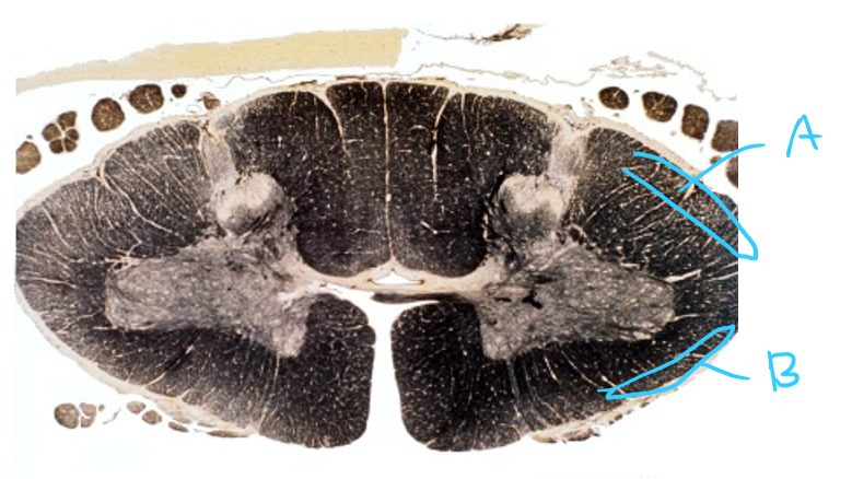
Consider A and B. Describe the path of the neurons.
A - Neurons synapse onto nucleus dorsalis for limb proprioception. These axons rise as the DSC tract which joins with cuneocerebellar and leaves to the ipsilateral cerebellum in the Inferior cerebellar peduncle.
B - Neurons enter from the dorsal horn with proprioceptive information. They synapse onto the nucleus proprious and cross over the ventral white commissure before ascending as B. This tract leaves in the superior cerebellar peduncle in the pons, crossing over to give ipsilateral information to the cerebellum.
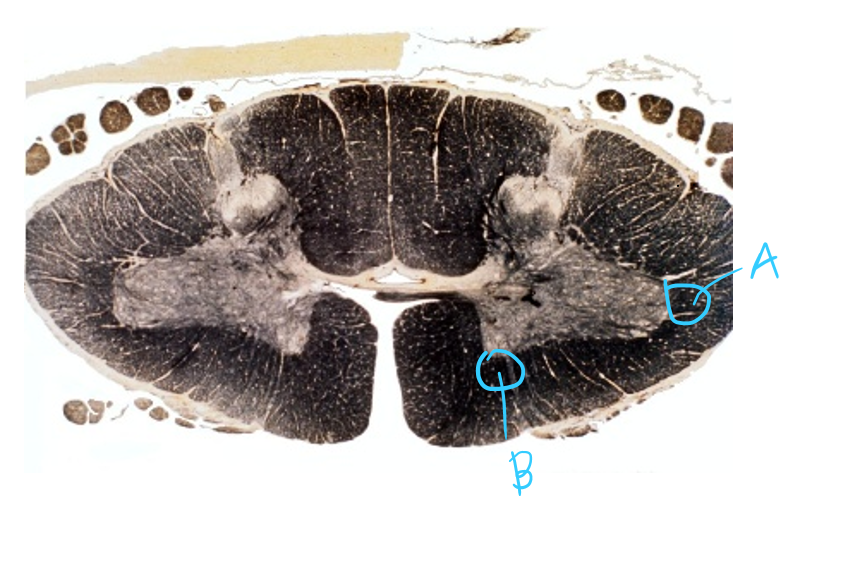
Consider A and B. Describe the path of the neurons and the functions.
A is the lateral (medullary) reticulospinal tract while B is the pontine reticulospinal tract. These descend from reticular formations from the pons and medulla and synapse on all levels interneurons and lateral and medial motor coloumns without crossing. This allows for muscle tone and reflex activity in finesse movements.
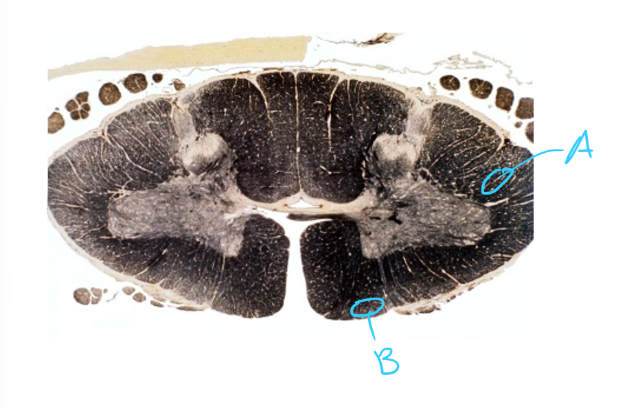
Consider A and B. Describe the path of the neurons and the functions.
A is the rubrospinal tract. It descends from the red nucleus in the rostral midbrain, crossing at its nuclei level and synapsing onto contralateral lateral motor column. It controls muscle tone and reflex activity in upper limbs.
B is the vestibulospinal tract. it comes from the vestibular nuclei in pons - medulla level, descending on one side to attach to the medial motor column in all levels. it allows for posture and balance.
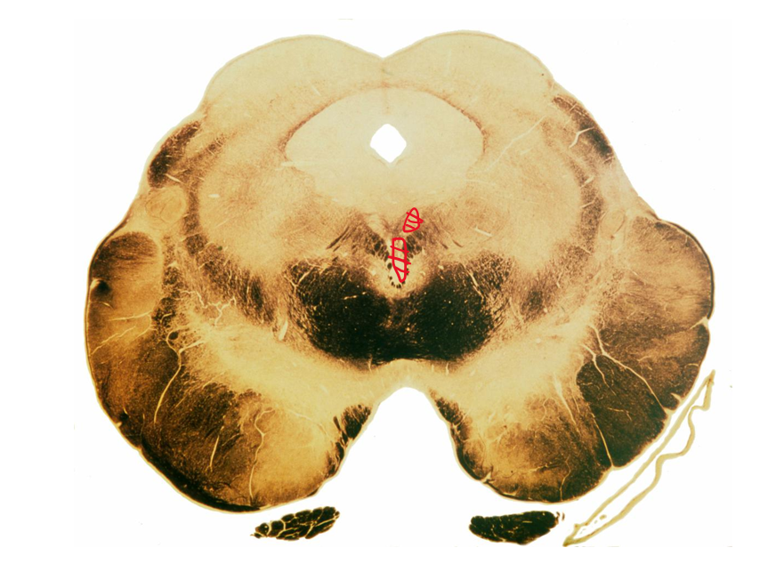
Damage to the following nuclei highlighted in red will result in what kinds of damage?
Inability to: elevate, depress, adduct eye, loss of elevation during adduction, inability to raise eyelid,
Permanently dilated pupil and inability to focus lens.
all symptoms are ipsilateral.
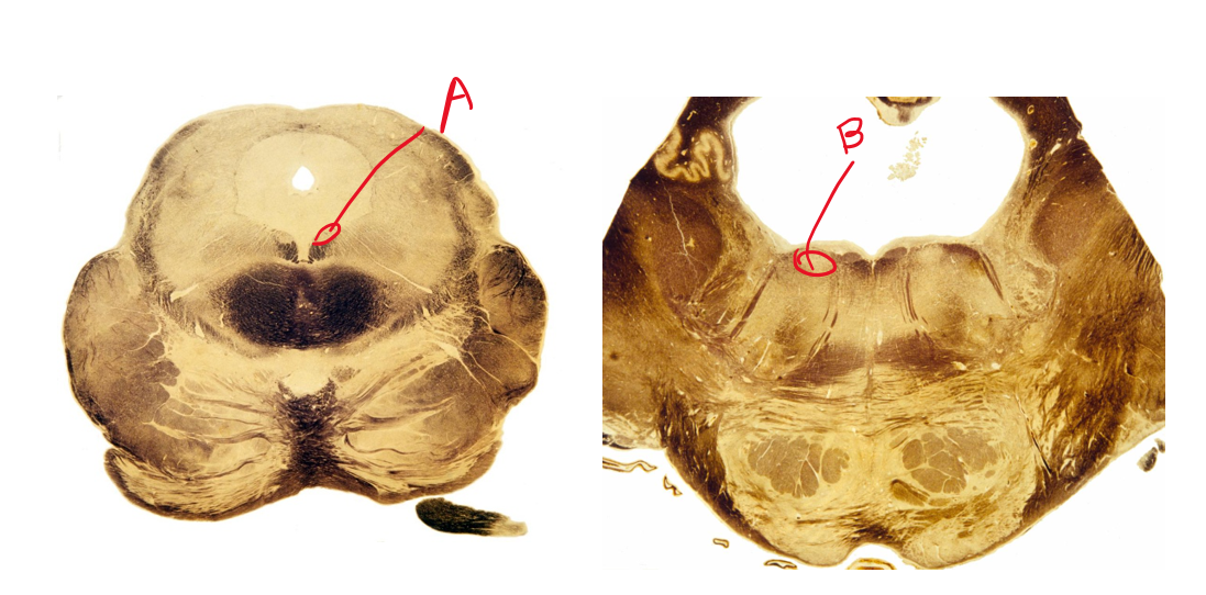
Describe the symptoms of damaging A and B
A: inability to depress and adduct eye on contralateral side of damage. B: inability to abduct eye on ipsilateral side of damage.
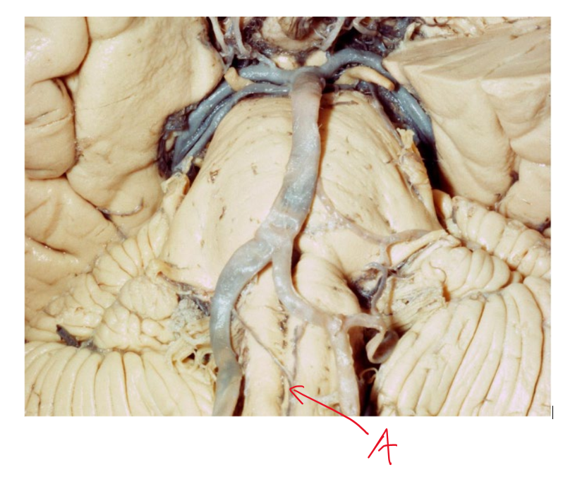
What kind of nuclei damage, and hence symptoms can we expect to see from damaging artery A? what is the name of this syndrome?
This is the anterior spinal artery. damaging it will cause medial medullary syndrome, damaging hypoglossal nuclei, medial lemniscus and corticospinal tract.
Symptoms include:
Ipsilateral tongue atrophy and paralysis, contralateral body loss of find touch, vibration and proprioception and contralateral muscle weakness and paralysis.
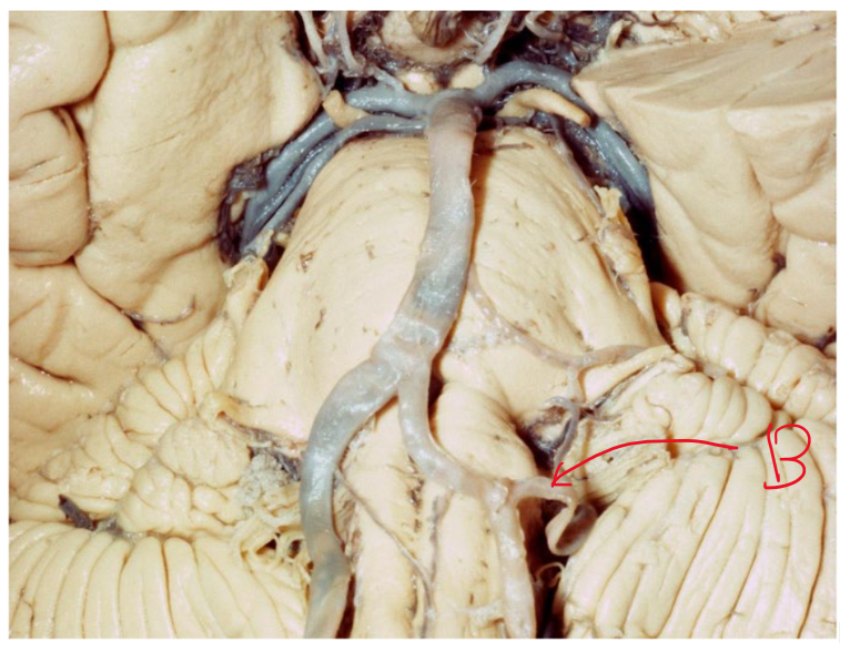
What kind of nuclei damage, and hence symptoms can we expect to see from damaging artery B? what is the name of this syndrome?
This is the posterior inferior cerebellar artery. Destroying it will remove bloodflow to the posterior rostral medulla, causing lateral medullary syndrome.
Affected nuclei include: spinothalamic tract, vestibular nuclei, inferior cerebellar peduncle, spinal trigeminal tract and nucleus.
Symptoms include: loss of pain and temperature over contralateral body, vertigo, ipsilateral loss of motor coordination, ipsilateral loss of face pain and temperature.
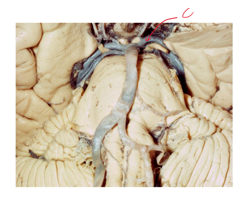
What kind of nuclei damage, and hence brainstem symptoms can we expect to see from damaging artery C? what is the name of this syndrome?
This is the Posterior cerebral artery. Damaging it can cause webers syndrome, complete destruction of midbrain nuclei, including the oculomotor and edingerwestphal nuclei, sensory tracts and cerebral peduncles (motor tracts)
symptoms are: contralateral loss of sensation, contralateral loss of motor and eye issues.
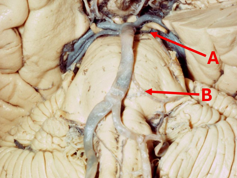
What kind of nuclei damage and hence symptoms can we see from damaging A and B?
A: This is the superior cerebellar artery. damaging it will cause loss of bloodflow to the pons at the rostral pons level. This will cause ipsilateral loss of coordination and vertigo.
B: This is the anterior inferior cerebellar artery, where damaging it will cause loss of bloodflow to the caudal pons, damaging nuclei such as vestibular nuclei, cerebellum and principal and spinal trigeminal nuclei. Hence, we can see vertigo, loss of coordination and ipsilateral loss of pain, temperature and fine touch.
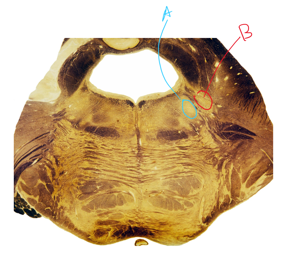
Describe the function and path of both A and B
A: This is the trigeminal motor nucleus. The corticobulbar tract descends down and gives motor input to this nucleus bilaterally. This nucleus then sends out nerves which innervate the muscles of mastication.
B: This is the Principal/Chief trigeminal nucleus. Sensory information about vibration and fine touch synapses here. The nucleus then sends out second order neurons which joins with other trigeminal nuclei for the trigeminothalamic tract. This trigeminothalamic tract follows the spinothalamic and medial lemniscus to the thalamus.
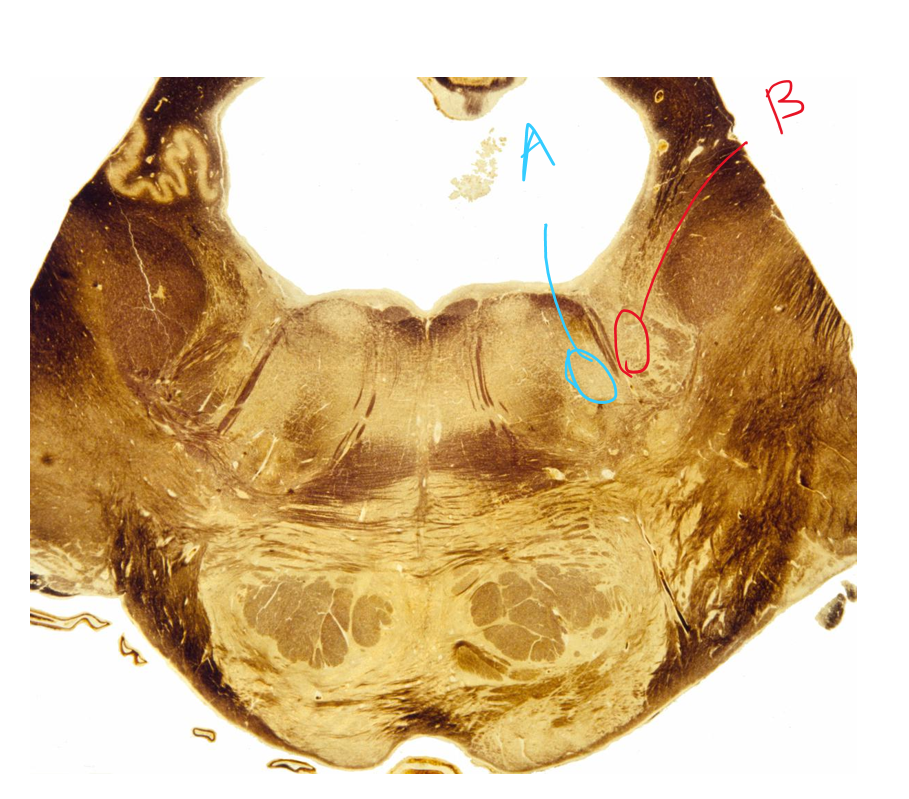
Describe the function and path of both A and B
A: Facial nuclei: The corticobulbar tract runs down from the motor cortex and gives bilateral input to the facial nucleus for the top half of the face and contralateral input for the second half of the face. Neurons then leave to give input on facial expression.
B: This is the spinal trigeminal nucleus. It receives pain and temperature from the face, where sensory neurons travel through the spinal trigeminal tract towards the correct level before synapsing onto the nucleus. After synapsing onto the nucleus, neurons cross and create the trigeminothalamic tract, travelling next to the medial lemniscus and spinothalamic tract to the thalamus.