Micro Anatomy Practical Images
1/70
There's no tags or description
Looks like no tags are added yet.
Name | Mastery | Learn | Test | Matching | Spaced |
|---|
No study sessions yet.
71 Terms
Simple Squamous Epithelium
What type of epithelium is shown?
Simple Squamous Epithelium
What type of epithelium is shown?
Simple Squamous Epithelium
What type of epithelium is shown?
Simple Squamous Mesothelium
What type of epithelium is shown?
Simple Sqaumous Mesothelium
What type of epithelium is shown?
Simple Cuboidal Epithelium
What type of epithelium is shown?
Simple cuboidal epithelium
What type of epithelium is shown surrounding this structure?
Hyaline Cartilage
Chondrocytes
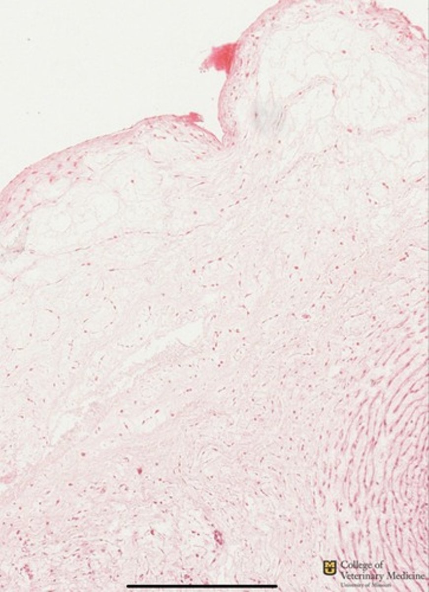
Skin
This tissues is an example of what type of Cartilage?
What type of cells are present?
What organ is this from?
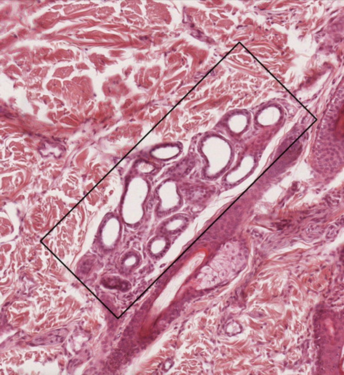
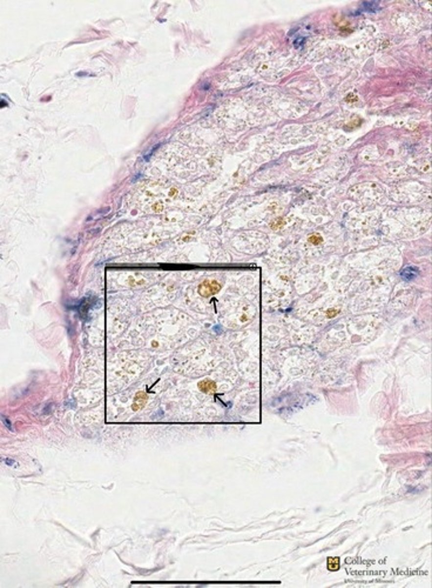
Sebaceous Gland or Holocrine Gland
Sebum
Skin
What type of gland is represented in the black box?
What does this gland secrete?
What organ is this gland from?

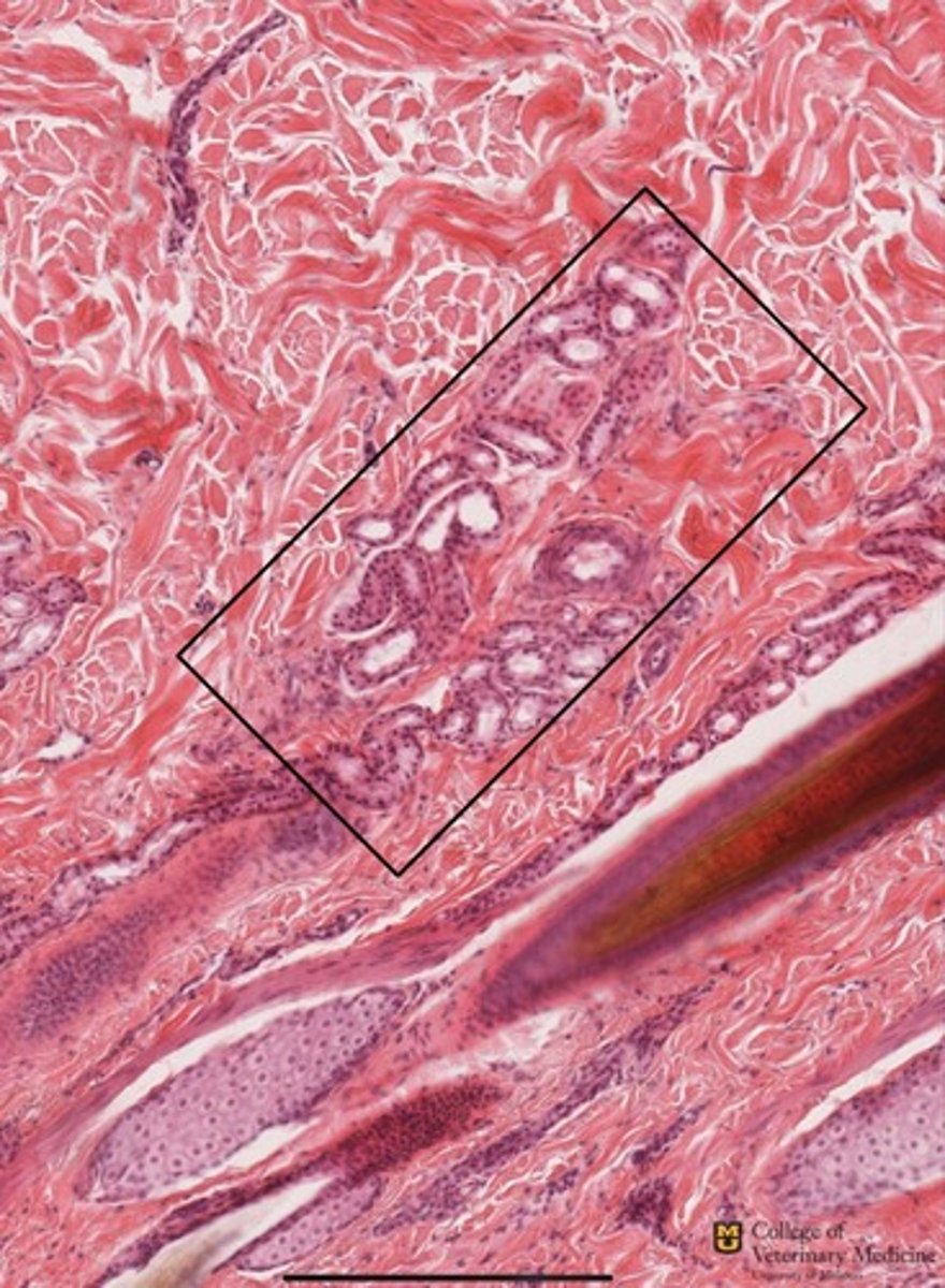
Merocrine Gland
Stratified cuboidal epithelium
What type of gland is located within the black box?
What type of epithelium surrounds this gland?
What organ is this from?
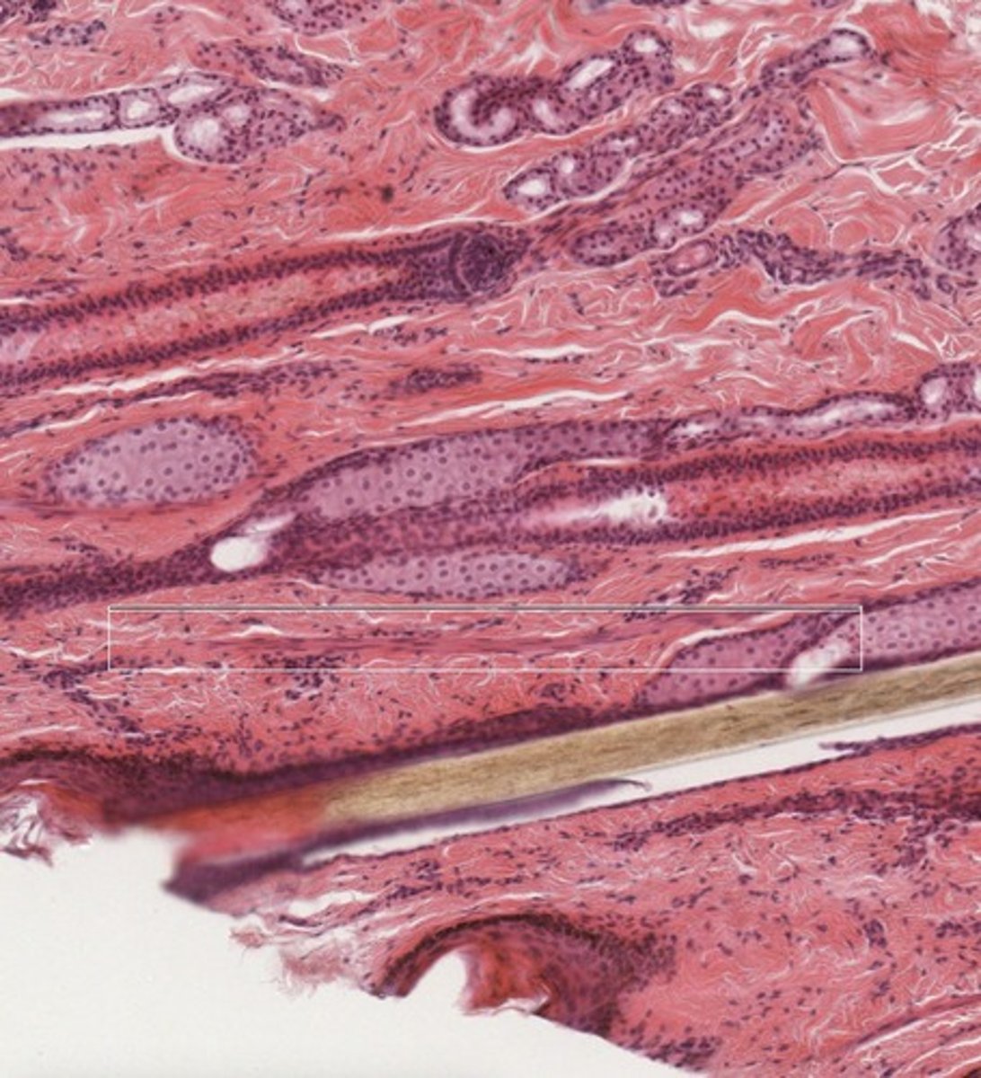
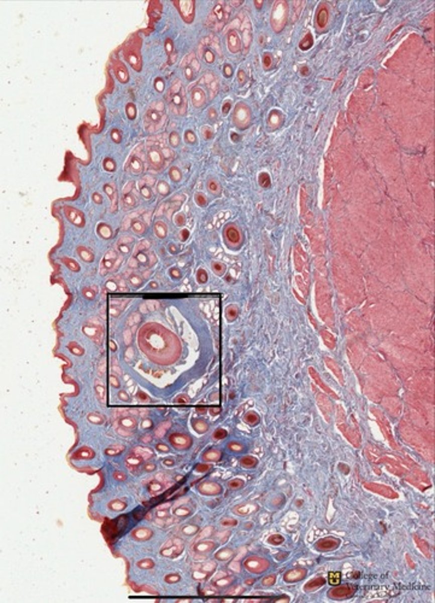
Arrector Pilli Muscle
Piloerection for insulation
Skin
What structure is within the white box?
What is its function?
What tissue structure is this from?
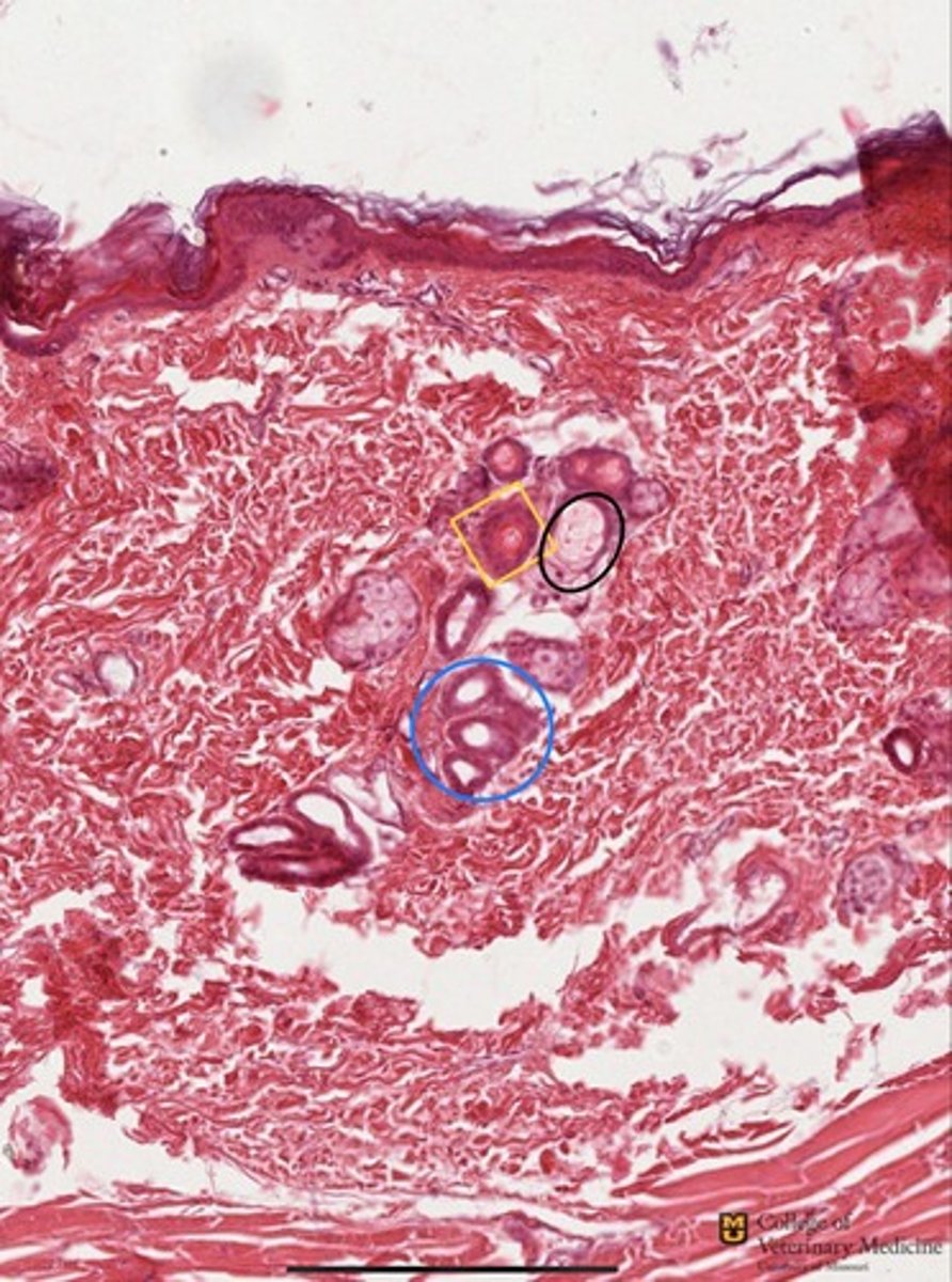
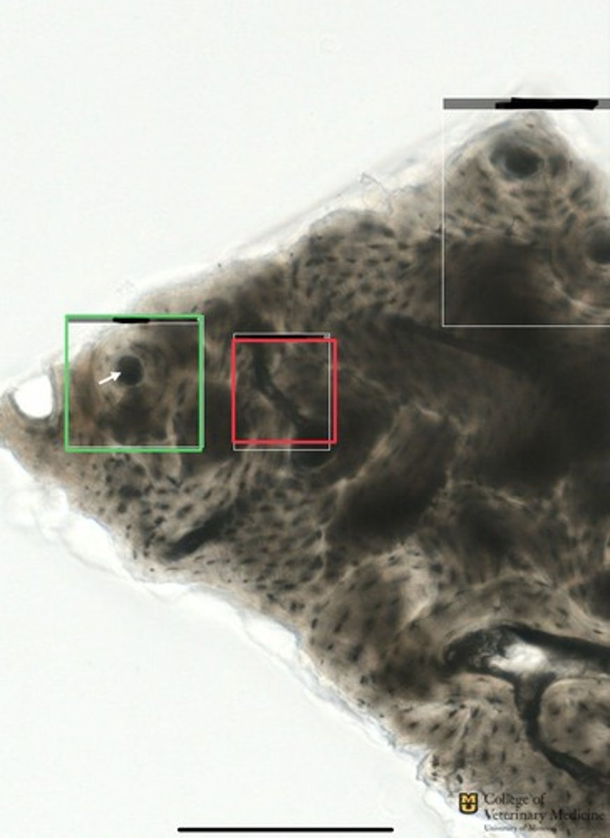
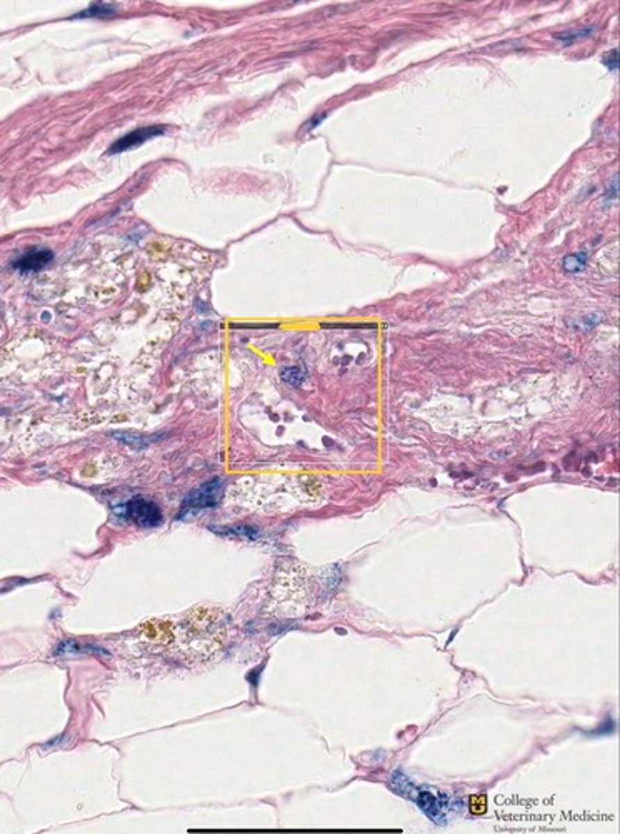
Apocrine Sweat Glands
Skin
Hair follicles
What type of gland is shown in the black box?
What tissue type does it come from?
Where do the glands empty?
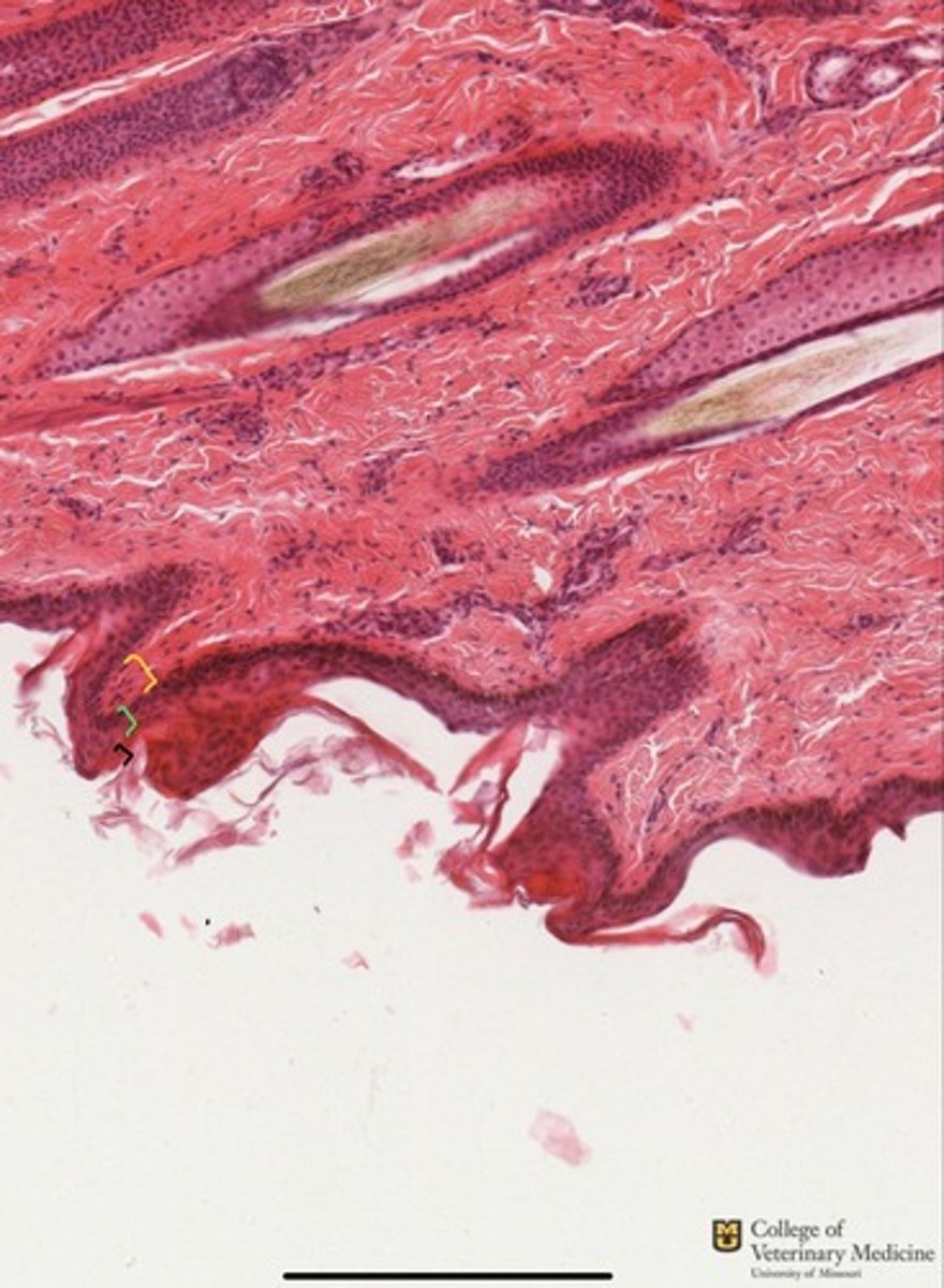
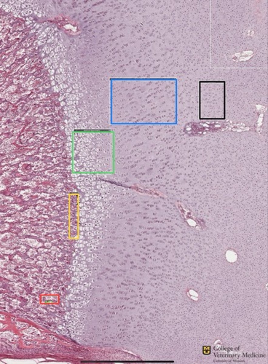
Skin
Y=stratum Basale
G=Stratum Spinosum
Blk=Stratum corneum
What tissue type is this from?
What three layers are present shown via the colored brackets?
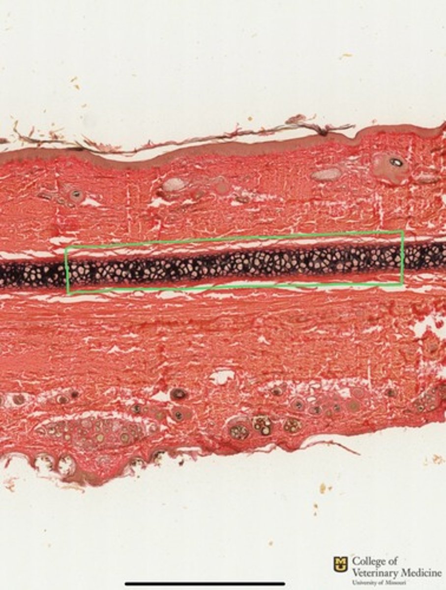
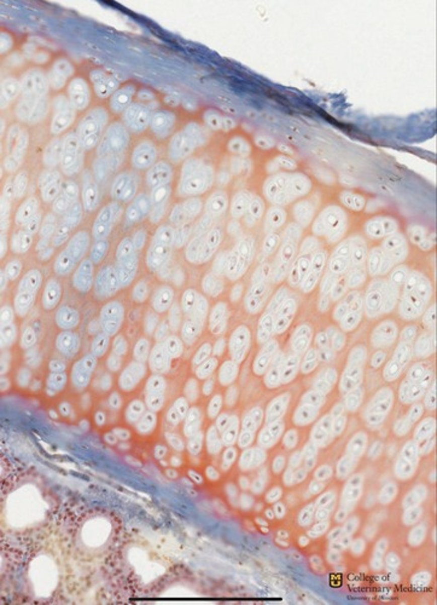
Elastic Cartilage
Ear canal
What type of cartilage is shown within the green box?
What tissue structure is this typically found in?
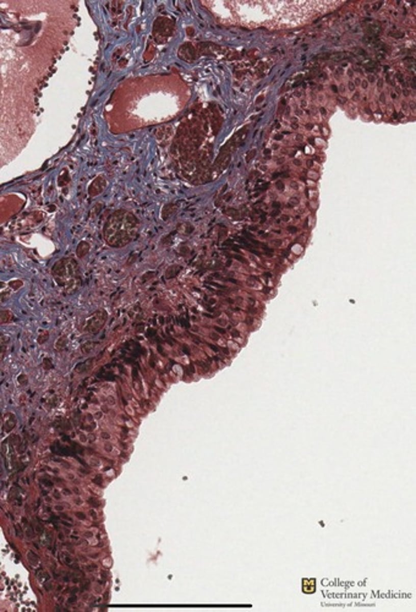
Compound Hair follicle
What type of hair structure is identified within the green box?
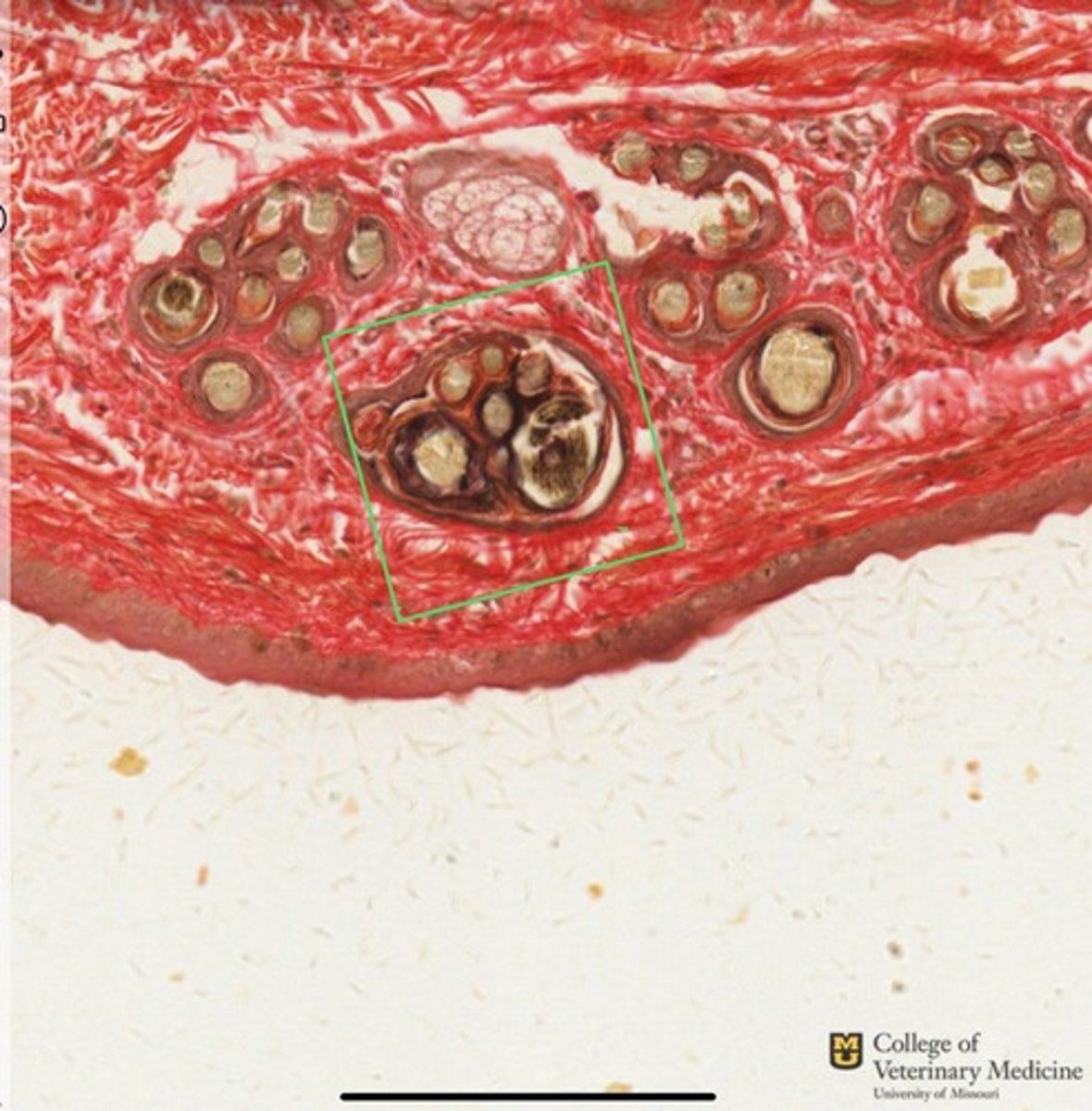
Blk=sebaceous gland
Y=hair follicle
Blu=Ceruminous glands
Ear canal
What structures are shown?
What tissue structure are these located within?
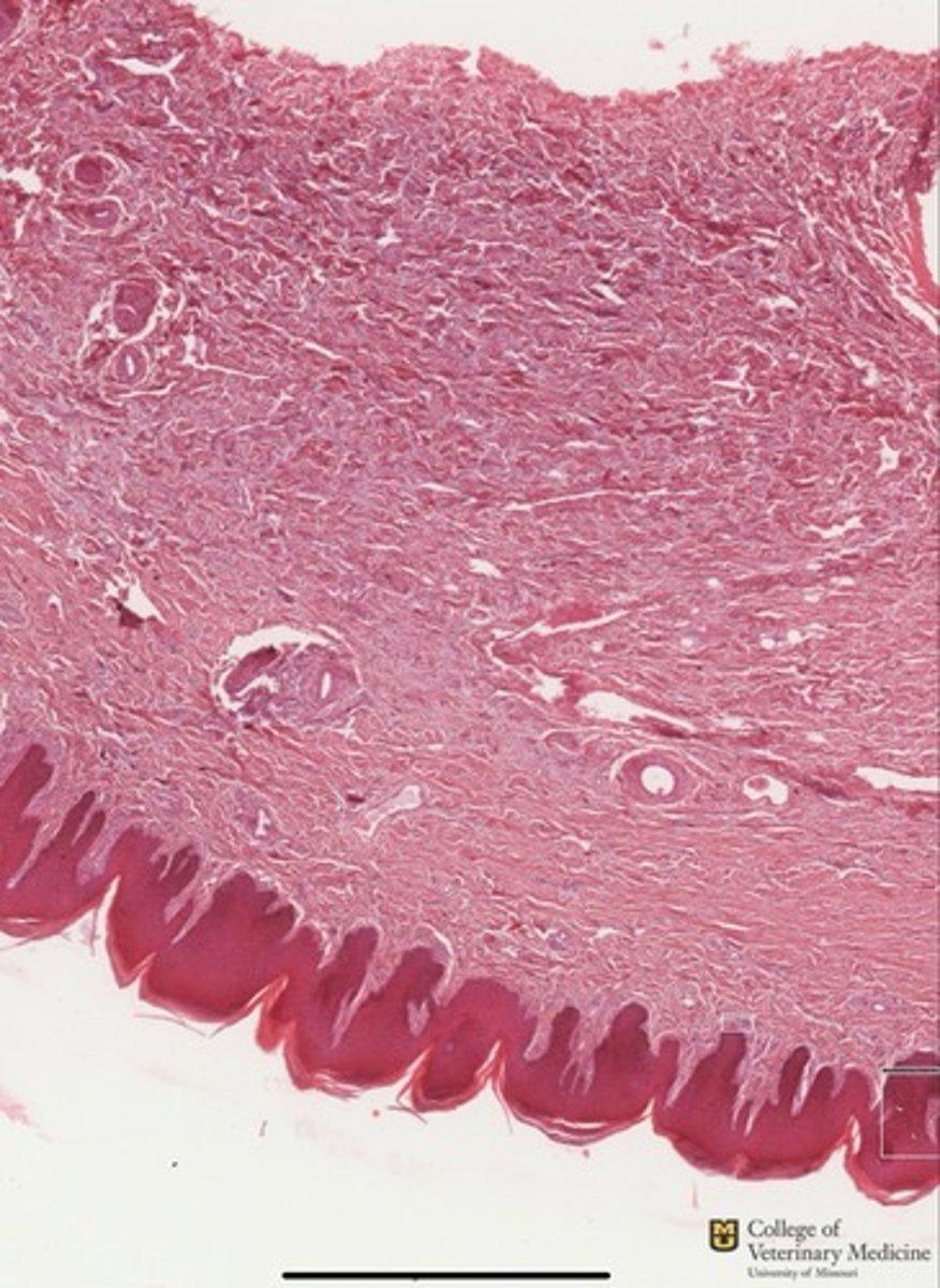
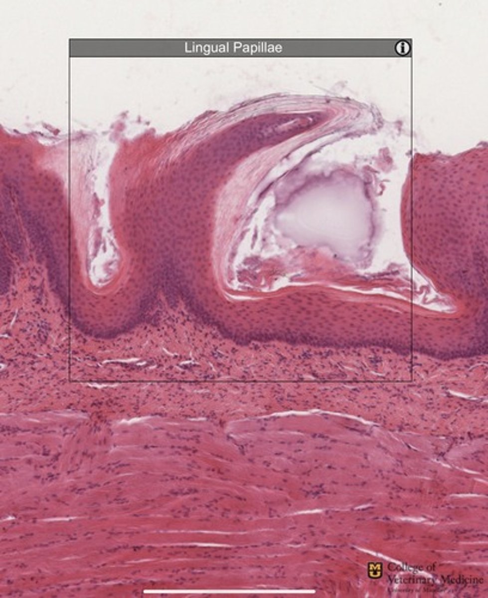
Tactile Hair Follicle (Whisker)
Skin
What structure is within the black box?
What tissue structure does it belong to?
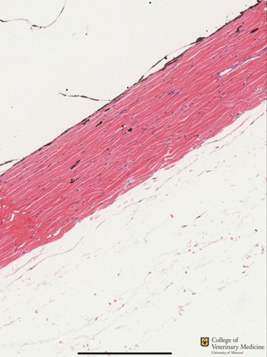
Osteon
Bone
What tissue structure is shown within the green box?
What tissue type is it from ?

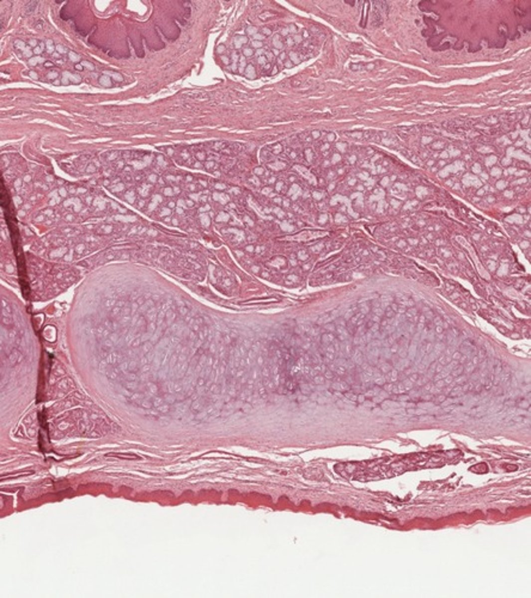
Perforating Canal
Bone
What structure is shown in the red box?
What tissue is it from?
Cortical Bone
What tissue type is depicted?
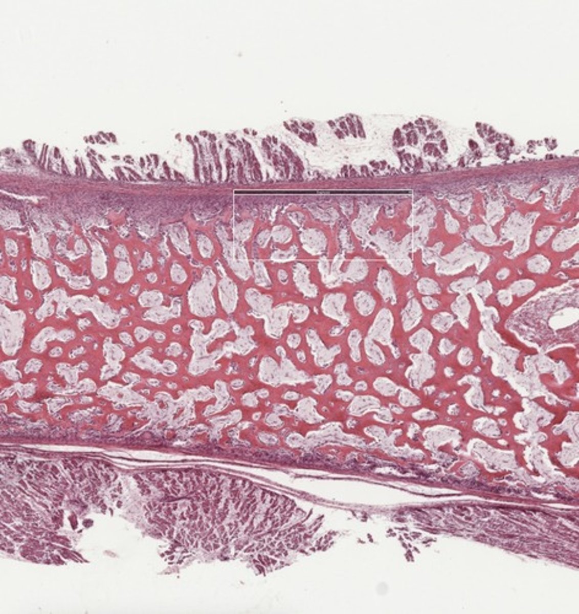
Trabecular Bone
What tissue type is depicted?
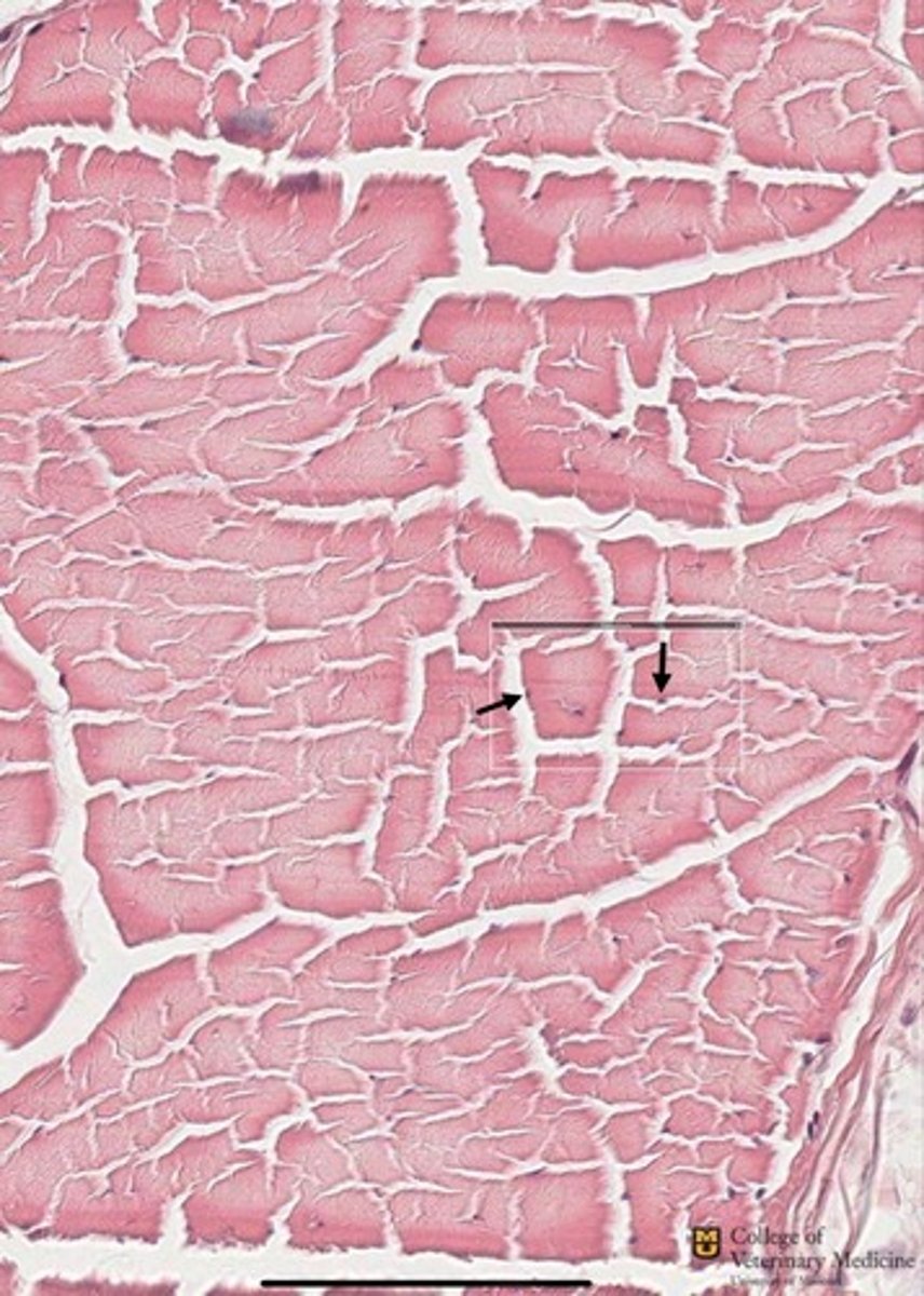
Lacunae
Bone
What stature is shown within the box?
What tissue is this from?
Black= Rest Zone
Blue=Zone of Proliferation
Green=Zone of Hypertrophy
Yellow=Zone of Calcification
Red=Zone of Ossification
Bone
What zones are depicted?
What tissue type are they from?
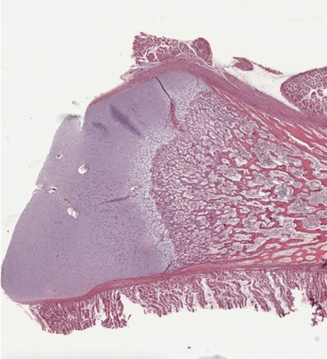
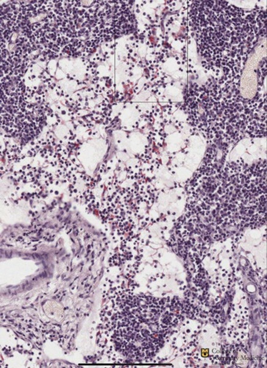
Mesenchyme
Early Trabecular bone
What tissue type is shown within the yellow box?
What will this tissue become?
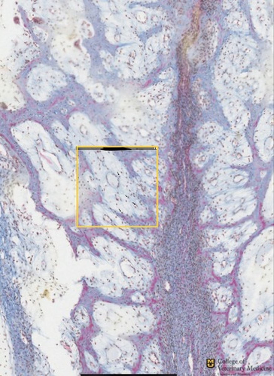
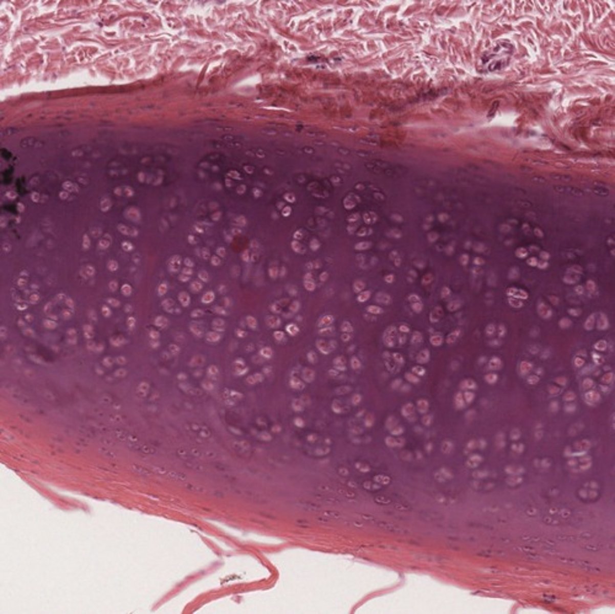
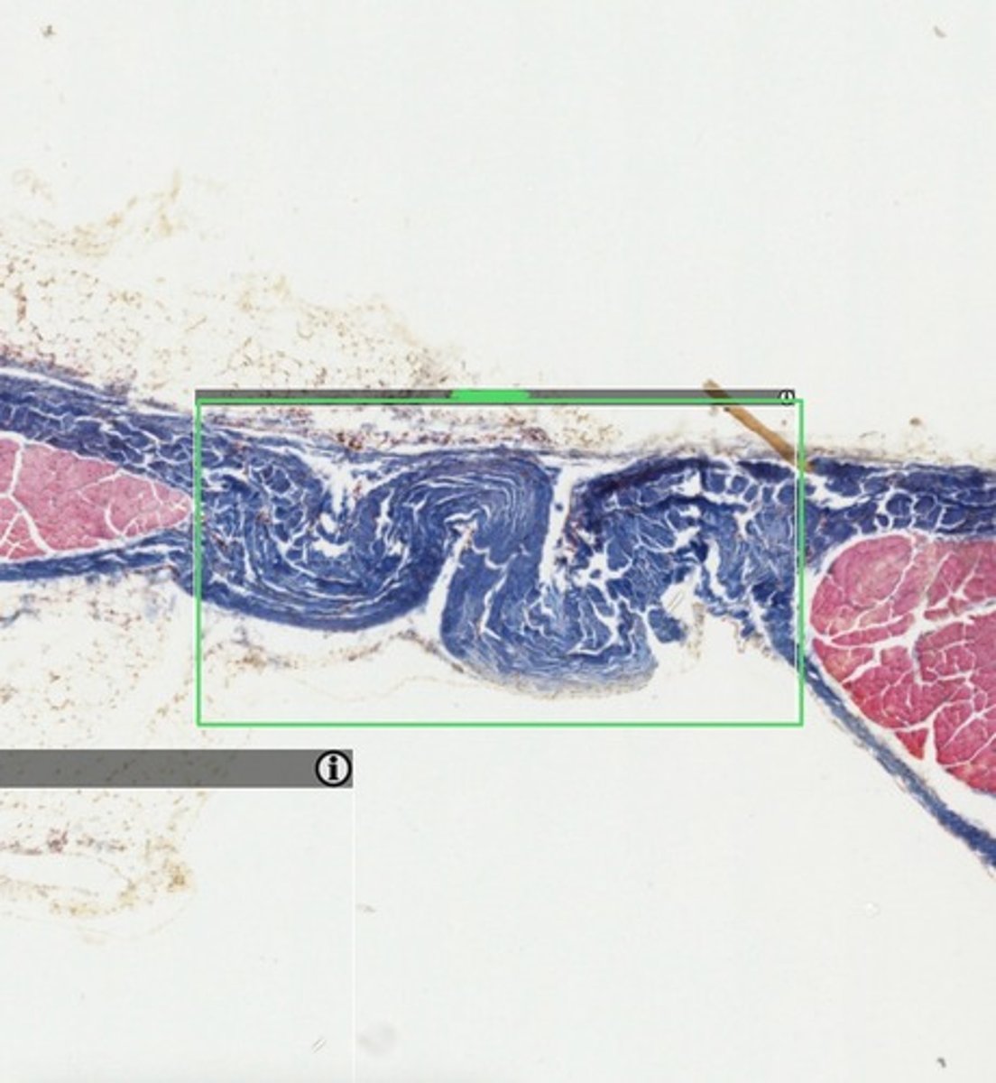
Fibrocartilage
What tissue type is depicted within the yellow box?
Elastic Cartilage
What cartilage type is depicted here?
Adipose Tissue-White (Unilocular)
What tissue type is depicted as a whole?
Hyaline Cartilage
What type of cartilage is depicted?
DICCT
Linea Alba
What tissue type is shown as a whole?
What structure does it compose?
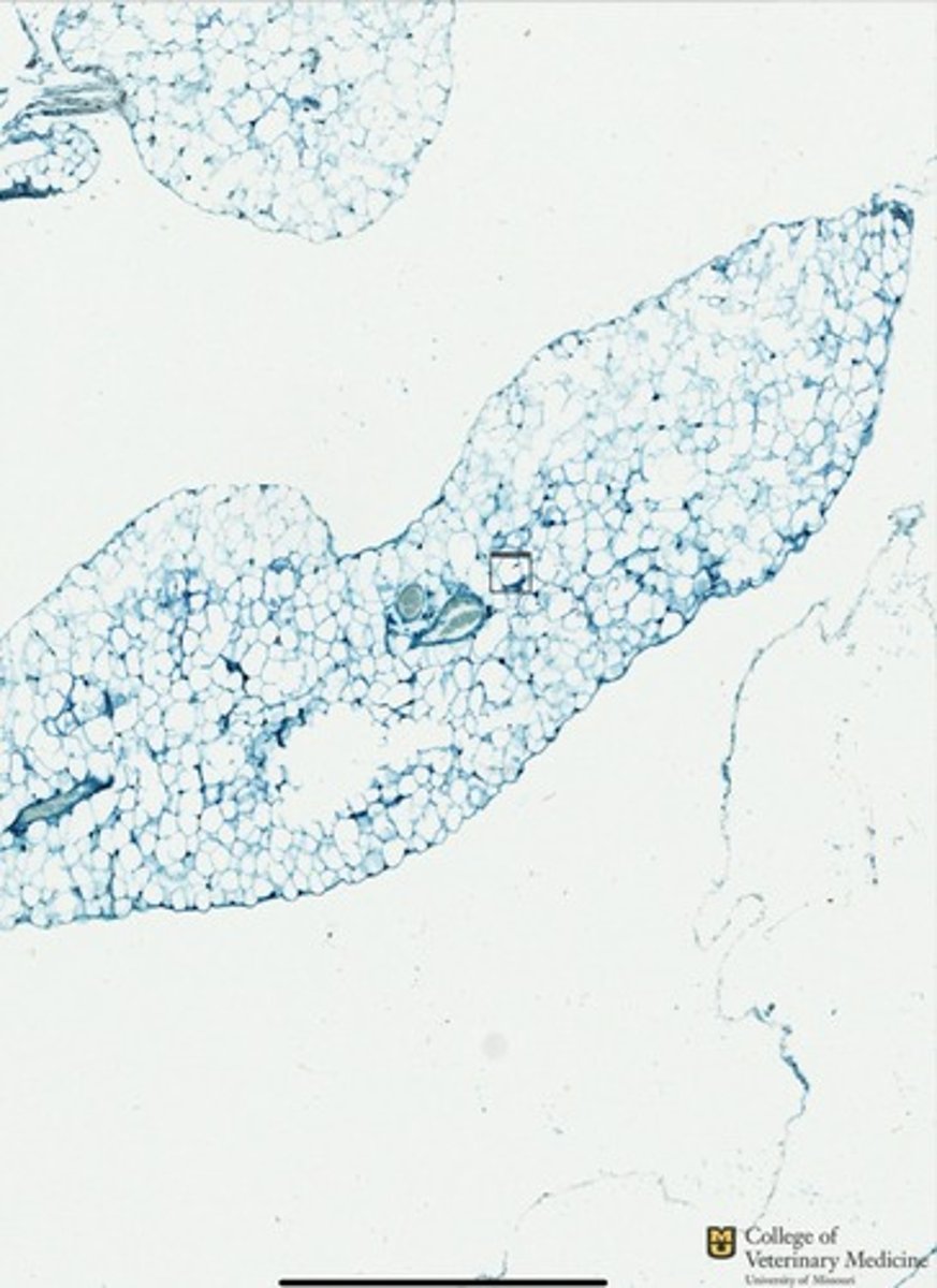
Adipose Tissue- Brown (Multilocular)
What tissue type is depicted as a whole?
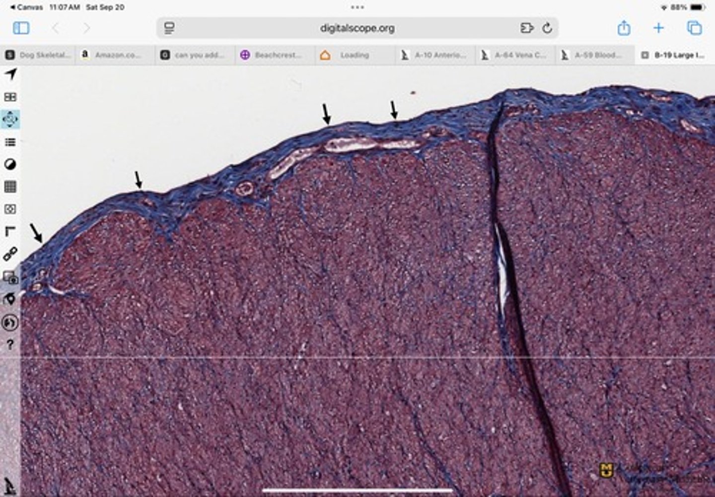
DICCT
Dermis-Corium of Skin
What tissue type as a whole is depicted?
What layer does it compose in this structure?
DICCT
Sclera of Eye
What tissue type is shown?
What structure does it compose ?
Elastic Cartilage
Loose Connective Tissue
What cartilage type is depicted?
What is the tissue type stained blue?
Elastic Cartilage
Loose CT
What cartilage type is depicted?
What is the tissue type stained blue?
Loose CT
Macrophage
What tissue type is depicted?
What cellular structure is shown in the box?
Loose CT
Mast Cell
What tissue type is depicted?
What cellular structure is shown?
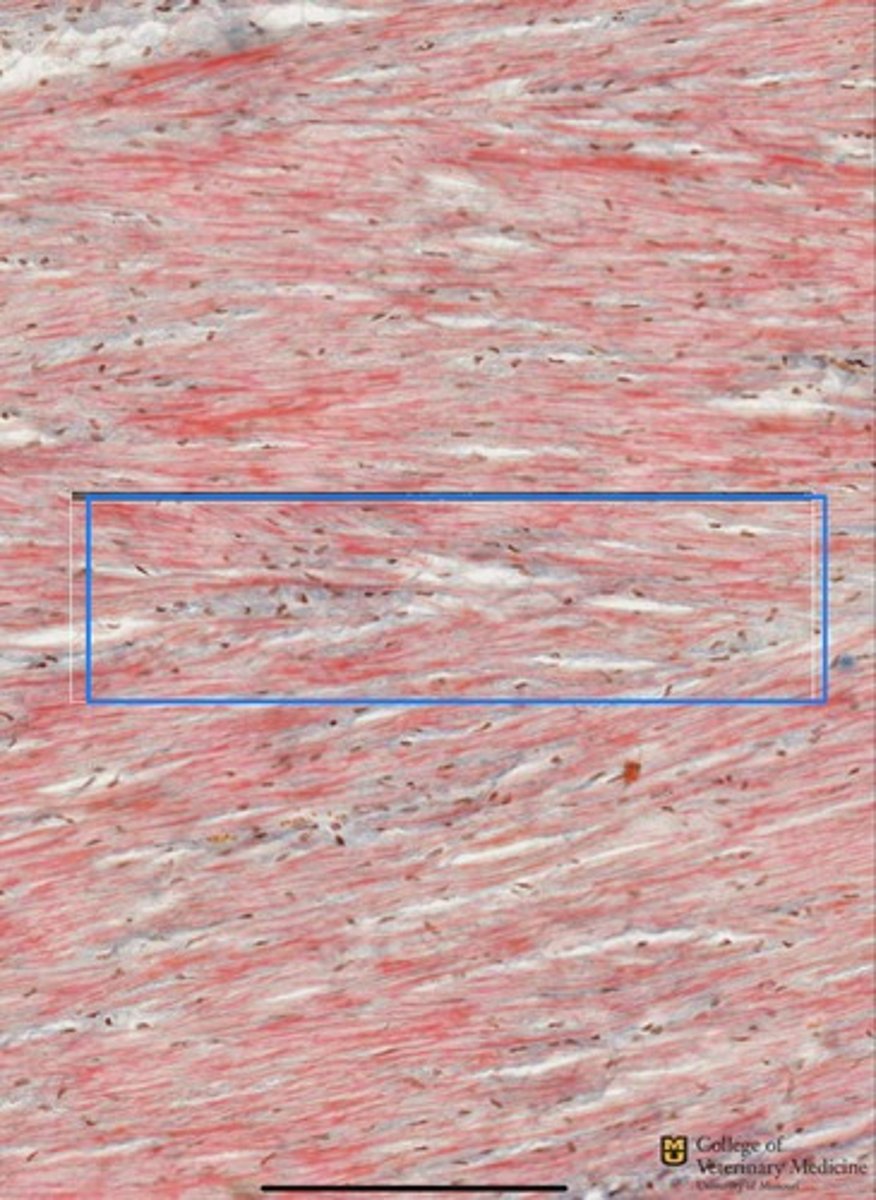
Dense Regular CCT
What tissue type is depicted as a whole?
Loose CT
Macrophages
What tissue type as whole is shown?
What cell type is shown within it?
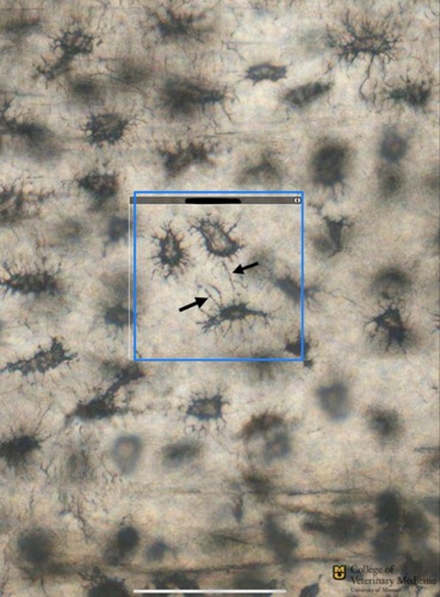
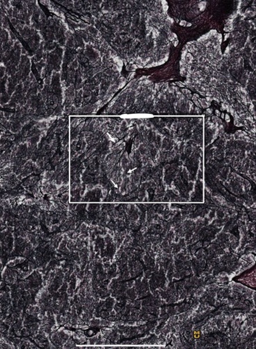
Reticular CT
Reticular Fibers
What tissue type is shown as a whole?
What structures are the white arrows pointing to?
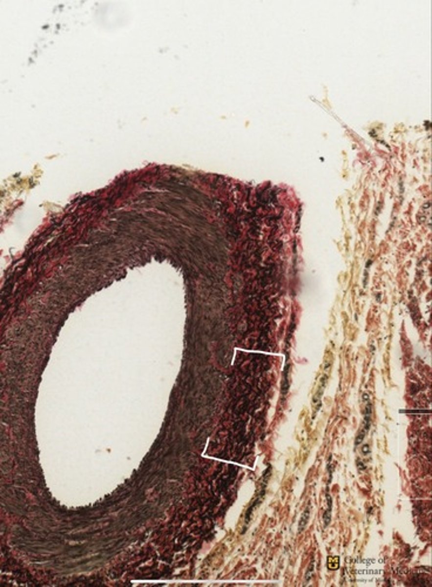
Elastic Fibers
Artery
What tissue type do the white brackets depict?
What structure is it surrounding?
Loose Ct
What tissue type is depicted as a whole?
DICCT
What tissue type is shown?
Mucous CT
Umbilical Cord
What tissue type is shown as a whole?
What structure is it typically found?
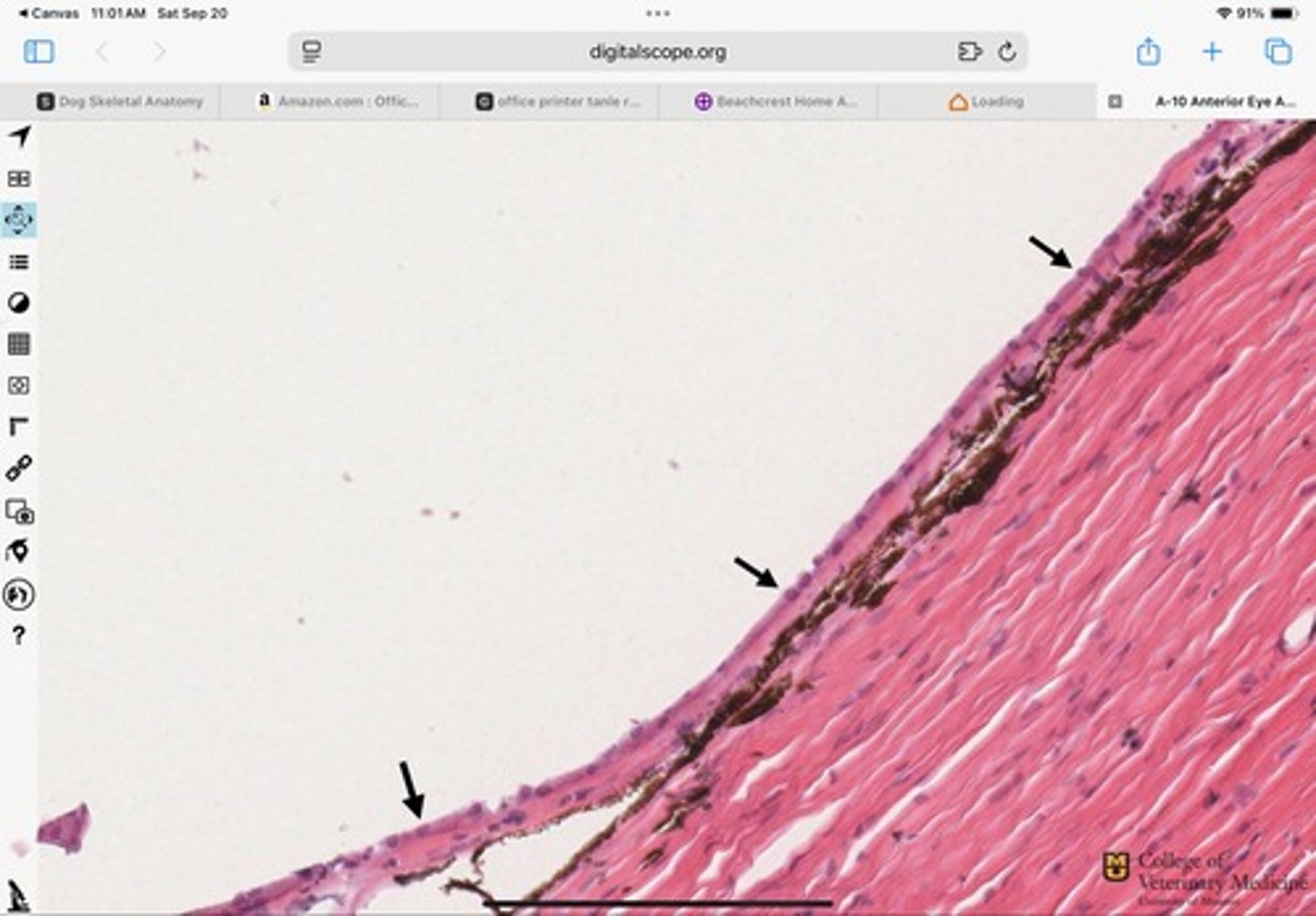
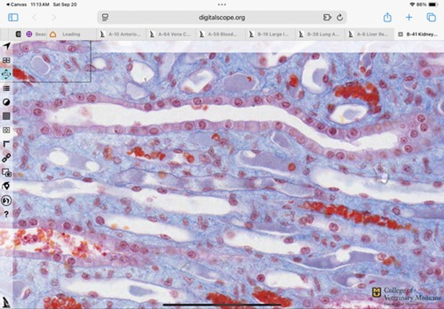
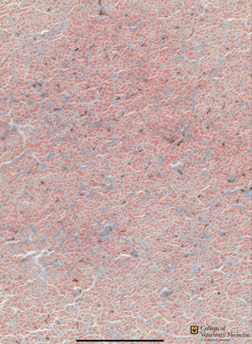
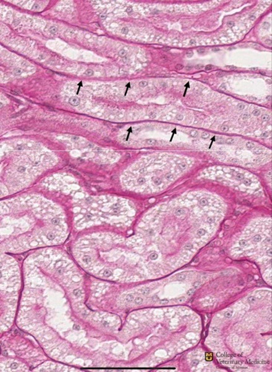
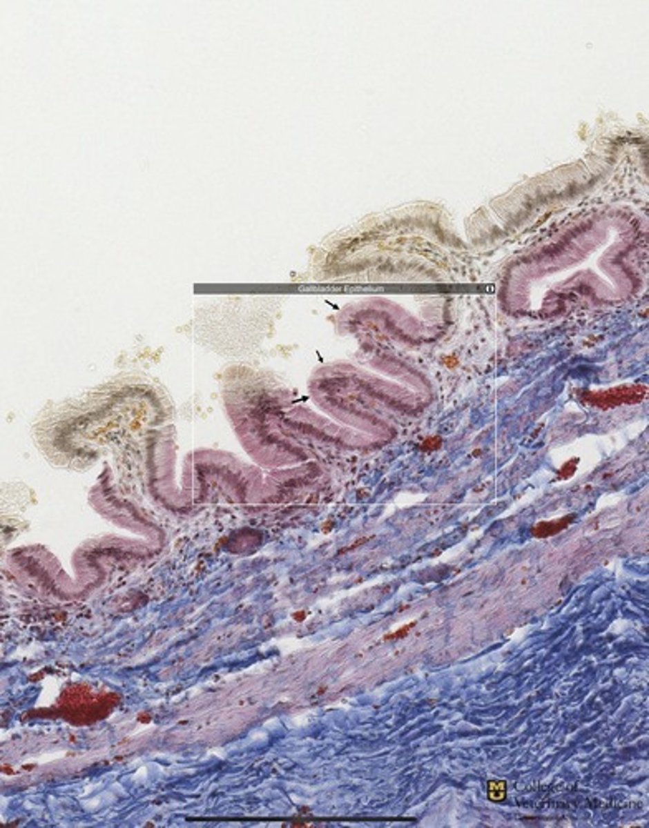
Basement Membrane
The black arrows are pointing to what cell structure?
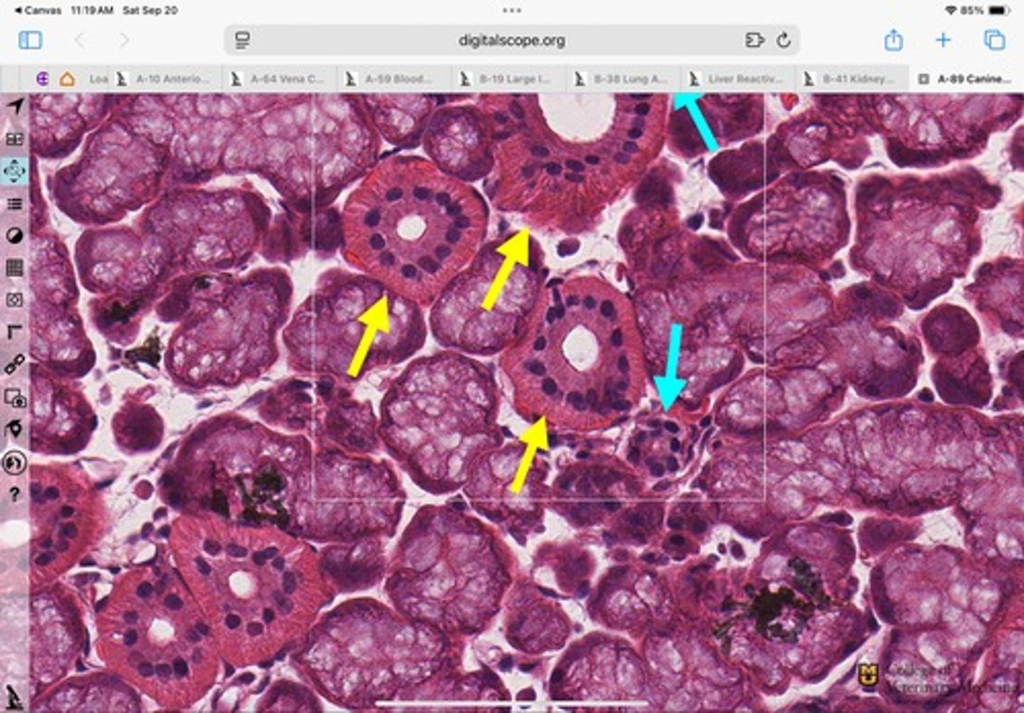
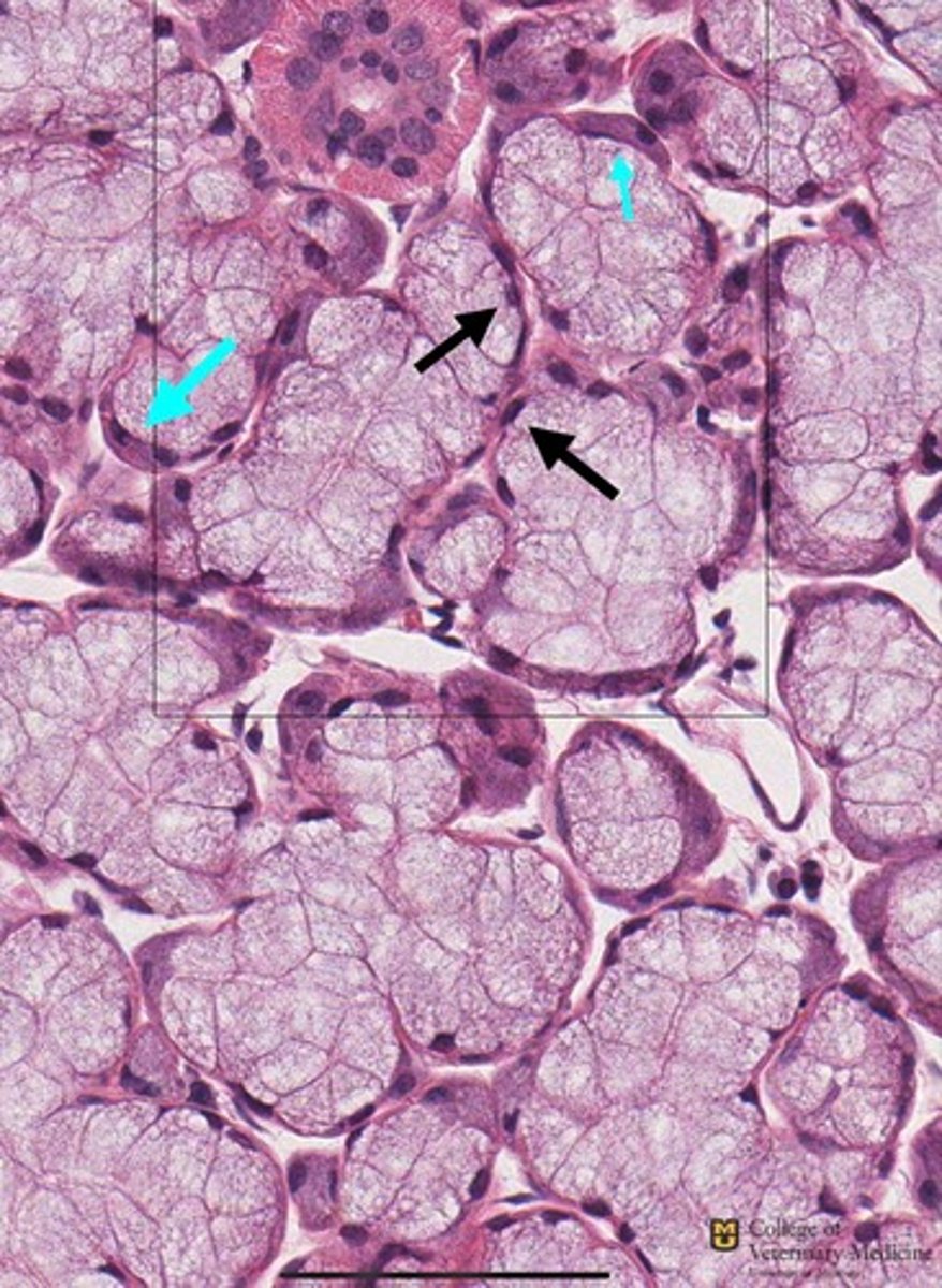
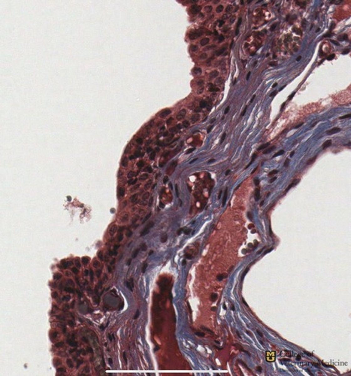
Mixed Gland- seromucous Acini
Blue-serous demilunes
Black-nuclei
What type of glandular epithelium is shown?
What structures are the respective arrows pointing to?
Serous Glandular Epithelium
Nuclei centrally located
What glandular epithelium type is shown in the yellow box?
How can you tell?
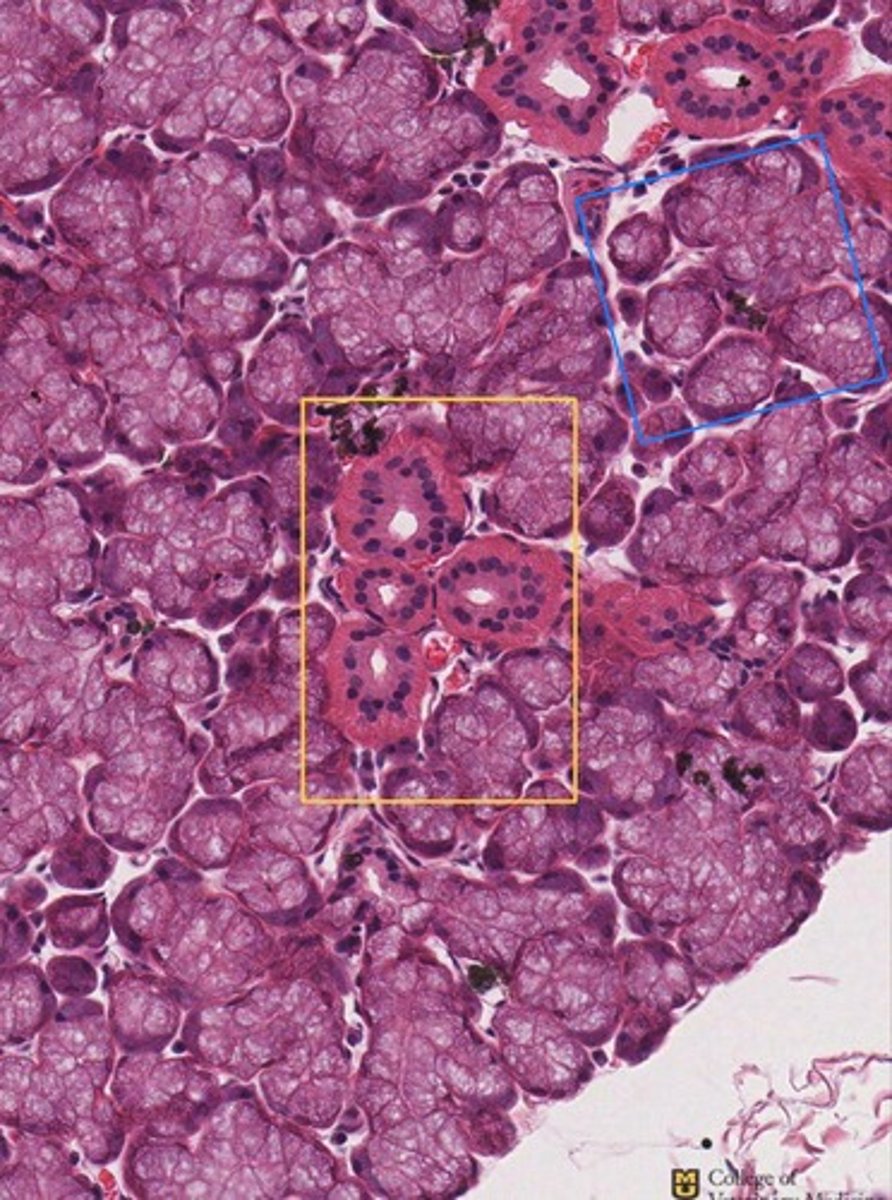
Mucous Glandular Epithelium
Nuclei pushed to outer edge
What glandular epithelium is shown in the blue box?
How can you tell?

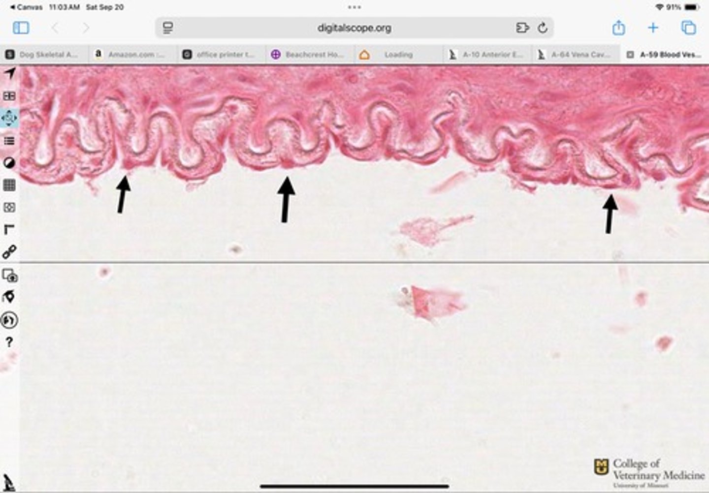
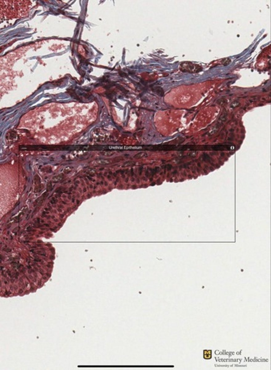
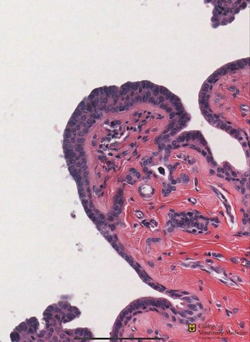
Transitional Epithelium
What tissue type is shown?
Transitional Epithelium
Ureter
What type of tissue is shown?
What structure is it lining?
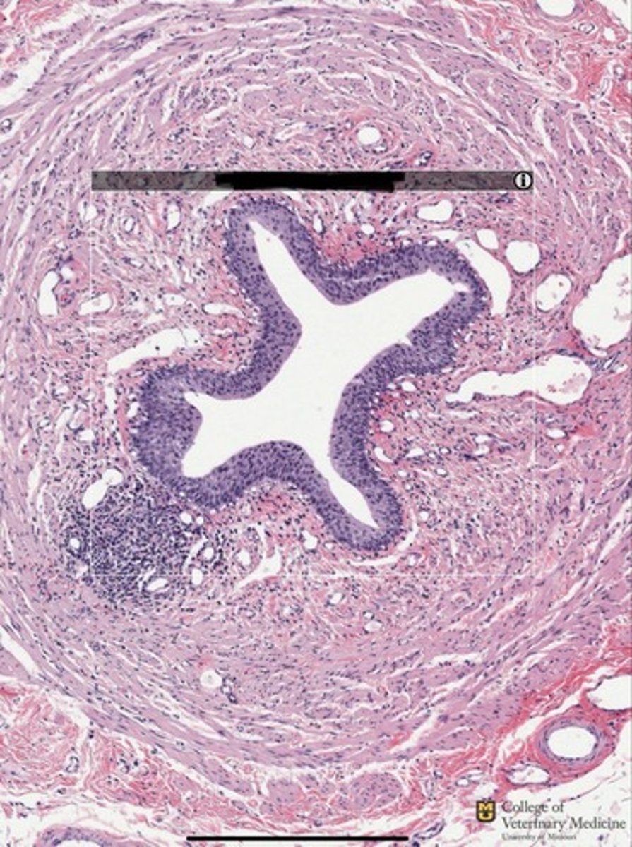
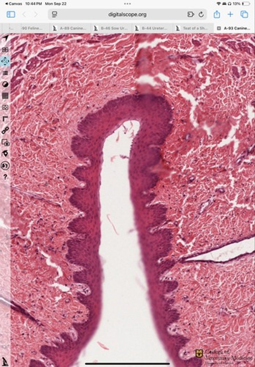
Transitional Epithelium
Bladder
What type of tissue is shown?
What structure is it lining?
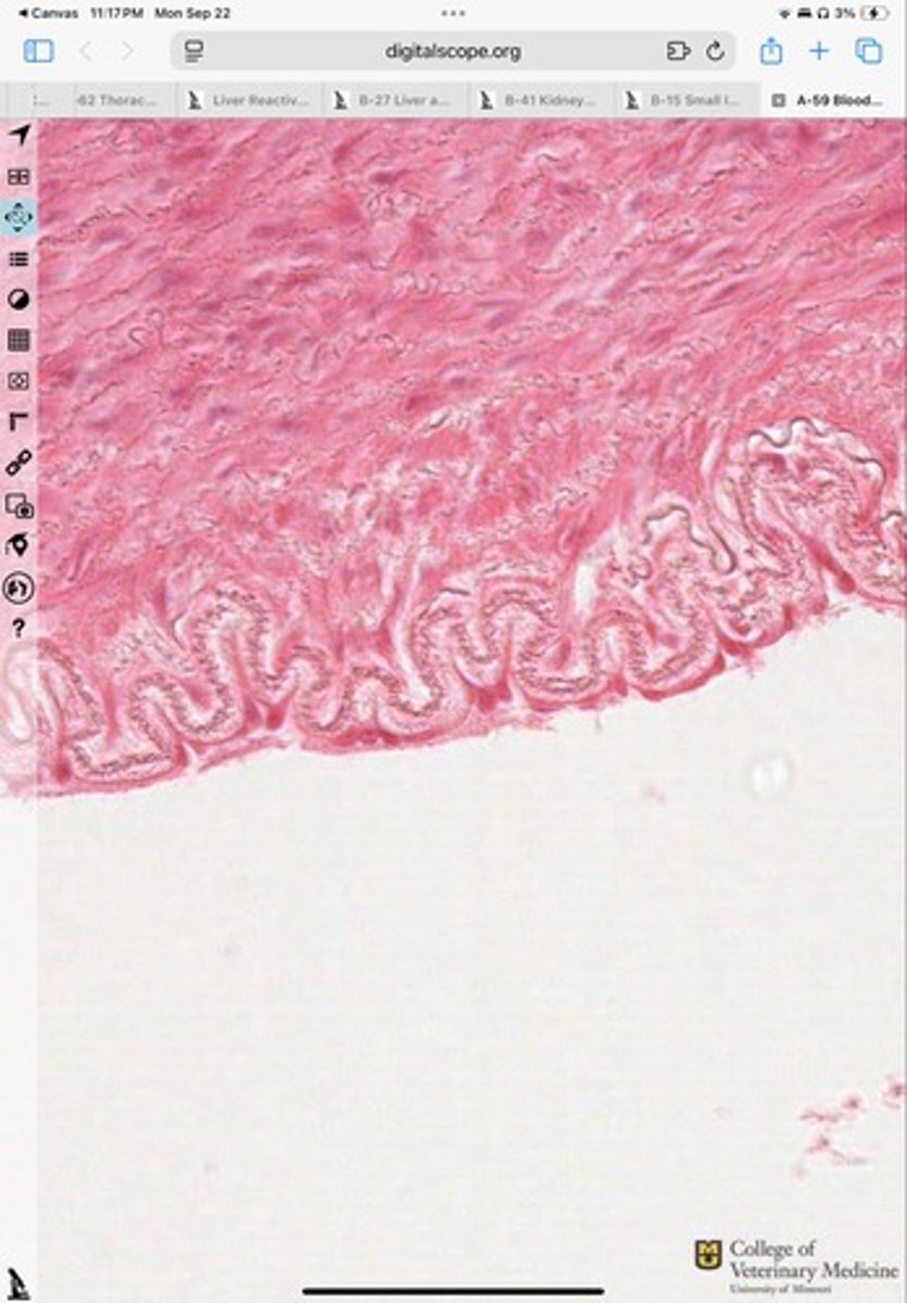
Simple Cuboidal epithelium
What tissue type is shown?
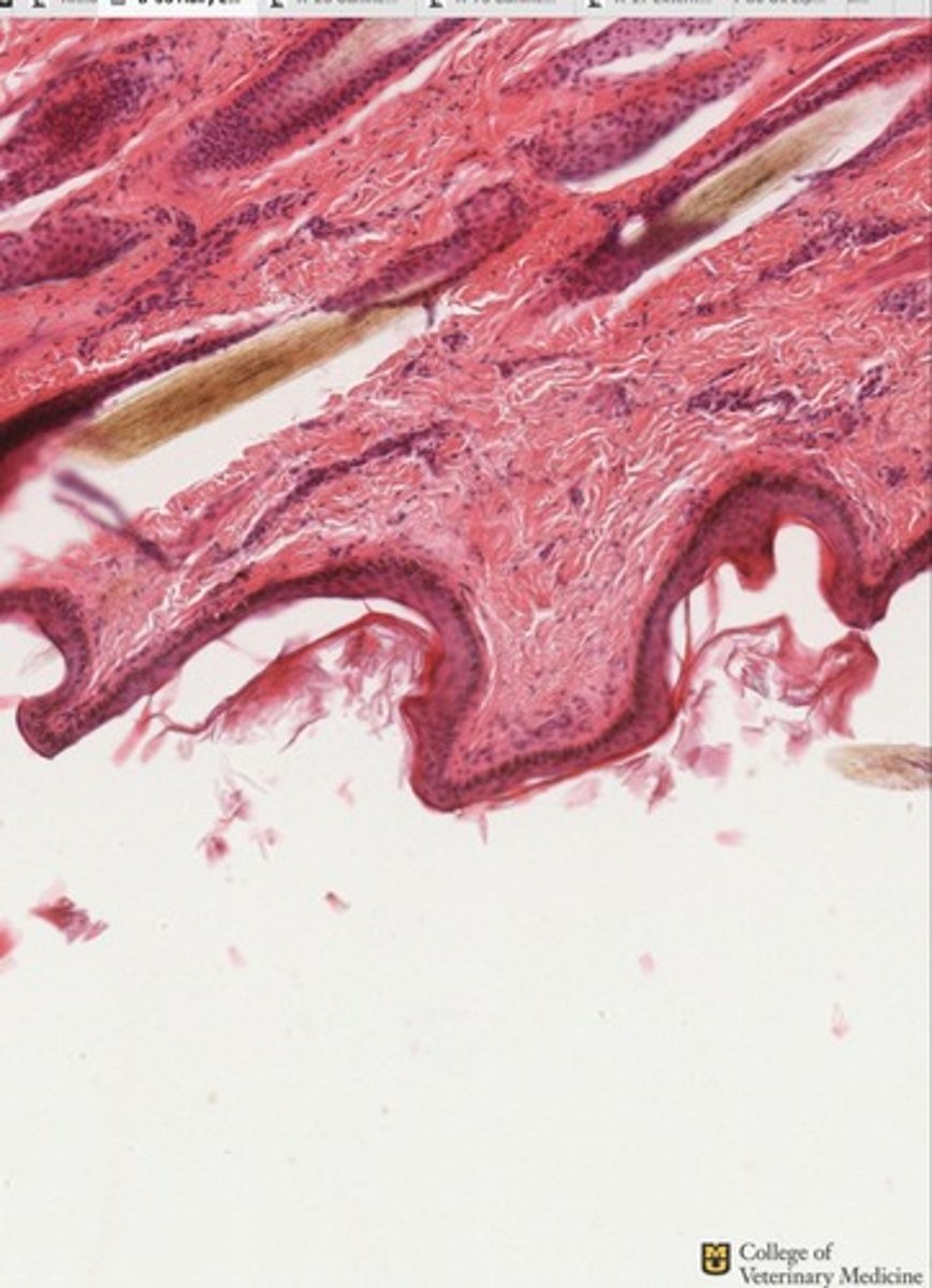
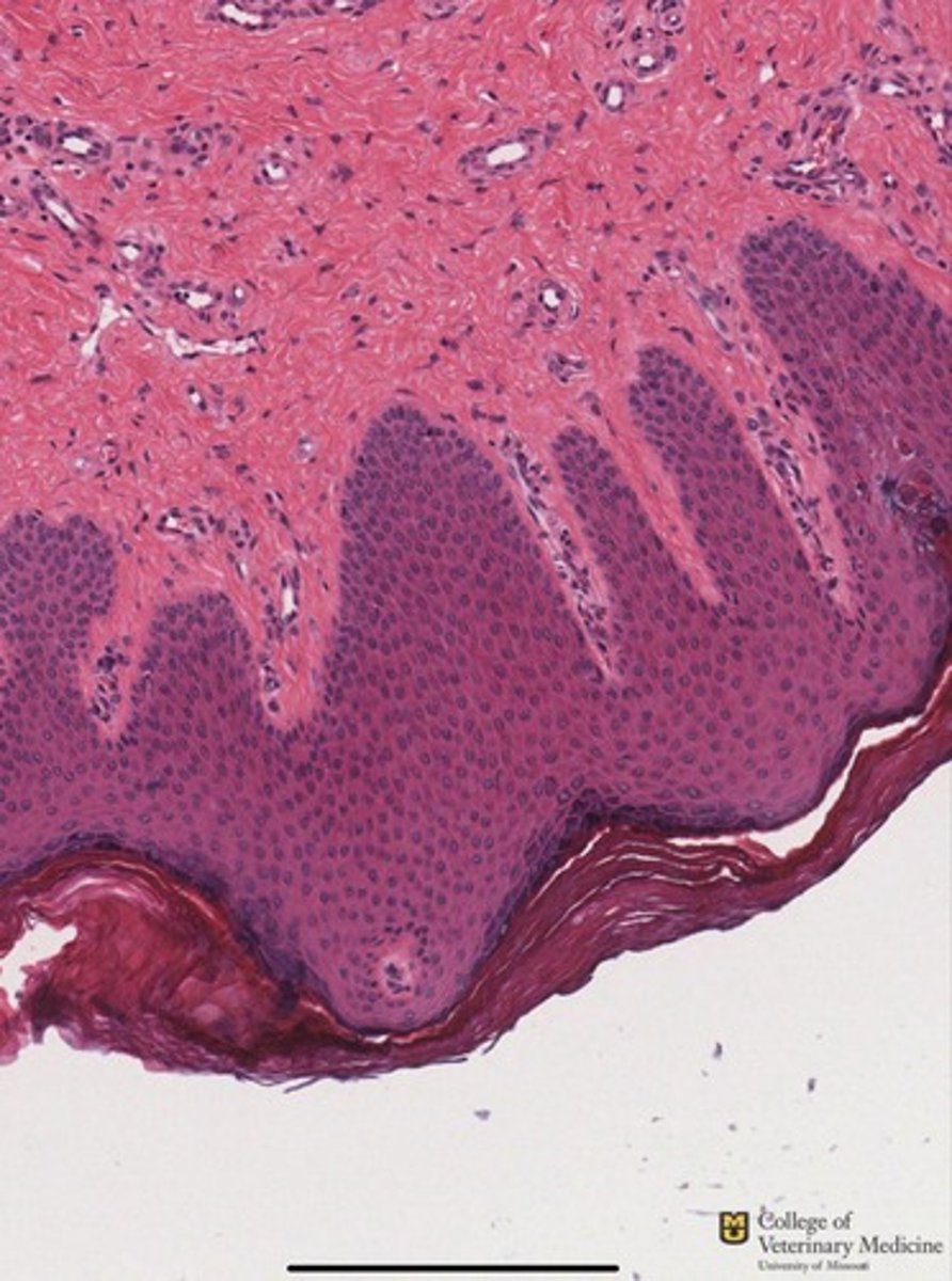
stratified squamous epithelium
What tissue is this?
stratified squamous epithelium-Keratinized
What epithelium type is shown?
Stratified Squamous Epithelium-Non Keratinized
What epithelium type is shown?
Stratified Cuboidal epithelium
What epithelial tissue is shown?
Stratified Columnar Epithelium
What type of epithelium is shown?
Stratified squamous epithelium-Keratinized
What epithelium type is shown?
Stratified squamous epithelium-Non Keratinized
6
What epithelium type is shown?
How many layers does it have?
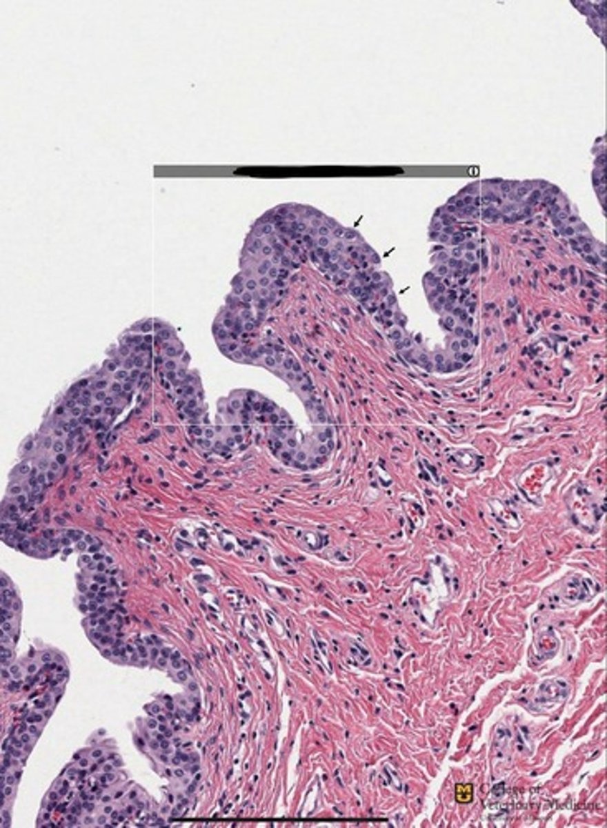
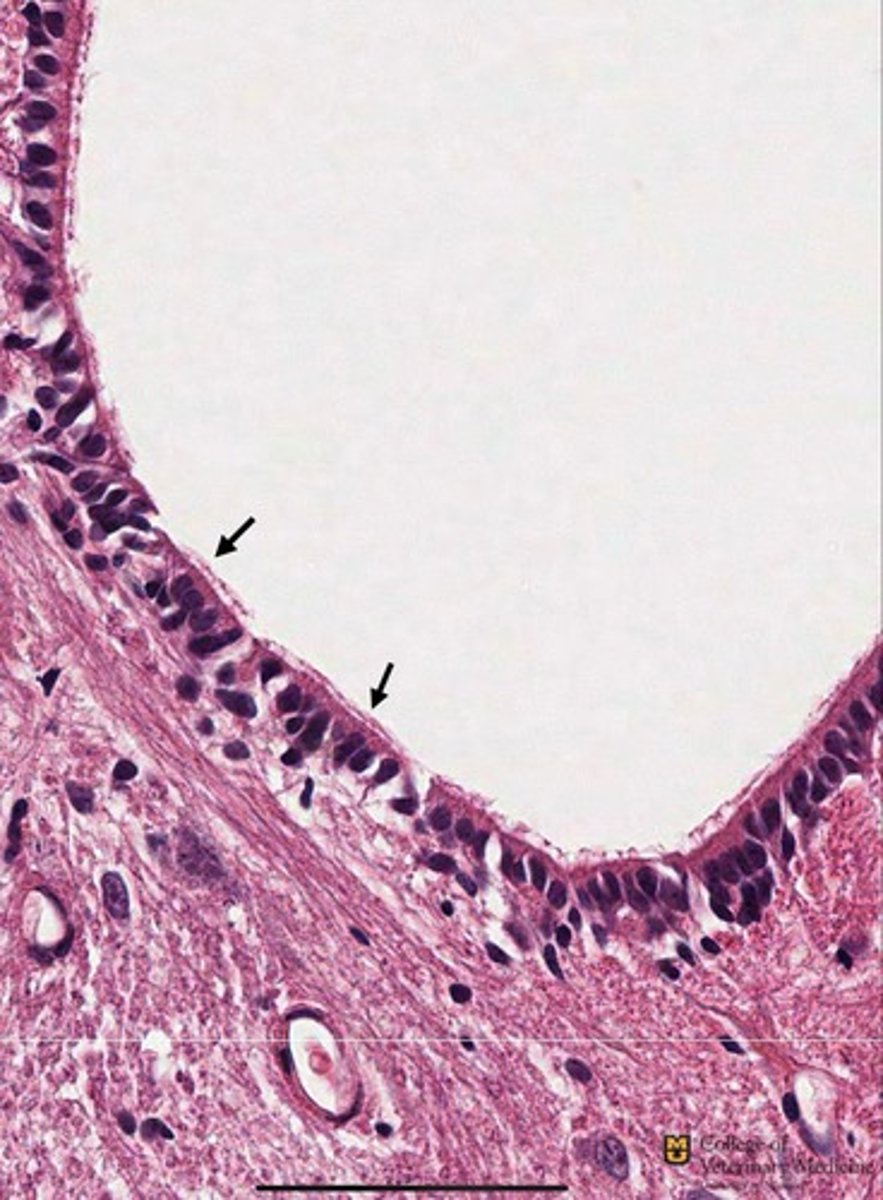
Simple cuboidal epithelium with Cilia
What epithelium type is shown? Note structures black arrows pointing to
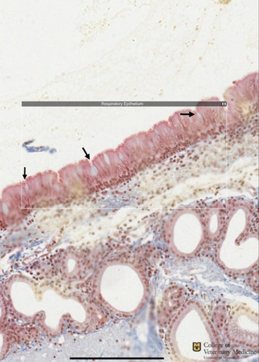
Pseudostratified columnar epithelium with cilia and Goblet cells
What epithelium type is shown and what structures accompany it?
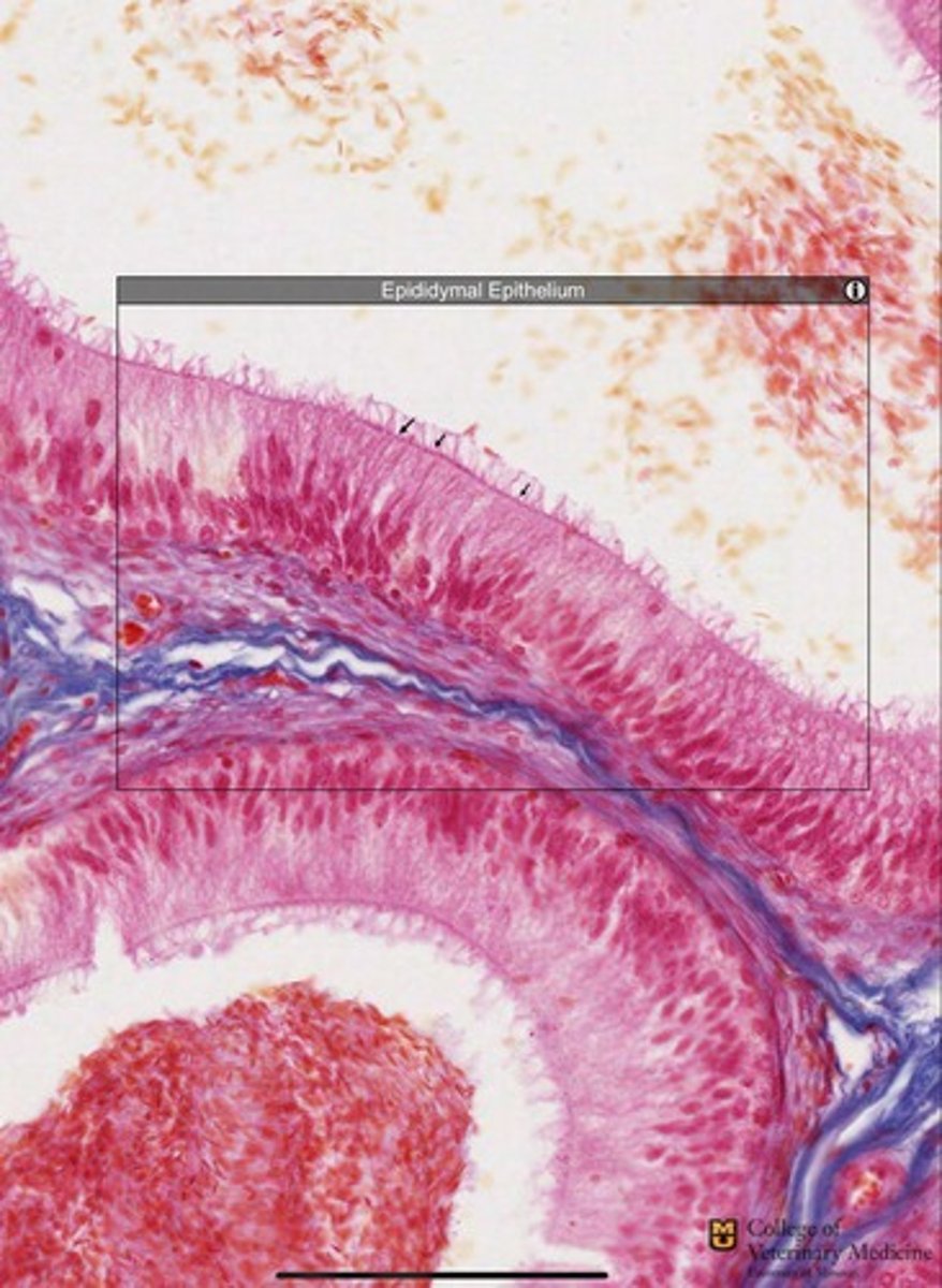
Pseudostratified epithelium with stereocilia
What epithelium type is present and what structure accompanies it?
Simple Squamous Epithelium
What epithelial tissue is shown?
Simple cuboidal epithelium
What tissue type is shown?
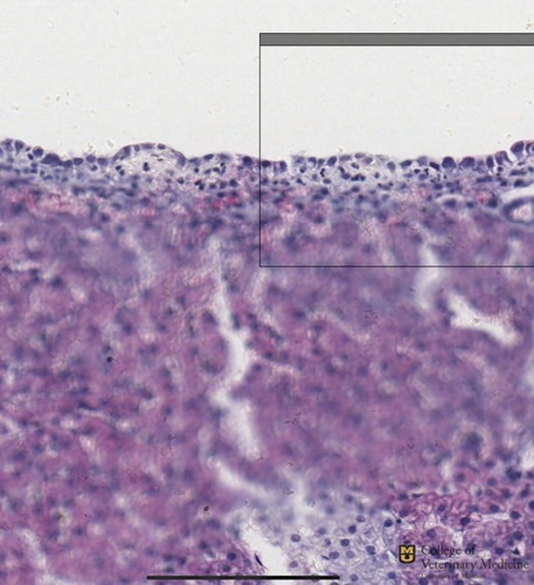
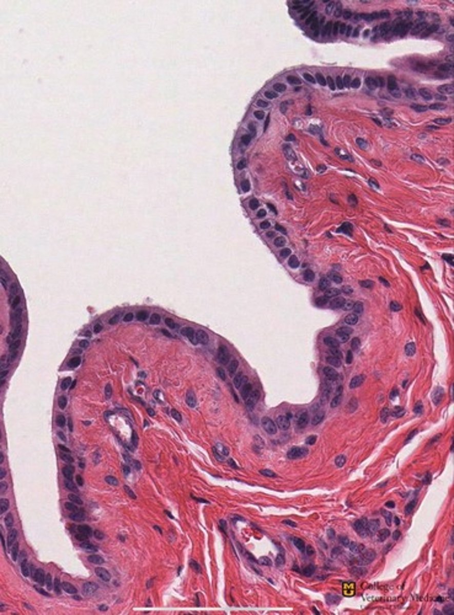
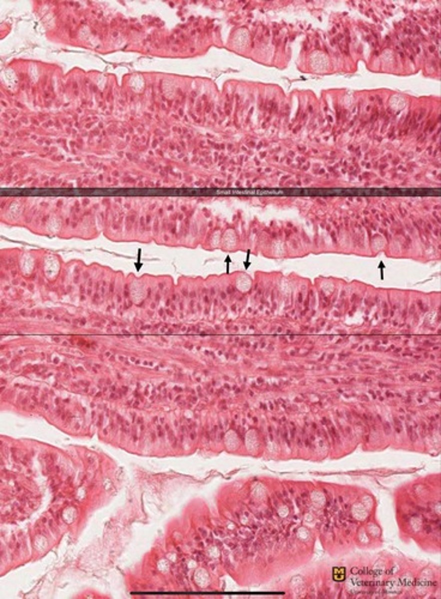
Simple columnar epithelium
What tissue type is shown?
Simple columnar epithelium
What tissue type is shown?
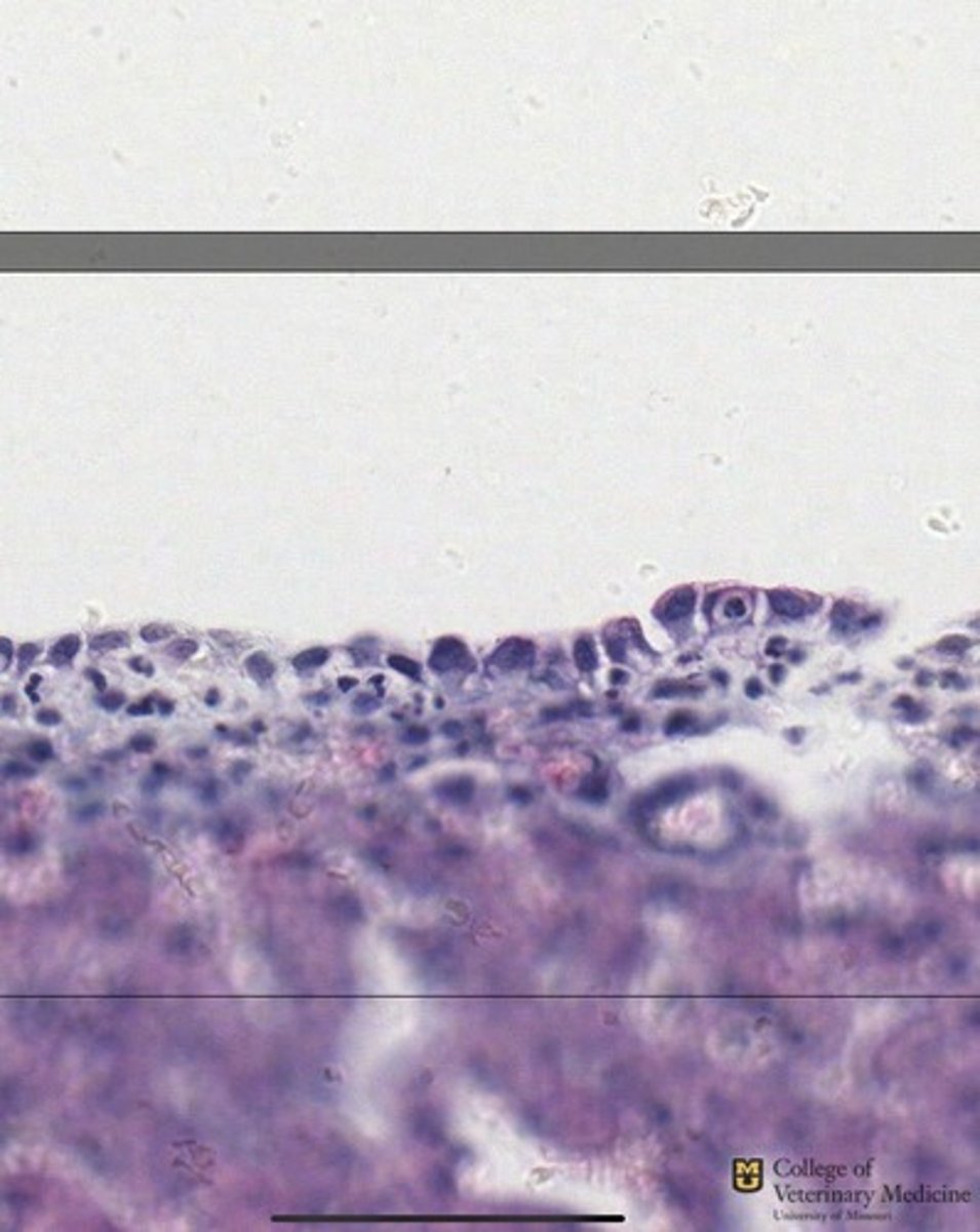
Simple columnar epithelium with Goblet cells
What tissue type is shown and what structures accompany it?
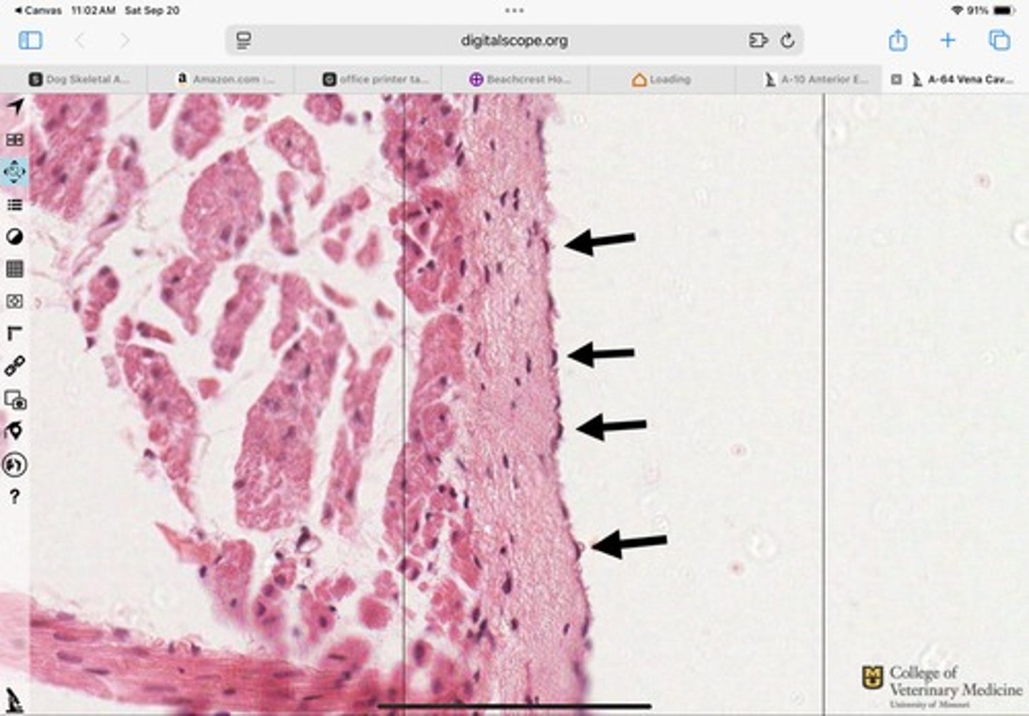
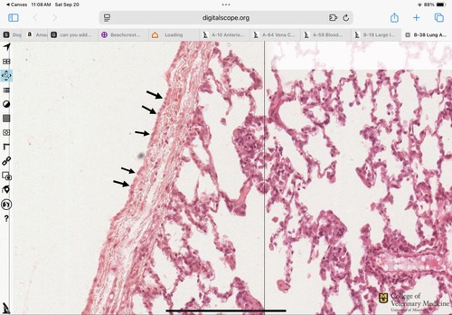
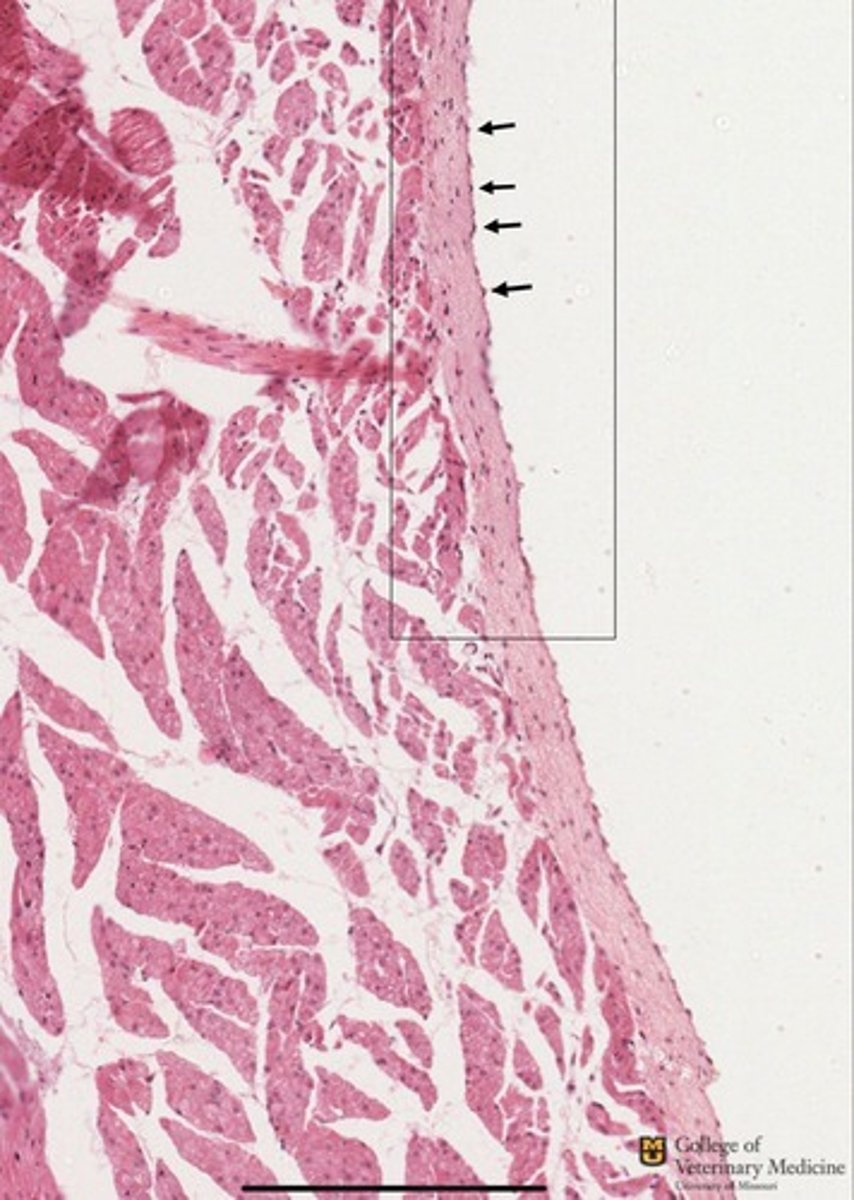
Endothelium
What tissue type is shown?
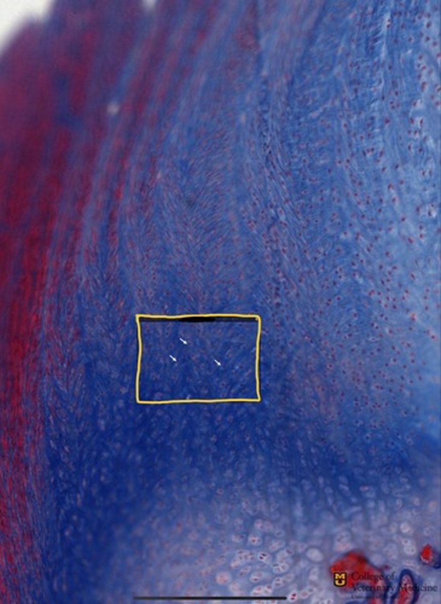
stratum spinosum
What layer of Keratinized SSE is shown by the RED bracket?

Stratum Granulosum
what layer of Keratinized SSE is shown by the GREEN bracket?

stratum lucidum
What layer of Keratinized SSE is shown by the BLUE bracket?
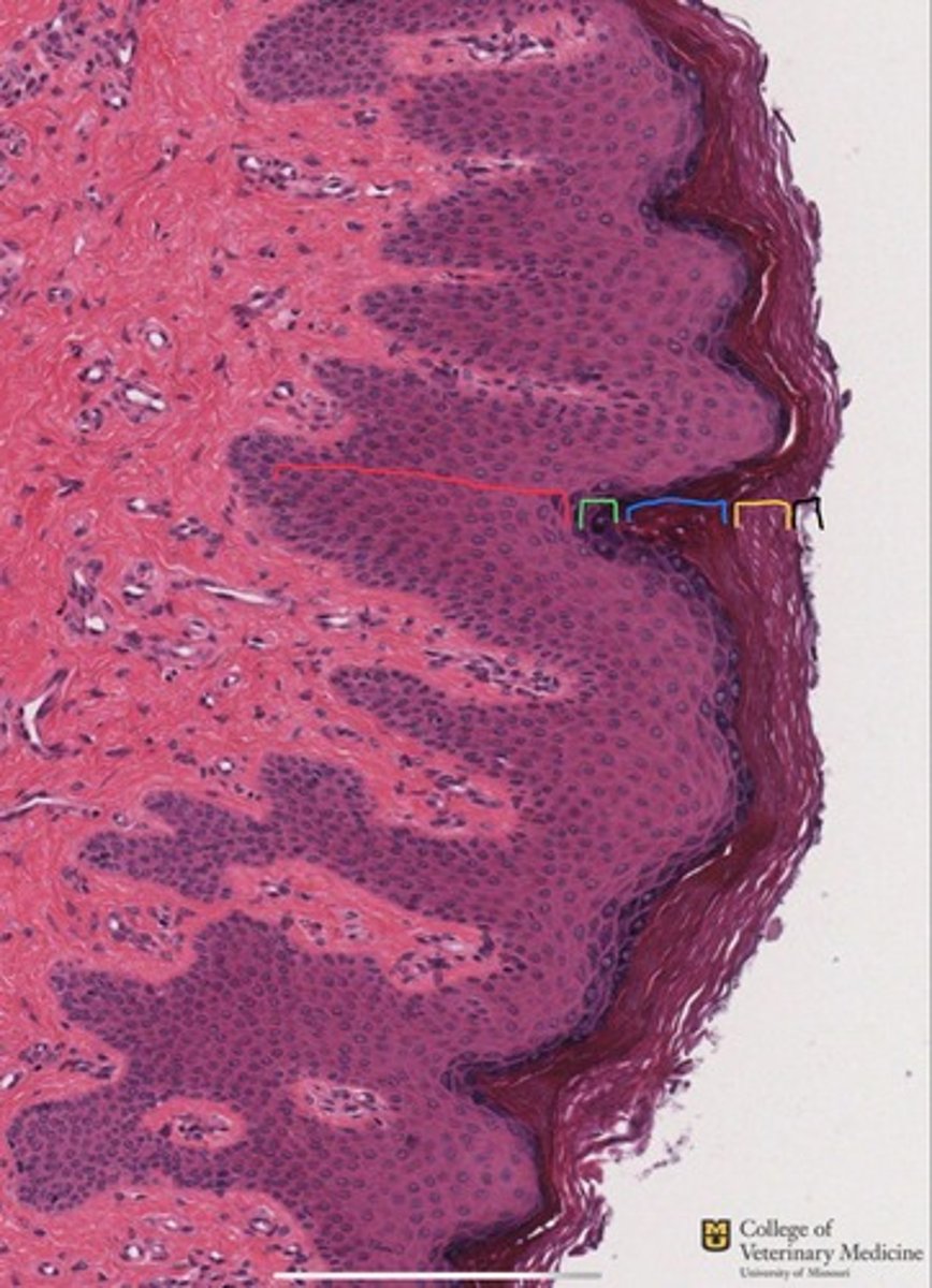
stratum corneum
What layer of Keratinized SSE is shown by the YELLOW bracket?
