MYCO | 1.3 Fungal Structure & Ultrastructure
1/128
There's no tags or description
Looks like no tags are added yet.
Name | Mastery | Learn | Test | Matching | Spaced |
|---|
No study sessions yet.
129 Terms
Intermediate stage between yeast cells and true hyphae
Pseudohypha
T/F: There are specific structures fungi produce depending on their habitus or type of environment they’re found
TRUE
This is bc the type of environment determines the evolution of their form, e.g., Thallus of Rhizophlyctis, Plasmodia in Plasmodiophora
Structure used by fungi to invade internal cellular structures of plants; for feeding, preparation for reproduction
(G) Plasmodia

Body (structure) composed of matted mycelia, then mycelial network emanating from it; for general growth and nutrient absorption
(F) Thallus

The evolutionary history of fungi reflects _
how they were able to conquer many habitats through a diversity of their forms
Fungi have colonized a wide range of habitats, fulfilling important roles in diverse ecosystems through the _, often rooted in the ability of the fungal hypha to grow and form a mycelium
diversity of their forms

Fungi have colonized a wide range of habitats, fulfilling important roles in diverse ecosystems through the diversity of their forms, often rooted in the _
ability of the fungal hypha to grow and form a mycelium

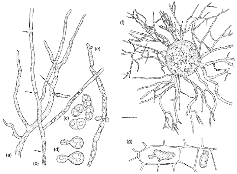
Describe the various growth forms of fungi as shown in the figure
(1/4 20 um) A = Aseptate hypha of Mucormucedo
B = Septate branched hypha of Trichoderma
(10 um) C = Dividing yeast cells of Schizosaccharomyces
D = Budding yeast cells of Dioszegia
(10 um) E = Pseudohypha of Candida (intermediate stage between yeast cells and true hyphae)
F = Thallus of Rhizophlyctis
E = Plasmodia of Plasmodiaphora
_ branches to form a mycelium
Hypha
2 basic forms of fungi
Hyphal (filamentous)
Yeast
T/F: There are fungi with no base yeast or hyphal form in terms of their cellular ultrastructure
FALSE
All fungi have a base yeast or hyphal form in terms of their ultrastructure

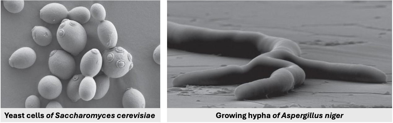
Identify
(A) Yeast cells of Saccharomyces cerevisiae
(B) Growing hypha of Aspergillus niger

Basic fungal form
Microscopic fungi consisting of solitary cells that reproduce by budding
Yeasts
Form of asexual reproduction where a small protrusion forms on parental cell’s surface, referred to as the bud, then the bud enlarges as cytoplasm and organelles are shared between parent and the bud, eventually forming a septum that leads to pinching off of the bud from the parent cell
Budding

The number of times a yeast cell reproduced can be estimated based on the _
number of scars from budding the parental cell has on its surface
Basic fungal form
AKA filamentous or mold form
Have long filaments called _, which grow by apical (tip) extension
Hyphal form
Yeasts reproduce via _
budding
Hyphae in hyphal fungi grow by _
apical (tip) extension
Elongated filaments with a moving protoplasm inside, growing via apical (tip) extention
Hyphae
Yeasts are considered fungi because they possess features of the kingdom, including _
cesaf
Chitin in cell walls
Extracellular digestion
Saprophytic nutrition
Asexual reproduction
Food stored as glycogen
Some fungi are _, meaning they can alternate cell growth between yeast and filamentous forms
dimorphic
Example of possible triggers for dimorphism in fungi
Temperature
Humidity
Presence of internal environment of a host
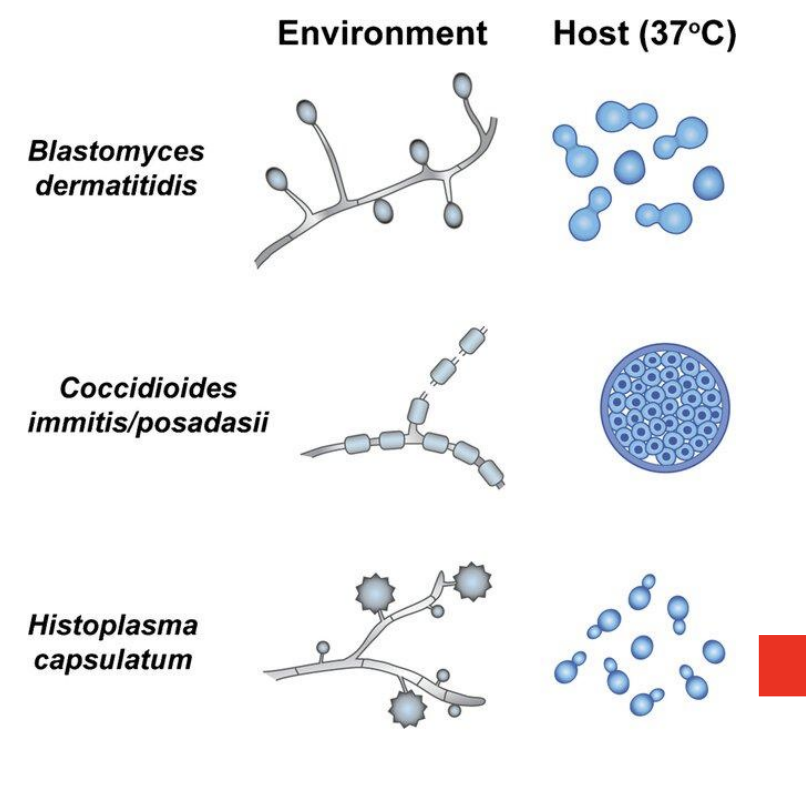
3 examples of dimorphic fungi
bch
Blastomyces dermatitidis
Environment (nutrient-poor) = Hyphal
Host (37C, nutrient-rich) = Yeast
Coccidioides immitis / posadasii
Environment (nutrient-poor) = Hyphal
Host (37C, nutrient-rich) = Yeast
Histoplasma capsulatum
Environment (nutrient-poor) = Hyphal
Host (37C, nutrient-rich) = Yeast
Why is it important to study dimorphism in fungi?
Medically, it is important because there are lots of fungi invading the human body that start out as filamentous, enter as yeast, then develop into filaments once inside the human body
and understanding the pathogenicity, immune evasion, and tissue invasion mechanisms of these fungi can play a critical role in developing effective diagnostic tools and antifungal therapies to combat fungal infections, particularly in immunocompromised individuals.
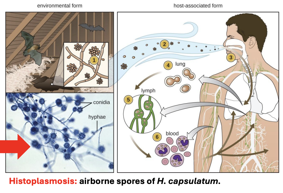
Explain mechanism of Histoplasma capsulatum that causes histoplasmosis mostly in construction workers
In the environment, H. capsulatum exists in its hyphal form, but this hyphal form has reproductive structures containing spores, spores are then released from repro structures, turning into yeasts once inside human body (due to human body temperature at 37C)
H. capsulatum is typically found in its hyphal form in bat droppings. If these droppings are agitated due to construction work and you’re not wearing PPE, these spores can get released and be inhaled by workers
Because spores are large, these are filtered out by nasal passage ways, but some spores are so tiny that they can actually reach the lung
Once in the lung, they grow as yeast (due to human body temp 37C), proliferating inside the body, eventually finding their way in lymphatic tissues and thus spreading all over to different organs and organ systems
Dimorphic fungus that grows in soil exposed to bird feces or bat feces
Histoplasma capsulatum
T/F: Candida albicans can start out as filamentous then become yeast inside the body, or start out as yeast then become filamentous inside
TRUE

What happens when Candida albicans reaches the lung?
It can cause emphysema-like symptoms (feel like drowning) because C. albicans is invasive to alveoli, potentially leading to pneumonia due to internal water accumulation

T/F: Ultrastructurally, yeasts appear like a typical eukaryotic cell but with chitinaceous cell wall
TRUE
Several fungi grow as _ that divide via budding
budding, uninucleate yeasts

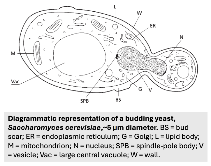
Describe the figure shown
Diagrammatic representation of a budding yeast, Saccharomyces cerevisiae, ~5 um in diameter

Yeasts are not different from hyphal fungi but represent a _
different growth form adapted to nutrient-rich environments
T/F: Hyphal growth is used in nutrient-poor conditions to move around and get as much nutrition as they could
TRUE
In a plate with isolated fungi, why is the central portion darker and outer portion lighter?
Central portion = most mature and contains reproductive structures
Outer portion = youngest, hyphae seeking nutrition
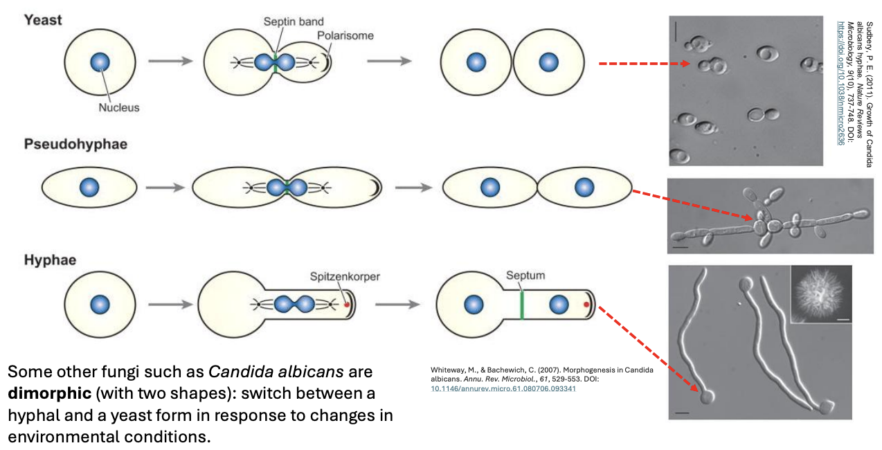
Some other fungi such as _ are dimorphic (with two shapes) and can thus switch between yeast and hyphal forms in response to changes in environmental conditions
Candida albicans
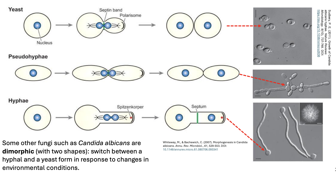
Depending on environmental conditions, temperature, etc., Candida albicans can grow as _
Yeasts
Pseudohyphae
Hyphae

_ look like hyphae because it’s elongated, but there are bunch of cells clustered together
Pseudohyphae

T/F: There are yeasts capable of developing as yeast, hyphae, or pseudohyphae separately or simultaneously depending on the situation
TRUE
How does a dimorphic yeast (e.g., C. albicans) become medically problematic?
If the yeast has different forms, then this allows it to traverse a variety of crevices in the human body
It can create growth forms that will allow it to colonize as much crevices in the body
Trigger for development of pseudohyphal form of C. albicans
Consequence of environmental gradient because it’s an intermediate form that allows yeast to colonize the environment, which might have a different temperature or pH, or not big enough to form hyphae but good enough to form pseudohyphae to colonize species
Some other fungi such as C. albicans are dimorphic (with two shapes) and can thus _
switch between a hyphal and a yeast form in response to changes in environmental conditions
_ is essentially a tube with a rigid wall, containing a moving slug of protoplasm
Hypha
Protoplasm vs. Cytoplasm
Cytoplasm = gel-like; excluding plasma membrane
Protoplasm = all living components; everything apart from the cell wall; including plasma membrane
The fungal hyphal tip contains an organizing center for _
hyphal growth and morphogenesis
_ is the organizing center for hyphal growth and morphogenesis located in the fungal hyphal tip
Apical vesicular cluster (AVC), organized as spitzenkorper in dikarya (Asco- & Basidiomycota)

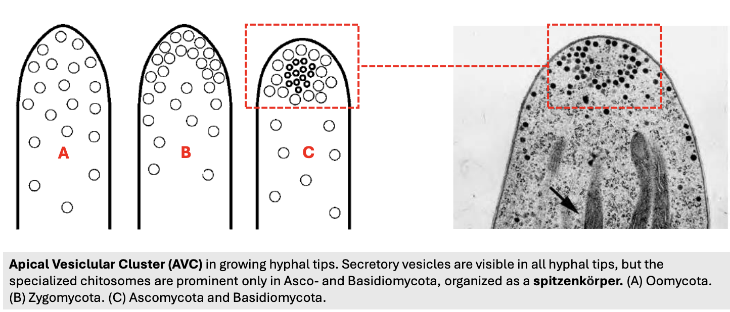
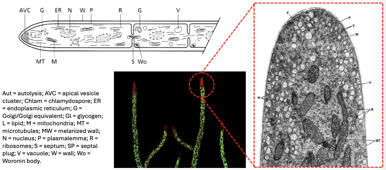
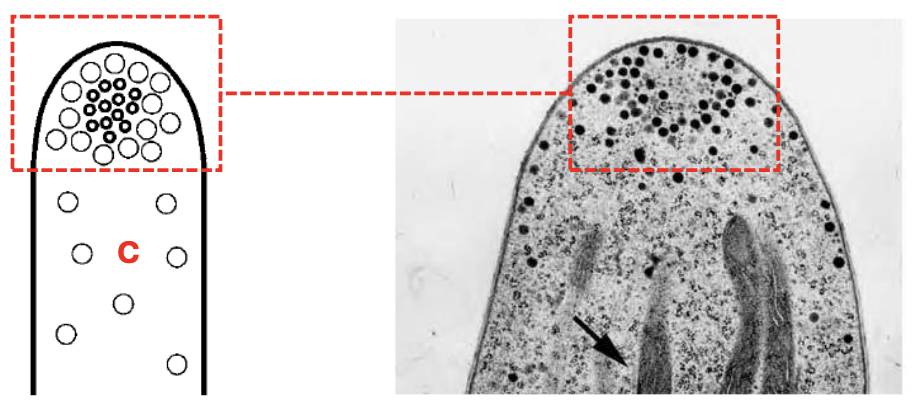
Describe the figure
Figure shows the apical vesicular cluster (AVC) in growing hyphal tips
Secretory vesicles are visible in all hyphal tips of (A) Oomycota (Water molds), (B) Zygomycota, (C) Asco- & Basidiomycota
However, specialized chitosomes are prominent only in Asco- & Basidiomycota, organized as a spitzenkorper
An apical vesicular cluster (AVC) is only considered a spitzenkorper:
dvc
Once it reaches a particular density
Has several vesicle types
Chitosomes are microscopically observable
T/F: The secretory vesicles in apical vesicular clusters (AVCs) are prominent only in Asco- & Basidiomycota
FALSE
The secretory vesicles in apical vesicular clusters (AVCs) are prominent in all hyphal tips of Oomycota, Zygomycota, and Asco- & Basidiomycota
Specialized chitosomes within apical vesicular clusters (AVCs) are prominent only in Asco- and Basidiomycota and thus are organized as _
spitzenkörper (SPK)
T/F: Spitzenkorper solely determines the growth pattern of hyphal fungi
FALSE
Not necessarily because fungus is also capable of recognizing different stimulus at the hyphal tip
_ occupies the extreme apex of the hypha and directs growth and branching
Spitzenkorper

Spitzenkorper occupies the extreme apex of the hypha and _ (function)
directs growth and hyphal branching
Spitzenkorper in German means _
“pointed body with secretory vesicles organized by microfilaments”
Spitzenkorper is present in _ but is termed AVC in all others; it helps determine the direction of the growth and facilitates hyphal branching
Dikarya (Asco- & Basidiomycota)

_ helps determine the direction of growth and facilitates hyphal branching
Spitzenkorper in Dikarya (AVC in others)
Major reservoir of chitin synthase (enzymes that produce chitin)
Chitosomes

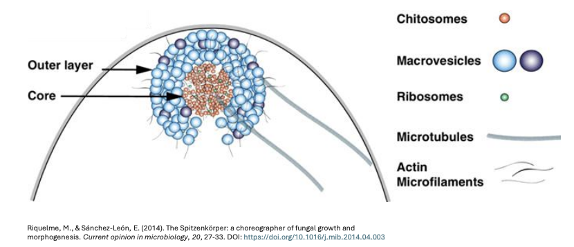
Describe the structure of a Spitzenkorper
2 layers
Outer layer = Macrovesicles
Core = Chitosomes, Ribosomes
Connecting the 2 = Microtubules, Actin microfilaments

What triggers the spitzenkorper to stop hyphal growth?
Tactile stimulus, e.g., when there’s no more room to grow, thus fungi stop sending growth signals and try to change the direction of growth instead
Chemical stimulus, e.g., encountering harmful chemicals potentially problematic for fungi to deal with, thus chemoreceptors stop growth
Nutrient deprivation
What happens if you cut the hyphal tip?
Plug the cut area with Woronin bodies to prevent protoplasmic leak
Nuclei then streams very close to the area, producing a new hyphal tip extension on another direction
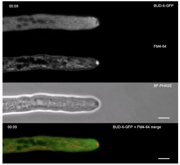
Describe the figure shown
Parallel panels of Neurospora crassa hypha, with Spitzenkorper clearly visible at the tip (stained red at bottom panel)

Hyphal types can be classified based on _
Septation
Cell arrangement
T/F: Fungal hypha cannot elongate without chitin produced by chitin synthase within chitosomes
TRUE
Self-assembling aggregates of the enzyme chitin synthase
Chitosomes
Each vesicle (of chitosome) contains sufficient _ to produce, after proteolytic activation (where protease cleaves portion of chitin synthase), a chitin microfibril composed of several chitin chains
inactive chitin synthase

Activation causes a _ to appear in the chitosome
The shell of the chitosome is opened/shed and an extended microfibril arises from the fibroid particle (R)
coiled microfibril (R = fibroid)

_ causes a coiled microfibril (R = fibroid) to appear in the chitosome
Proteolytic activation
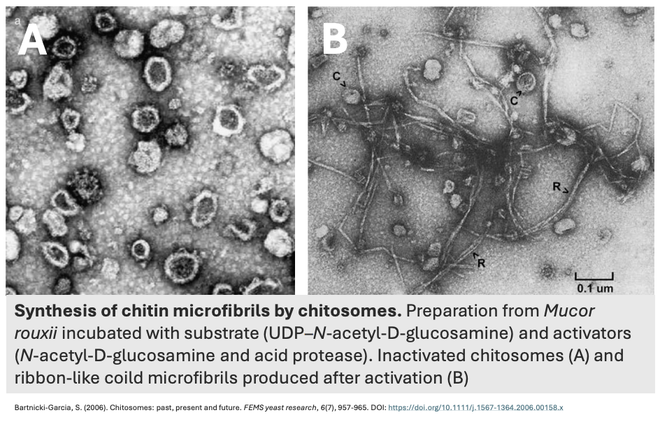
Describe the figure shown
Synthesis of chitin microfibrils by chitosomes
Preparation from Mucor rouxii, incubated with substrates (UDP-N-acetyl-D-glucosamine) and activators (N-acetyl-D-glucosamine, acid protease)
(A) Inactivated chitosomes
(B) Ribbon-like chitin microfibrils produced after activation
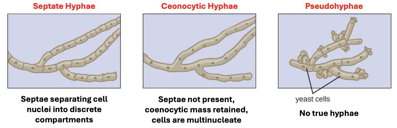
Hyphal type
Septae is not present
Coenocytic mass is retained
Cells are multinucleate
Coenocytic hyphae

T/F: More often than not, septate fungi is only a single cell with multiple compartments
TRUE
It’s a case-to-case basis. There are cases where they can develop into separate cells.
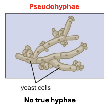
Hyphal type
No true hyphae
Has elongated yeast cells clustered together
Pseudohyphae
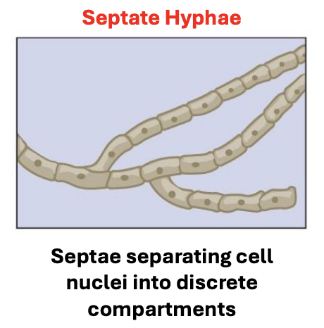
Hyphal type
Septae separates cell nuclei into discrete compartments
Septate hyphae
Each vesicle of chitosome contains sufficient inactive chitin synthase to produce, after proteolytic activation, a _ composed of several chitin chains
chitin microfibril
The evolution of fungal hypha is thought to have begun from a _ form, choreographed by key innovations in eccda elongation, compartmentalization, communication, differentiation, & adhesion
coenocytic
Main reason why fungi evolved to have a hyphal form
To be able to colonize a variety of different environments, as each environment would have a different supply and cycling of nutrients, thus having hyphae that can grow, move, and traverse to different areas allows fungi to survive in some of the most extreme environments
T/F: Vesicles responsible for enzyme secretion are part of SPK
FALSE
Though, there are vesicles in SPK that facilitate delivery of these enzymes to hyphal tip
The evolution of a fungal hypha is thought to have begun from a coenocytic form, choreographed by key innovations in _ (5)
eccda
Elongation
Compartmentalization
Communication
Differentiation
Adhesion
_ are perforated crosswalls that compartmentalize the hyphal cells in septate fungi
Septa
T/F: Each vesicle of chitosome contains sufficient chitin synthase to produce, before proteolytic activation, a chitin microfibril composed of several chitin chains
FALSE
Each vesicle of chitosome contains sufficient chitin synthase to produce, after proteolytic activation, a chitin microfibril composed of several chitin chains
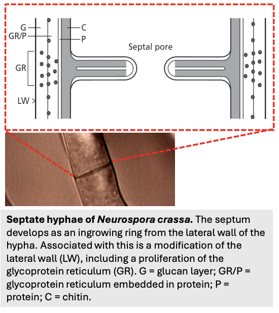
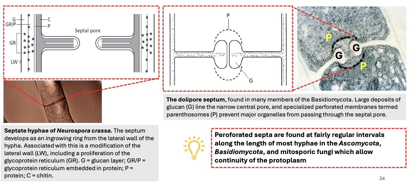
_ develop as ingrowing rings from the lateral wall of the hypha
Associated with this is the modification of the lateral wall, including _
Septum
Proliferation of glycoprotein reticulum (GR), glucan layer (G), proteins (P), chitin (C)
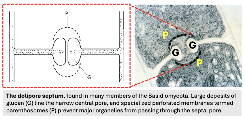
Found in many members of Basidiomycota
Characterized by
Large deposits of glucan lining narrow central pore,
Specialized perforated membranes called parenthosomes preventing major organelles from passing through septal pore
Dolipore septum
_ are found at fairly regular intervals along the length of most hyphae in the Ascomycota, Basidiomycota, & mitosporic fungi, which allow the continuity of the protoplasm
Perforated septa
T/F: Nuclei can pass through dolipore septum
FALSE
Nuclei is too big to pass through dolipore septum, but it can pass through normal hyphal septa
2 characteristics of dolipore septum
Large glucan deposits lining narrow central pore
Parenthosomes preventing major organelles from passing through the septal pore
Advantages of having septa in fungi
Isolation of damage or damage control
If part of hypha is damaged, the septa can compartmentalize the injury to prevent protoplasmic leakage and maintain the integrity of the remaining hyphal network
Con: movement of material is slower in septate fungi
Structural support
Provides additional structural reinforcement to hyphae, making it more resilient against mechanical stress
_ prevents cytoplasmic loss at septal pores of Ascomycota
Woronin bodies
T/F: The dolipore septum of Basidiomycota prevents protoplasmic leakage by utilizing Woronin bodies to seal its specialized pore in case of damage
FALSE
Woronin bodies are exclusive to Ascomycota. Protoplasmic leakage in Basidiomycota is prevented by parenthosomes
Specialized organelle derived from peroxisomes found near the septae of filamentous Ascomycota; unit membrane + protein core (HEX-1) attached via Leashin to Woronin Sorting Complex
Woronin body

Components of a Woronin body
Unit membrane + Protein core (HEX-1) attached via Leashin to pore Woronin Sorting Complex
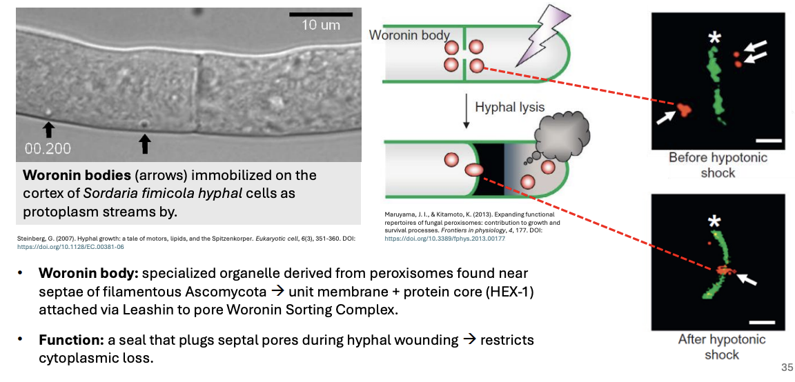
Function of Woronin bodies
Act as a seal that plugs septal pores during hyphal wounding, thus restricting cytoplasmic loss
Perforated septa are found at fairly regular intervals along length of most hyphae in _, which allow continuity of protoplasm
Ascomycota, Basidiomycota, mitosporic fungi
T/F: Dolipore septum is found in many members of Ascomycota
FALSE
Dolipore septum is found in many members of Basidiomycota
Specialized perforated membranes that prevent major organelles from passing through the septal pore
Parenthosomes
What happens when there is damage in the hyphal tip of an ascomycete?
The portion of hypha damaged then lyses to ensure that the damaging agent remains and does not spread to other areas
Woronin bodies then plug the septal pore
Structure in plants similar to Woronin bodies in Ascomycetes
Tyloses
Condition where cells are exposed to significantly lower external solute concentration than inside the cell, leading to water influx due to osmosis
Hypotonic shock
_ typically develops from a single germinating spore that produces a young hypha called the germ tube, which eventually develops into a mycelium
Hypha
T/F: Hypha is not an individual but almost always part of a colony
TRUE
T/F: There are hyphae that act as holdfasts (roots), stems, that generate reproductive structures
TRUE
Typical asexual life cycle of fungi
sghm
Spores (conidia) are produced by conidiophore (spore-producing organ)
Upon encountering a proper environment, spore germinates, producing germ tube
Germ tube then develops into hyphae
Hyphae then differentiates, forming a network called mycelia

T/F: Yeasts are very rarely produced from spores
TRUE
Spores must grow into hyphae first before they can form yeast