KIN 223 Chapter 14: Spinal Cord and Spinal Nerves
1/80
There's no tags or description
Looks like no tags are added yet.
Name | Mastery | Learn | Test | Matching | Spaced | Call with Kai |
|---|
No analytics yet
Send a link to your students to track their progress
81 Terms
Spinal reflexes ______ require the involvement of the brain.
do not
A typical adult spinal cord ranges between ______ in length.
42 and 45 cm
A spinal nerve contains ______ axons.
motor and sensory
The spinal cord in an adult is ______ than the vertebral canal that houses it.
shorter
The spinal cord is partitioned into a ______ gray matter region and a ______ white matter region.
deep; superficial
The spinal cord and spinal nerves are responsible for ______, which are our quickest reactions to a stimulus.
reflexes
The spinal cord extends inferiorly from the brain through the foramen magnum, then through the vertebral canal, and ends at the level of the ______ vertebra.
L1
Gray matter is primarily composed of ______.
cell bodies and dendrites
Each anterior root and its corresponding posterior root unite within the ______ to become a spinal nerve.
intervertebral foramen
During development, the individual vertebrae ______ spinal cord growth is complete.
continue to grow after
The gray matter of the spinal cord is dominated by the:
dendrites and cell bodies of neurons
A posterior funiculus lies between the ______ gray horns on the posterior side of the cord and the posterior median sulcus.
posterior
In the spinal cord, the gray matter may be subdivided into the following components: anterior, posterior, and ______ horns, and the gray commissure.
lateral
Ascending pathways are ______ pathways.
sensory
A spinal nerve is classified as a ______ nerve.
mixed
A first-order neuron:
is the first neuron to transmit sensory information from the periphery of the body to the brain
The lumbar part of the spinal cord is closest to:
inferior thoracic vertebrae
A second-order neuron:
is an interneuron
A third-order neuron is also known as a:
tertiary
White matter that lies between the posterior gray horns is a:
posterior funiculus
Identify the anatomical component of motor pathways.
upper and lower motor neurons
Sensory nervous system pathways transmit:
information from somatic and special sense receptors to the central nervous system
An impulse from a lower motor neuron ______ a skeletal muscle.
stimulates or excites
A first-order neuron is also known as a:
primary
Cell bodies of upper motor neurons are located in the:
cerebral cortex
A second-order neuron is also known as a:
secondary neuron
Impulses transmitted through the direct, pyramidal, or corticospinal pathway, pass directly from upper to lower motor neurons (no ______ involved) and are responsible for ______ control of skeletal muscles.
interneurons; conscious
A third-order neuron extends from the secondary neuron to the ______.
cerebrum
Primary motor neurons of the direct motor pathway issue impulses for ______ control of skeletal muscles; primary motor neurons of the indirect motor pathway issue impulses for ______ control of skeletal muscles.
voluntary; involuntary
Motor pathways are ______ pathways.
descending
The spinal nerves connect the central nervous system to ______ and receptors.
muscles, glands
The cell bodies of ______ motor neurons are found within brainstem cranial nerve nuclei or in the anterior horn of the spinal cord.
lower
After leaving the intervertebral foramen, a typical spinal nerve almost immediately splits into branches, called ______.
rami
An impulse from an upper motor neuron ______ a lower motor neuron.
excited or inhibits
A nerve plexus is a network of interweaving ______ of spinal nerves.
anterior rami
A descending motor tract of the direct pathway is the ______ tract.
corticospinal
Primary motor neurons of the ______ motor pathway originating in the primary motor cortex, whereas primary motor neurons of the ______ motor pathway originate in the brainstem.
direct; indirect
There are ______ pairs of spinal nerves.
31;
- 8 cervical
- 12 thoracic
- 5 lumbar
- 5 sacral
- 1 coccygeal
Spinal nerve T12 is called a subcostal nerve because it arises ______ the ribs.
below
The two main branches of a spinal nerve are the ventral and ______ rami.
dorsal
The left and right cervical plexuses are formed primarily by the anterior rami of spinal nerves:
C1 - C4
The posterior rami of spinal nerves tend to follow a ______ pattern as they innervate the deep muscles and skin of the neck and back.
segmental
Each brachial plexus is formed by the anterior rami of spinal nerves ______.
C5 - T1
The left and right lumbar plexuses are formed from the ______ rami of spinal nerves L1-L4 located lateral to the L1-L4 vertebrae and along with the ______ muscle in the posterior abdominal wall.
anterior; psoas major
The anterior rami of spinal nerves ______ are called intercostal nerves because they travel in the intercostal space sandwiched between ______.
T1-T11; two adjacent ribs
The nerves emerging from a sacral plexus innervate the ______ region, pelvis, perineum, posterior thigh, and almost all of the ______.
gluteal; leg and foot
The left and right cervical plexuses are located deep on each side of the neck, immediately lateral to cervical vertebrae ______.
C1 - C4
Awareness of the stimulus occurs after the _______ action has been completed, in time to correct or avoid a potentially dangerous situation.
reflex
The left and right brachial plexuses are networks of nerves that supply:
the upper limb
A reflex arc is the neural wiring of a ______ reflex.
single
The anterior rami of spinal nerves L1-L4 form the ______ plexus.
lumbar
An ______ reflex is one that is developed after birth.
acquired
A stretch reflex is a ______ reflex that monitors and regulates skeletal muscle length.
monosynaptic
The left and right sacral plexuses are formed from the ______ rami of spinal nerves ______ and are located immediately inferior to the lumbar plexuses.
anterior; L4-S4
In which reflex does the elbow flex?
biceps reflex
Reflexes are rapid, automatic, involuntary reactions of ______ to a stimulus.
muscles or glands
The central nervous system forms primarily from the embryonic ______.
neural tube
A reflex arc always begins at a receptor in the ______, communicates with the ______, and ends at a peripheral effector, such as a muscle or gland cell.
PNS; CNS
Spinal Reflex
the integration center in this type of reflex is the spinal cord
Visceral Reflex
a gland may be the effector in this type of reflex.
Polysynaptic Reflex
this reflex includes one or more interneurons
Ipsilateral Reflex
The receptor and effector in this reflex are located on the same side of the body
Stretch in a muscle is monitored by a stretch receptor called a muscle ______.
spindle
Biceps reflex (C5, C6)
flexes elbow
Triceps Reflex (C6, C7)
extends elbow
Cremasteric Reflex (L1, L2)
elevates testis
Patellar Reflex (L2 - L4)
extends knee
Plantar Reflex (L5, S1)
flexes toes
During development, the cranial and spinal nerves form primarily from ______.
alar and basal plates
The initial component of a reflex arc is a(n) ______.
receptor
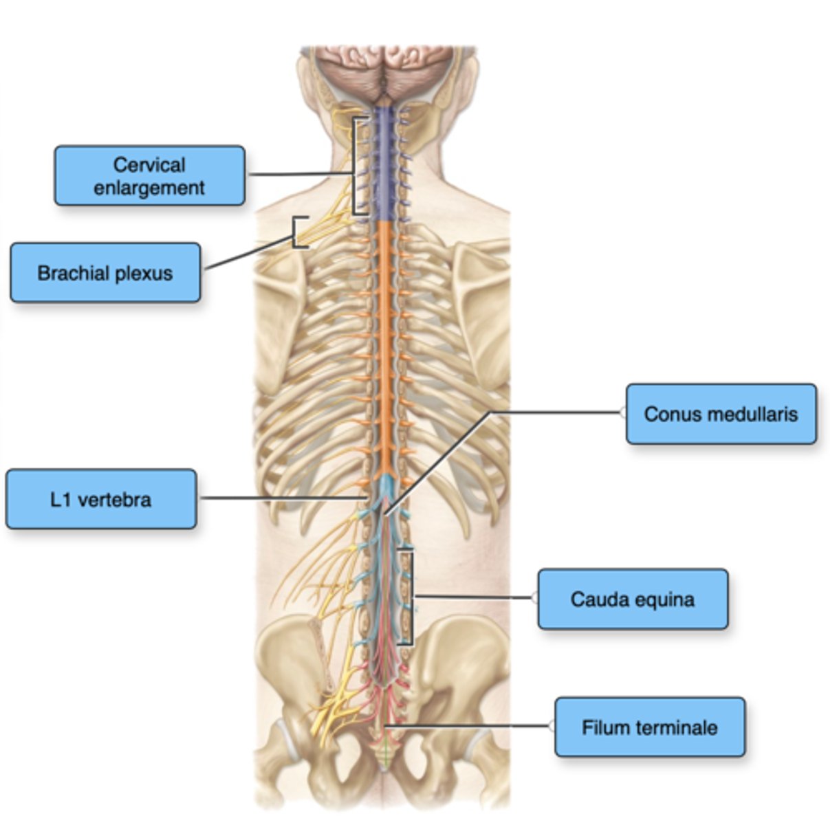
Label the structures of the spinal cord.
The cell bodies of sensory neurons are found:
in posterior root ganglia
Motor neurons contain their cell bodies in the ______ and axons in the ______.
anterior horn; anterior root
There are ___ pairs of spinal nerves that exit the vertebral column.
31
After leaving the _______, a typical spinal nerve almost immediately splits into branches called ______.
intervertebral foramen; rami
The ______ ramus is the smaller of the two main branches. It innervates the _______ and the skin of the back.
posterior; deep muscles of the back
The ______ ramus is the larger of the two main branches. This splits into multiple other branches, which innervate the ______, the upper limbs, and the lower limbs.
anterior; anterior and lateral portions of the trunk
Many of the anterior rami go on to form ______.
nerve plexuses
True of False: All reflexes share the property of being an involuntary response.
True
Nerve plexuses are organized such that axons from each ________ ramus extend to body structures through several different branches.
anterior
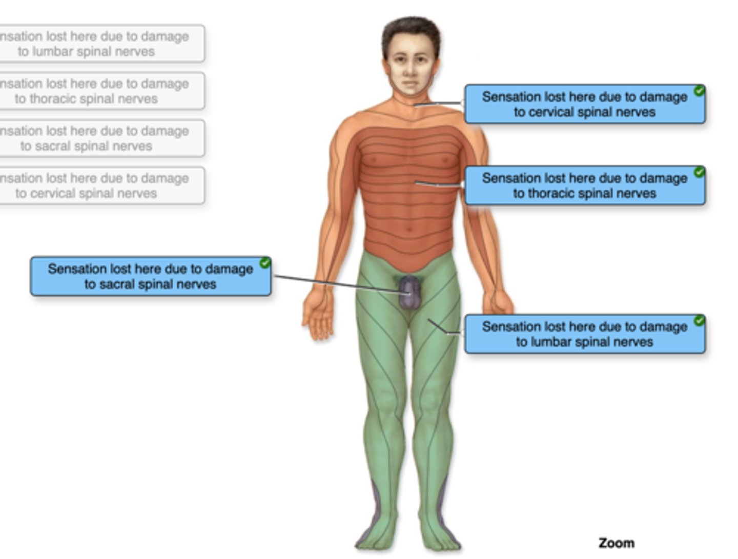
Correctly identify and label the dermatome(s) represented by the statement(s) associated with them.