Unit 2 Lecture Notes
 Call Kai
 Learn
Learn Practice Test
 Spaced Repetition
 Match
Match Flashcards
 Knowt Play
1/51

There's no tags or description
Looks like no tags are added yet.
Name | Mastery | Learn | Test | Matching | Spaced | Call with Kai | Chat |
|---|
No analytics yet
Send a link to your students to track their progress
52 Terms
types of cartilage
hyalin, elastic, fibrocartilage
whats in cartilage?
chondrocytes (maintain and support cartilage without making new matrix), abundant ECM, Lacuna (where chondrocytes live)
hyaline cartilage
l: ends of bones in synovial joints (articular cartilage), costal cartilages between ribs and sternum (sternal cartilage), along portions of respiratory tract, part of septum
f: stiff but flexible support
*resists compression due to large amounts of water

elastic cartilage
l: epiglottis, ear, voice box/auditory tube
f: provide support while tolerating distortion
*collagen AND elastic fibers

fibrocartilage
l: menisci, pubic symphysis, intervertebral discs
f: resists compression, prevents bone on bone friction (articular discs- wrist, jaw, knee), limits relative movement
*thick collagen fiber
*torsion is bad

functions of bones
support
movement
protection
mineral storage
blood cell and fat storage
energy metabolism
kinds of bones
long, short, flat, irregular, sutural
sesamoid bone
grows inside of a tendon creating power
big toe
thumb
patella
compact bone histology
osteon - ringed cylinder
central canal with veins, arteries, and nerves
perferating canal - 90 angles connecting central canals
lacunae - ECM
canaliculi - links osteocytes together, spreading nutrients
circumferential lamellae - holds osteons together

spongy bone
holds red marrow (red blood cell production), inside linings and ends

organic and inorganic composition of bones
organic - collagen made by osteoblasts (bending + flexibility)
inorganic - calcium phosphate (hard + resists compression)
cell types found in bones
osteogenic cell - bone stem cell, becomes osteoblasts
osteoblast - builds
osteocyte - mature osteoblast
osteoclast - reshapes
when does ossification begin? whats the two types?
8 weeks
intramembranous + endochondral
intramembranous ossifcation
bone formed by fibrous membrane
fibrous connective tissue turns to bone
skull and clavicle
endochondral ossification
bone formed by hyaline cartilage
bone grows from inside out
interstitial - grows length wise
appositional - grows in thickness
kinds of fractures
comminuted - 3+ pieces
compression - vertebrae, crushed bone
spiral - torsion, sports fracture
epiphyseal - through epiphyseal plate, stunting growth (kids)
depressed - skull fracture
greenstick - partial break partial bend (kids)
1 & 2 = common in elderly w/ osteoporosis
osteoporosis
low bone mass → brittle, porous, thin bones
vulnerable in lumbar region and neck of femur
women over 65, caucasians and asians, men with bad diets
dexascan for diagnosis (body composition)
treated with medicine (osteoblast stimulant or osteoclast inhibitor), calcium supplements, diet change, and restrictive exercises (stress turns on osteoblasts)
pagets disease
chronic bone disease, too much osteoblast work means thick bones
rickets disease
form of osteomalacia in kids
bones arent fully calcified → soft bones → cant hold weight
why? 1- low vit D 2 - low calcium phosphate
joints
bone on bone
bone on muscle
bone on teeth
*stability and mobility! (inversely related)
functional joint classification
synarthrosis (no moving)
amphiarthrosis (some moving)
diarthrosis (free moving)
structural joint classification
fibrous
cartilaginous
synovial
fibrous joint
connected by fibrous CT
no joint cavity
synarthrotic + amphiarthrotic
sutures - only in skull; synostoses = closed sutures
syndesmosis - ligaments only, motion depends on ligament length
gomphosis - tooth in socket, periodontal ligament
cartilaginous joint
bones united by cartilage
no joint cavity
synarthrotic + amphiarthrotic
synchondroses - hylaine cartilage (epiphyseal plates and first rib x manubrium)
symphyses - fibrocartilage (pubic symphysis and intervertebral discs)
synovial joint
diarthrotic
fluid filled joint cavity
joint cavity!
fibrous outside - dense irregular ct
synovial inside - hyalin cartilage
some have additional articular discs (TMJ, wrist, knee)
busae (fluid-filled cushion) + tendon sheaths decrease friction
types: plantar, hinge, pivot, condyloid, saddle, ball and socket
when are fontanelles closed by?
2 years
plantar synovial joint
nonaxial
intercarpal joints, intertarsal joints, joints between articular surfaces
hinge synovial joint
uniaxial
elbow joints, interphalangeal joints
pivot synovial joint
uniaxial
proximal radioulnar joints, atlantoaxial joint

condyloid synovial joint
biaxial
metaphalangeal joints, wrist joints

saddle synovial joint
biaxial
carpometacarpal joints of the thumbs
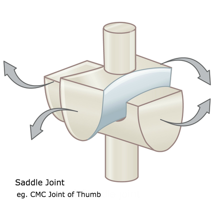
ball and socket synovial joint
multiaxial
shoulder joints and hip bones
angular movement
flexion/extension - dec/inc joint angle
abduction/adduction - away/towards midline
circumduction - propeller

rotation movement
turning around long axis
external vs internal

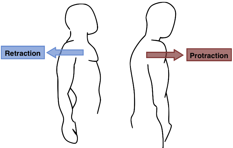
protraction and retraction
forward and back/ anterior and posterior
opposition movement
putting your thumb to each finger
dorsiflexion and plantar flexion

inversion and eversion

knee joint
largest joint
hinge joint
ligaments
patellar
fibular/lateral collateral
tibial/medial collateral
anterior cruciate - prevents anterior sliding of tibia
posterior cruciate - prevents posterior sliding of tibia

sinuses
frontal, ethmoid, sphenoid, maxillary
lined with mucus membrane
lighten skull
sound resonation

cleft palate
left and right parts of palate don’t join medially
food can aspirate into nasal cavity → pneumonia
fixed surgically
spinal curvature
cervical and lumbar → anertior; develops with gravity/after walking
thoracic → posterior; present at birth

spinal curve abnormalities
scoliosis → s-shape or c-shaped; treatment: nothing, physical therapy, brace, rods
kyphosis → hunched over
lordosis → cervical & lumbar curvatures are too extreme, compresses facets, pregnancy + obesity
pectoral girdle
clavicle + scapula
high mobility & shallow glenoid cavity = unstable
pelvic girdle
two hip bones
pelvis (& F vs M)
hip bones & sacrum
F: rounded pubic arch, wide and shallow outlet/inlet
M: narrow heart-shaped inlet/outlet
transverse ligament of atlas
holds c1 and c2 together (risk of serverence by dens)
condyles + c1 =
nodding
c1 + c2 =
shaking
hip dysplasia
femur head not in acetabulum → bracing, PT, surgery
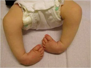
club feet
foot inversion
foot arches
medial longitudinal
lateral longitudinal
transverse