Lecture 5: Histology of the Esophagus, Stomach, and Intestines | Quizlet
 Call Kai
 Learn
Learn Practice Test
 Spaced Repetition
 Match
Match Flashcards
 Knowt Play
1/85

There's no tags or description
Looks like no tags are added yet.
Name | Mastery | Learn | Test | Matching | Spaced | Call with Kai | Chat |
|---|
No analytics yet
Send a link to your students to track their progress
86 Terms
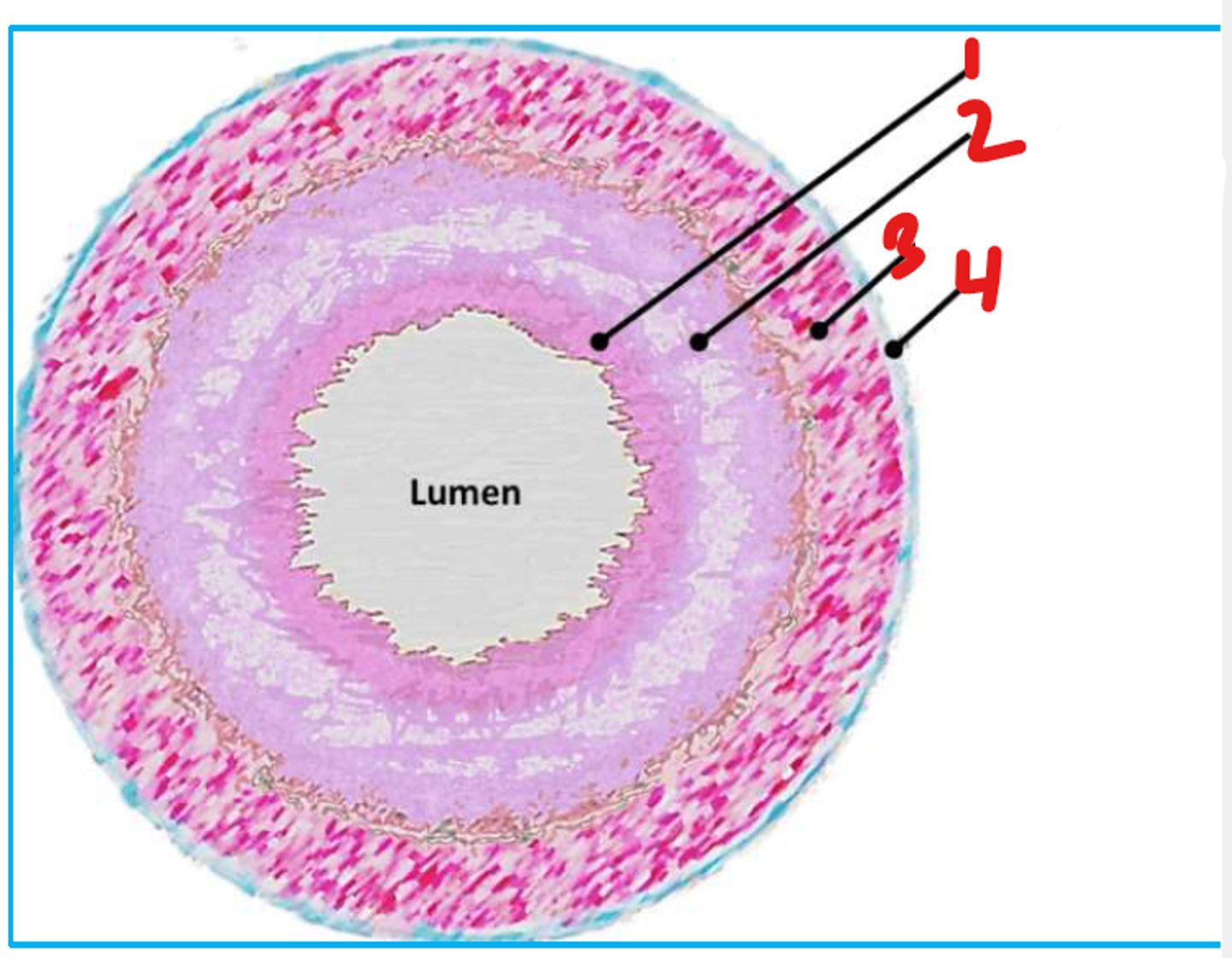
Muscularis Mucsoa
- thin layer of smooth muscle
- maintains constant state of gentle agitation
- helps expel secretions
Mucosa
1
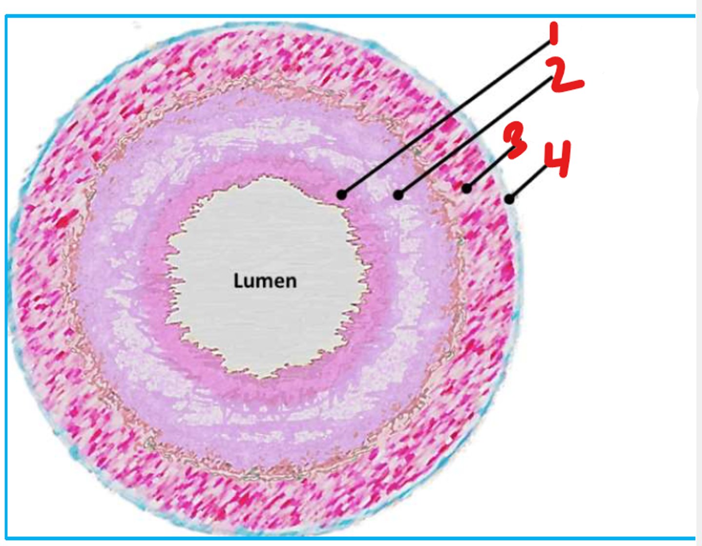
Submucosa
2
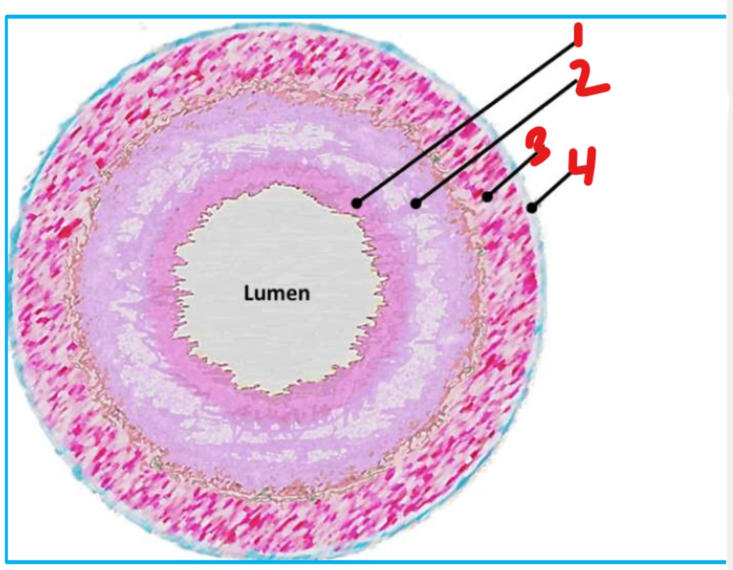
Muscularis Propria (Muscularis Externa)
3
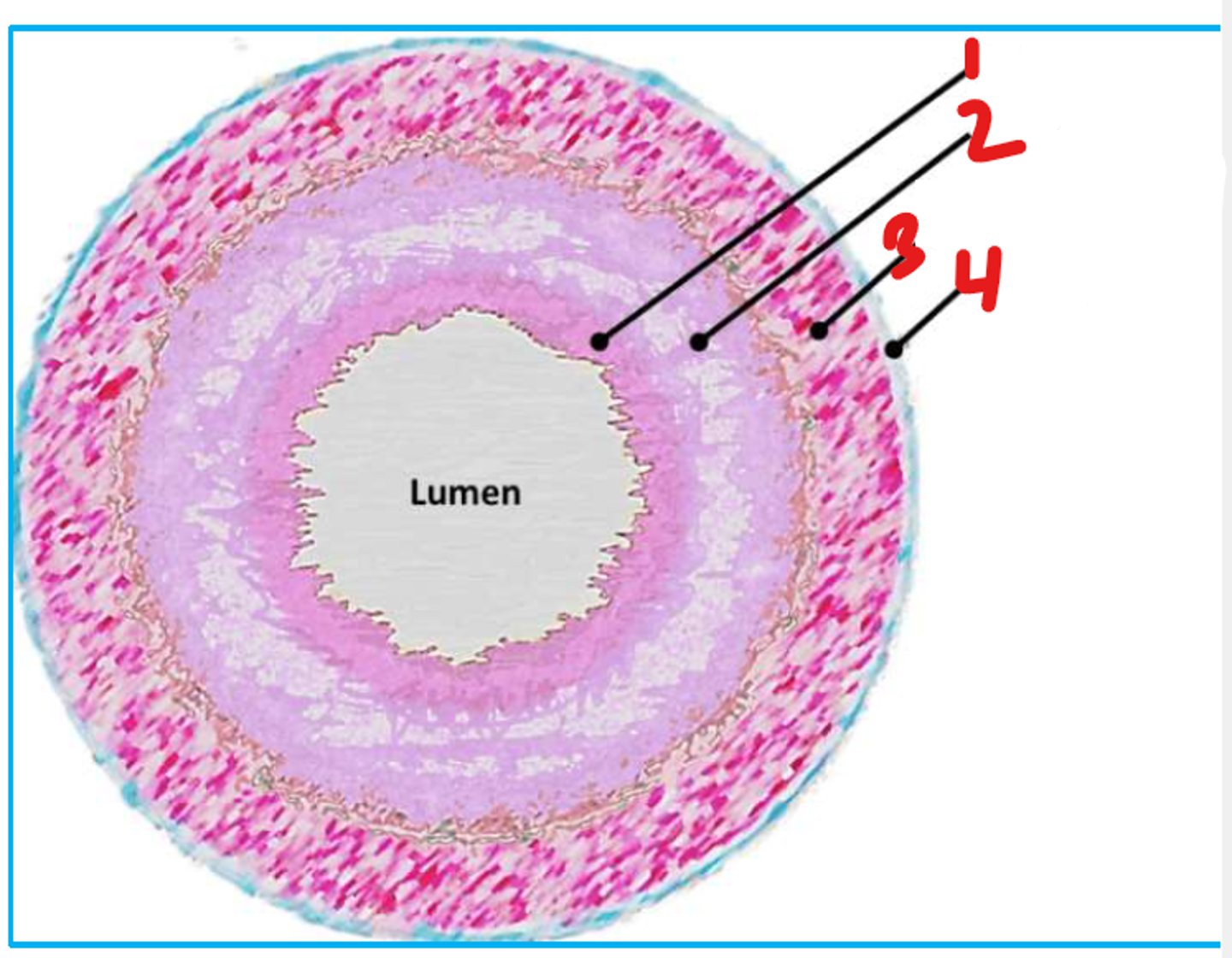
Serosa
4
Lamina Propria
-in mucosa of GI tract
-loose irregular connective tissue (CT)
- may contain immune cells (diffuse lymphoid tissue (MALT))
Non-keratinized Stratified Squamous
or
Simple Columnar
type of epithelium in the mucosa of the GI tract
Submucosa
• loose CT (collagen, adipose)
• contains nerves, lymphatics, vasculature, glands
- Meissner's plexus
Muscularis Propria (Muscularis Externa)
• typically 2 layers of smooth muscle (inner-circular; outer-longitudinal)
• peristalsis
• inner layer enlarged to form sphincters
• contains myenteric (Auerbach's) plexus
Adventitia
-is loose CT that blends with surrounding structures
- exs. retroperitoneal organs, esophagus
• contains large nerves, lymphatics, vasculature
Serosa
• contains large nerves, lymphatics, vasculature
-is loose CT with simple squamous epithelium (mesothelium)
- ex. intraperitoneal organ
Mucsoa
1
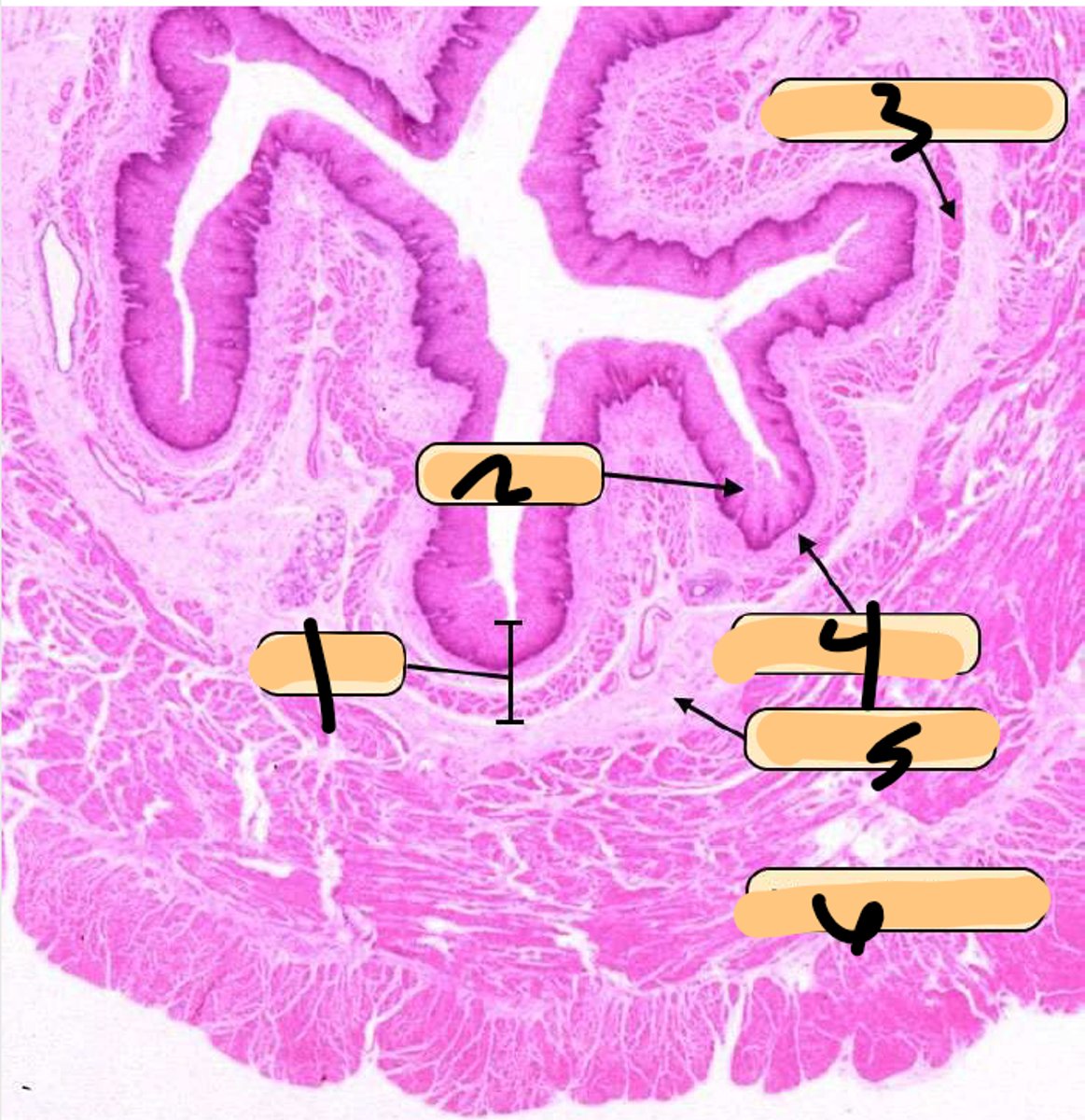
Epithelium
2
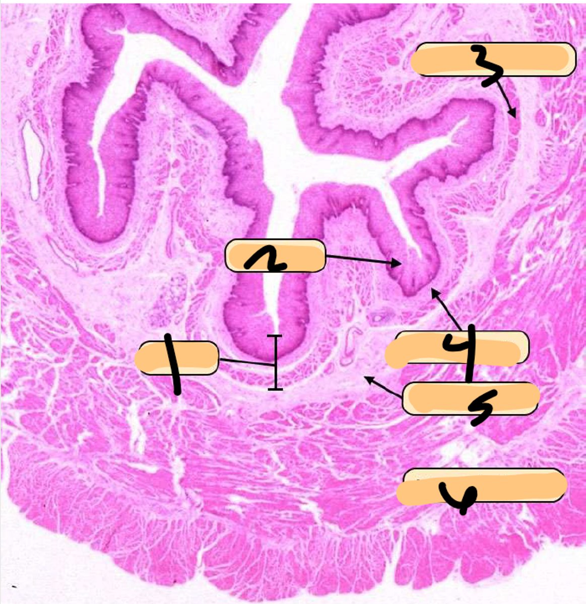
Muscularis Mucosa
3
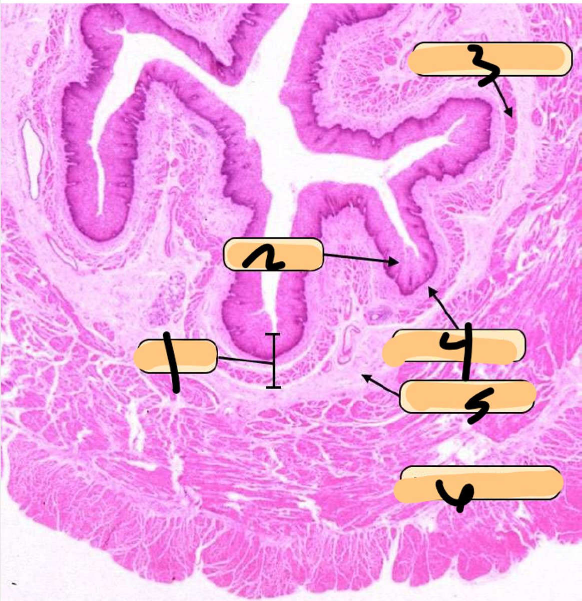
Lamina Propria
4
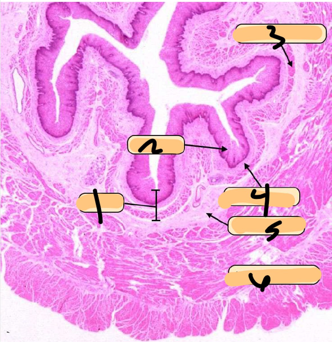
Submucosa
5
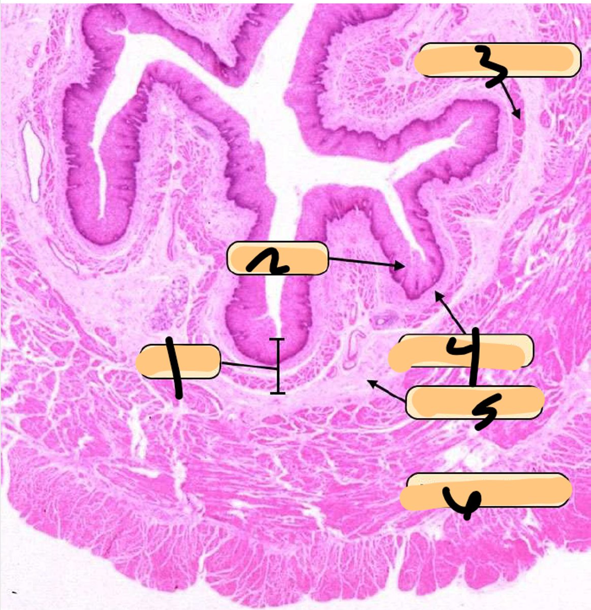
Muscularis Propria (Externa)
6
Submucosa (Meissner) Plexus
Tiny parasympathetic ganglia PG are scattered throughout the submucosa, forming the _________________________ from which postganglionic fibers supply the muscularis
Myenteric (Auerbach) Plexus
Between the layers of MUSCULARIS EXTERNA (MP), there are clumps of pale-stained parasympathetic ganglion cells of the ___________________________
Non-Keratinized Stratified Squamous
epithelium of the esophagus
-striated (proximal 1/3)
-smooth (distal 1/3)
-mixed (middle 1/3)
Types of muscle in the Muscularis Propria (Externa) of the esophagus?
Cardia
-region of the stomach
-narrow
-mucus production primarily
Fundus and Body
-region of the stomach
-releases acid to form gastric juice
Pylorus
-region of the stomach
-mucus production primarily
Simple Columnar
type of epithelium of the stomach
Gastric Pits
invaginations of the stomach that open into the lumen
Surface Mucous Cells
-line the lumen and gastric pits of the stomach
-hihgly viscous secretion
-protects against abrasion form food and self corrosion from stomach acid
Stem Cells
-located in the isthmus between gastric puts and gastric glands
Mucous Neck Cells
-located in the isthmus/neck of stomach
-apical granules and round nuclei
-acidic mucin
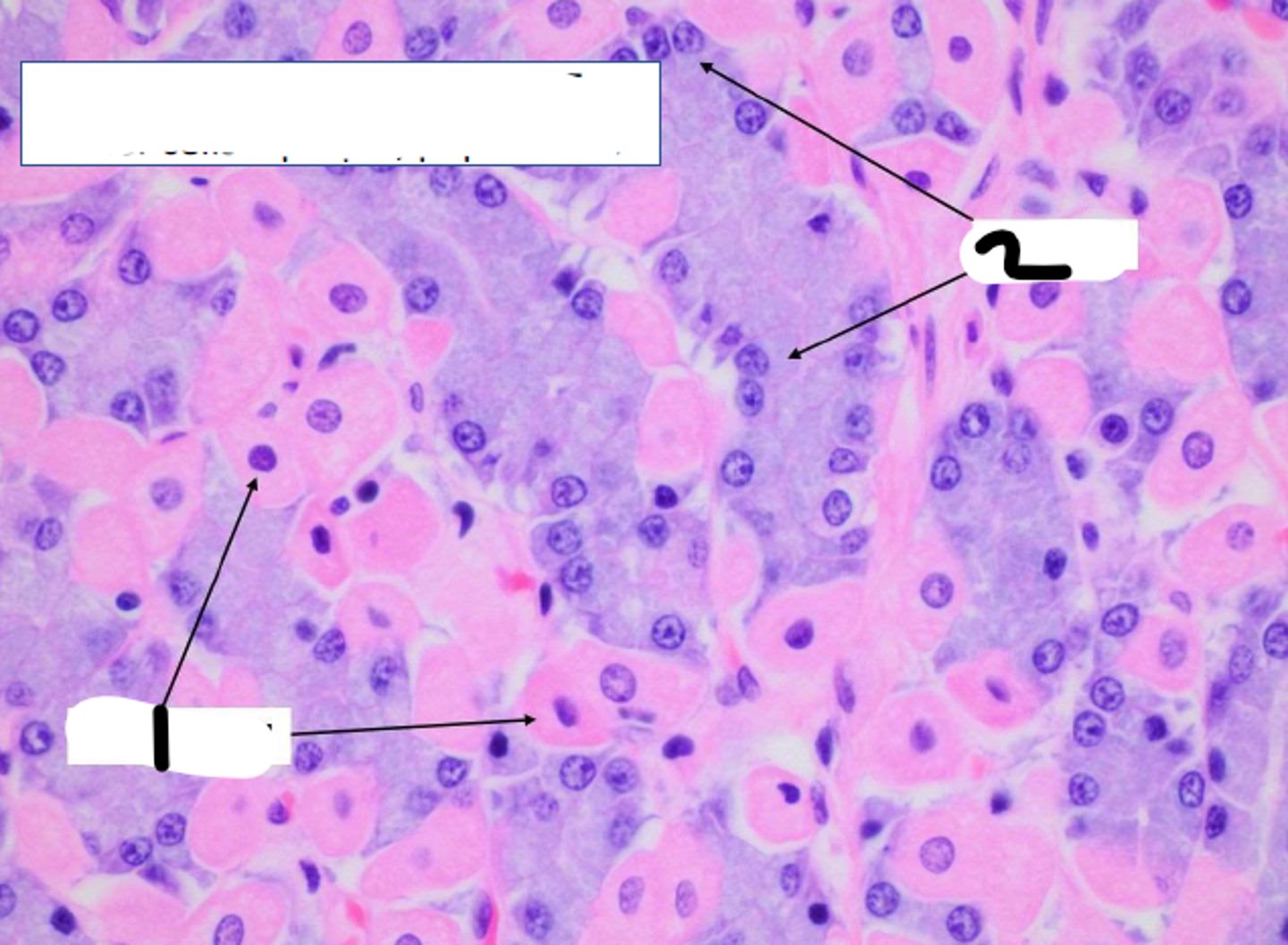
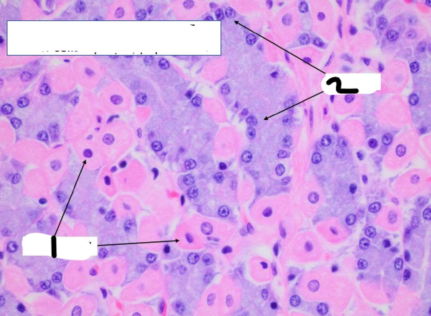
Parietal Cells
-large, often pyramidal-shaped
-located deep within the gland
-eosinophilic
-invagination of the PM on apical side that forms an intracellular canaliculus with many microvilli that produce HCl and secretes intrinsic factor
Chief (Zymogenic) Cells
-located deep within the gastric gland
-secrete pepsinogen and gastric lipase
Enteroendocrine (G) Cells
-scattered in stomach
-secrete hormones: 5-HT, histamine, gastrin, somatostatin, etc.
Gastric Pit
Gastric Gland:
1
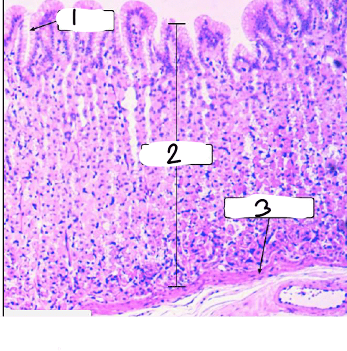
Gastric Glands
2
Muscularis Mucosa
Gastric Gland:
3
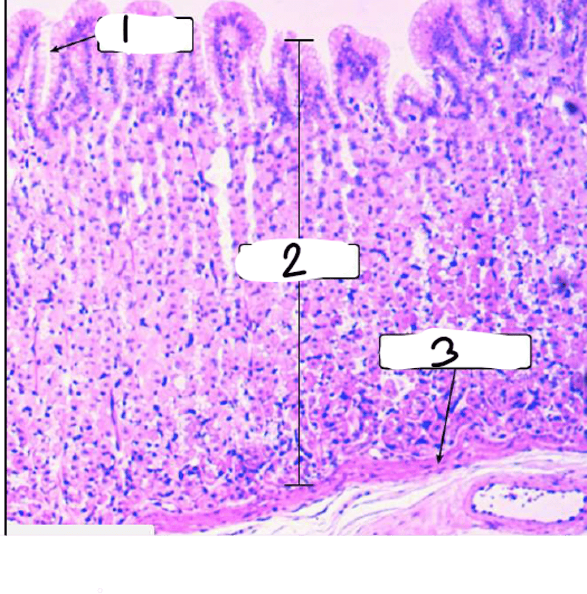
Submucosa
connective tissue containing large blood and lymphatic vessels and many lymphoid and mast cells
Muscularis Propria (Externa)
3 poorly defined smooth muscle layers - oblique (inner), circular (middle), longitudinal (outer)
Pyloric Sphincter
the middle layer fo the Muscularis Propria (Externa) thickens greatly to form the ___________________
Gastric Pits
2
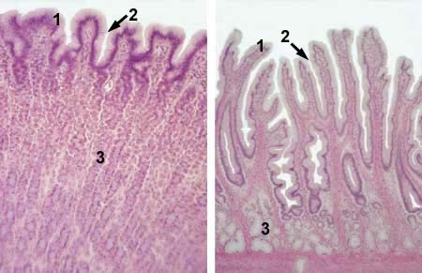
Gastric Surface Mucous Cells
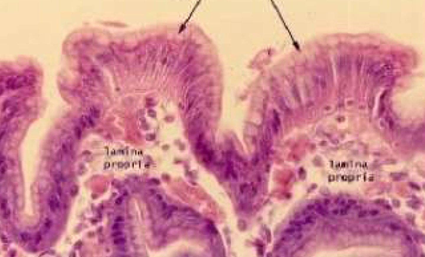
Parietal Cells
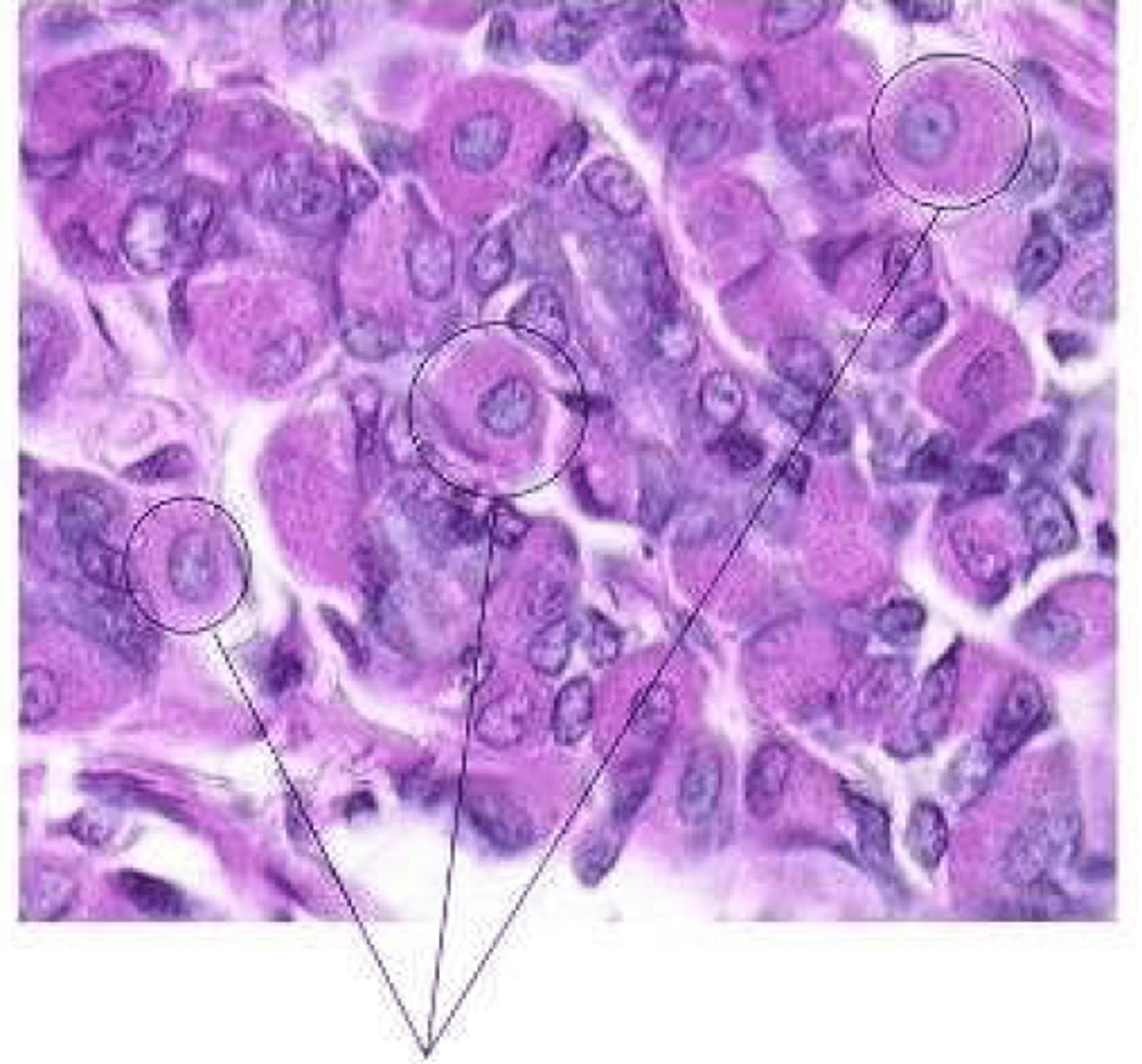
Parietal Cells
1
Chief (Zymogenic) Cells
2
Surface Mucous Cells
1
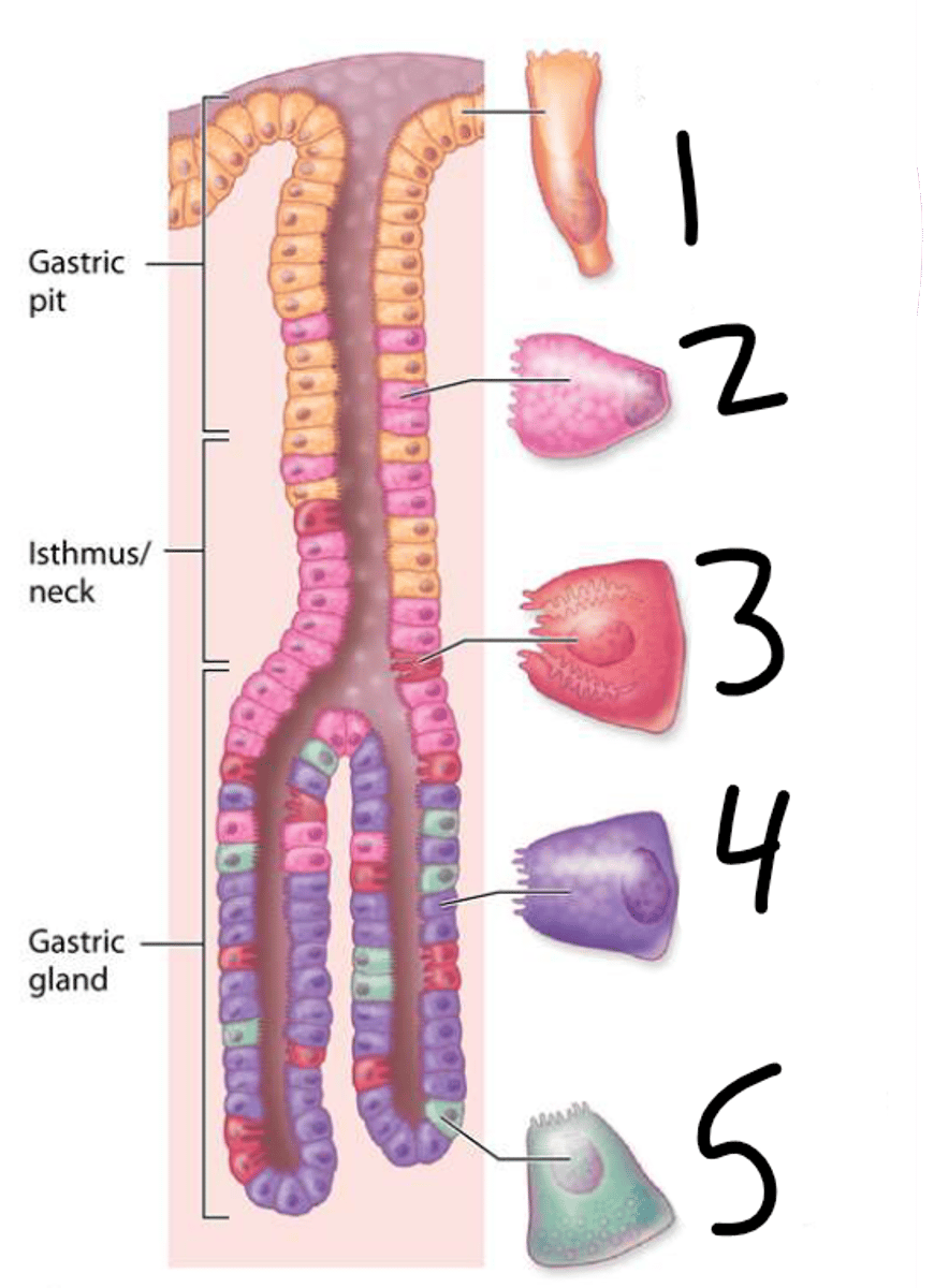
Mucous Neck Cells
2
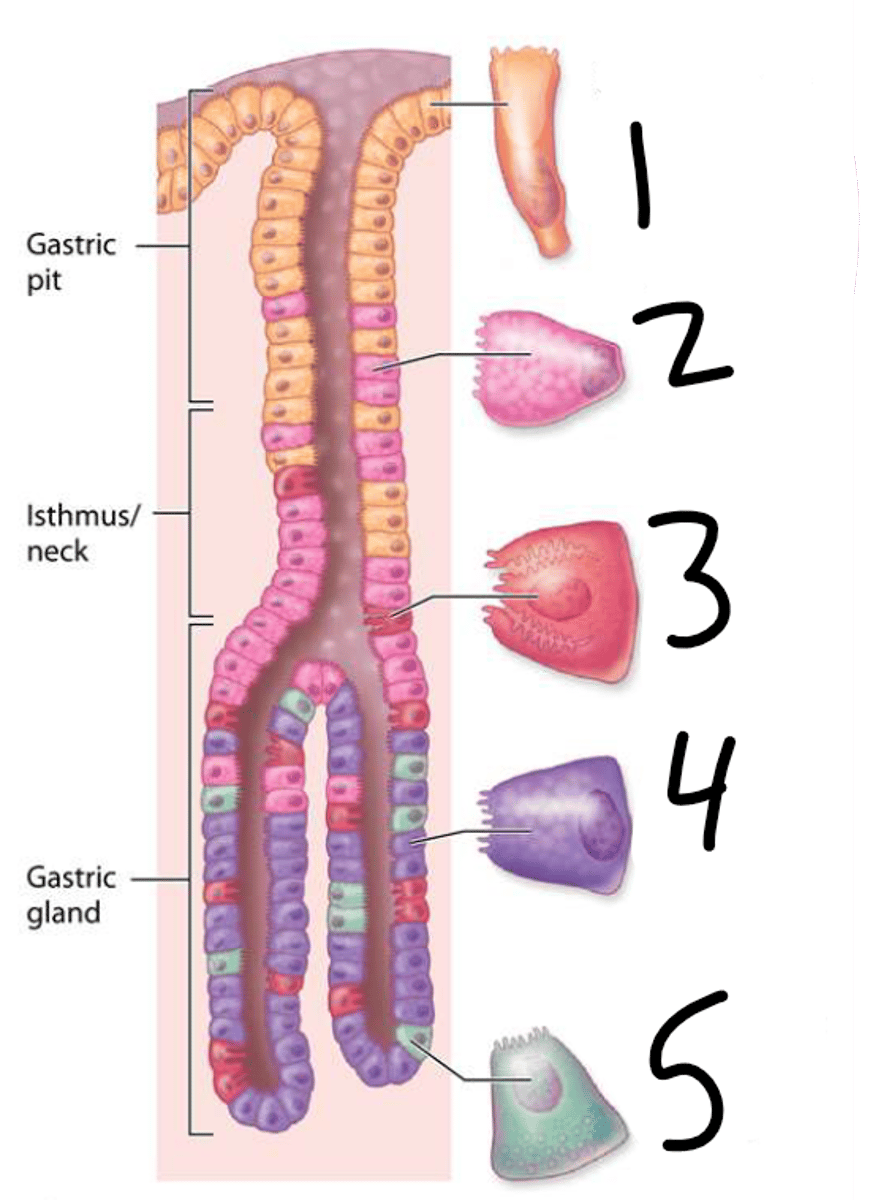
Parietal Cells
3
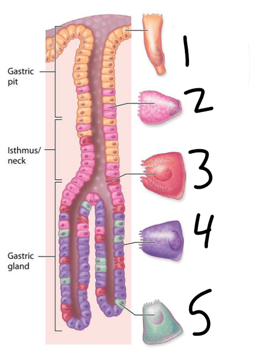
Chief (Zymogenic) Cells
4
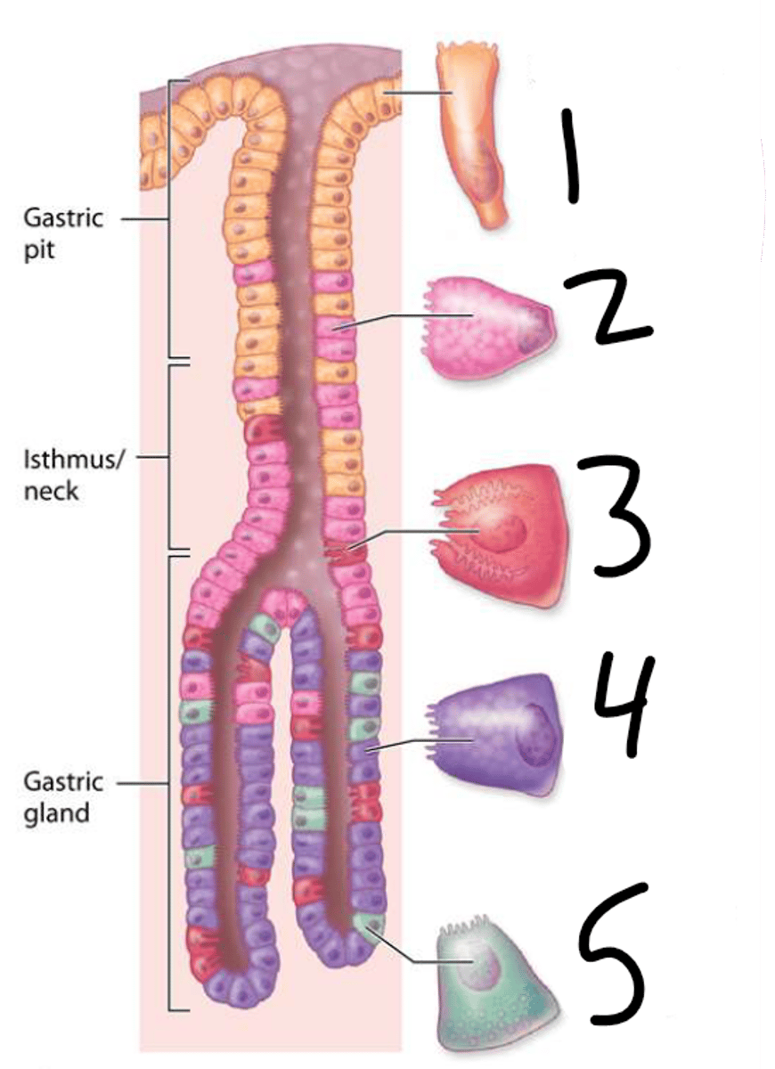
Enteroendocrine (G) Cells
5
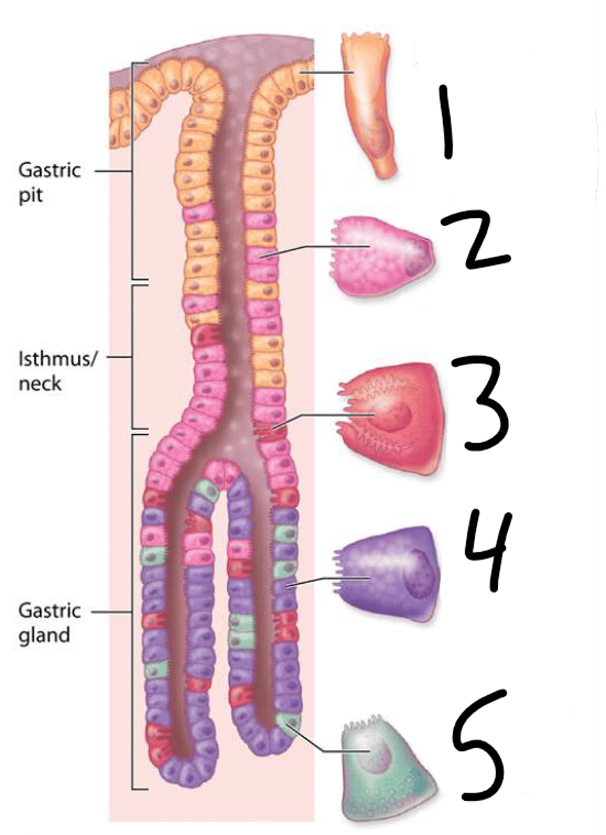
Enteroendocrine (G) Cells
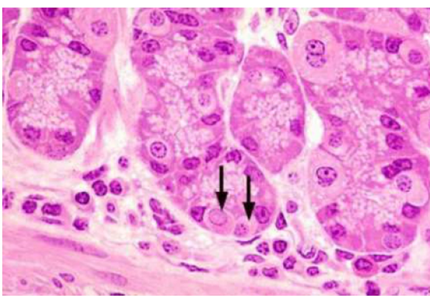
Enteroendocrine (G) Cells
small pyramidal cells with clear cytoplasm found interspersed among chief cells
Cardiac Gland
Which type of Gastric Gland:
-primarily secrete mucus
-fewer in number and more shallow in the stomach wall
Fundic Gland
Which type of Gastric Gland:
-simple, straight, with two or more opening into a single duct
-mainly secrete HCl
-release intrinsic factor
Pyloric Glands
Which type of Gastric Gland:
-secrete gastrin produced by G cells
Duodenum
-receives acidic chyme form the sotmach so it must neutralize gastric acid and pepsin as well as carry out further digestion
Jejunum
-the longest section of SI
-functions in digestion and absorption
Ileum
-last section of small intestines
-functions in absorption
Plicae Circularis
-circular folds
- most prominent in jejunum
- formed of mucosa and submucosa
- permanent (unlike stomach rugae)
Small Intestines
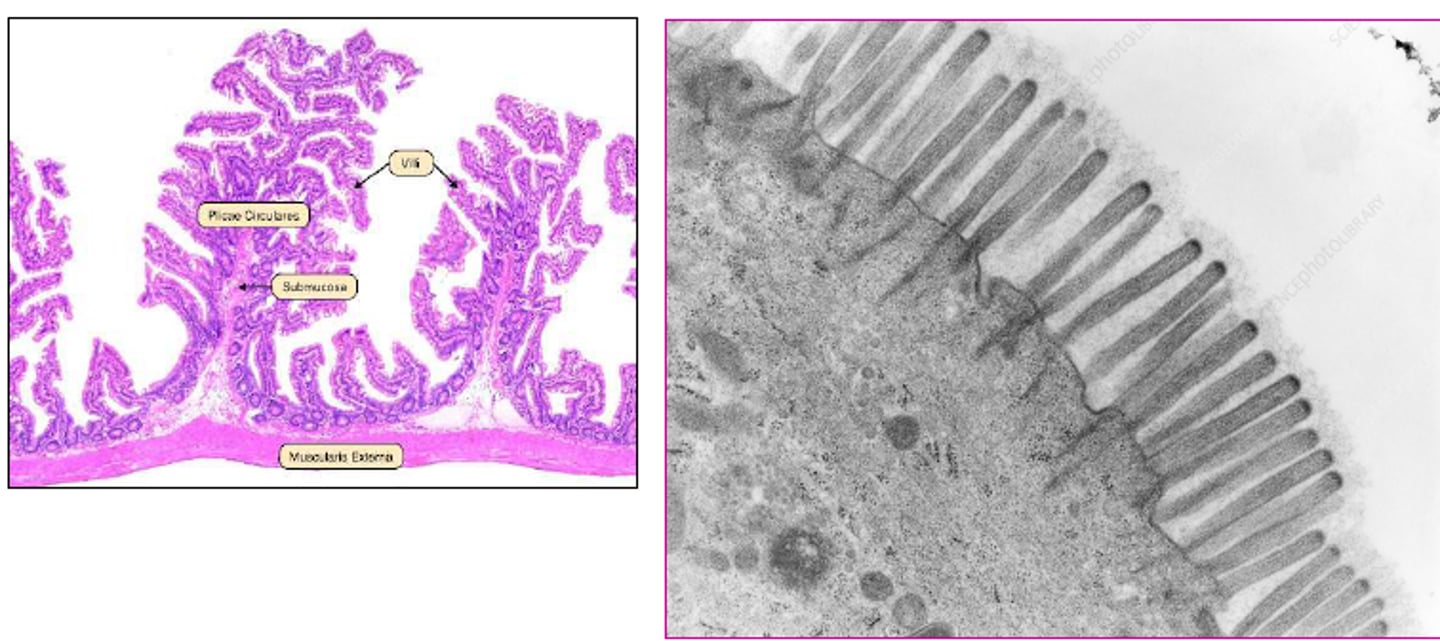
Brush Border
small intestines
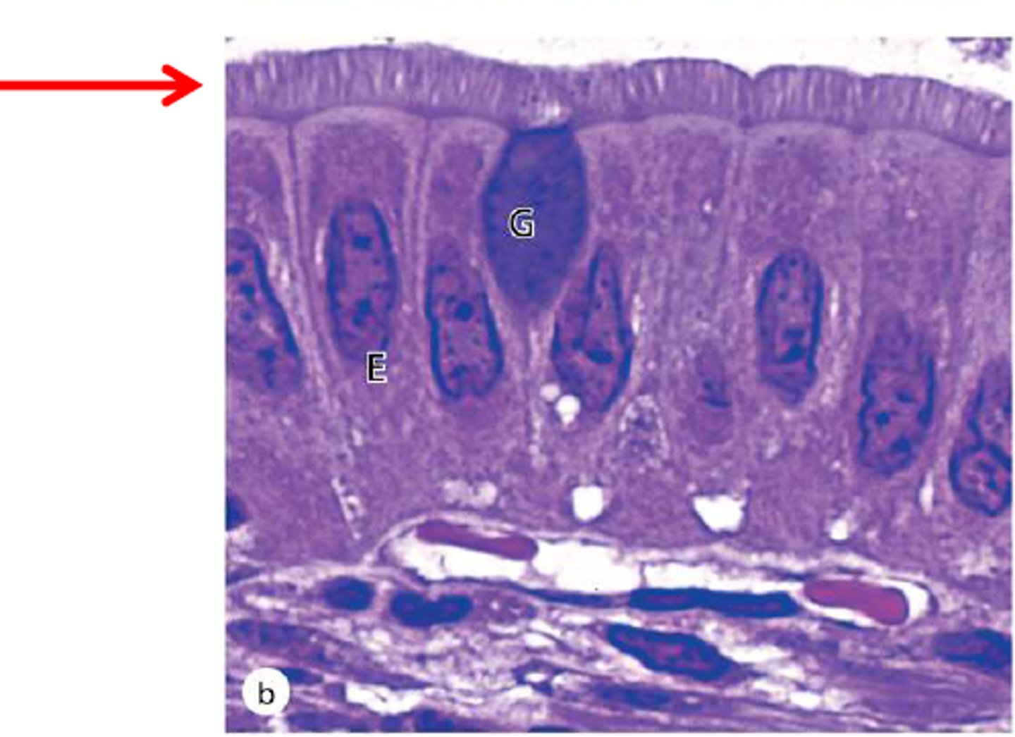
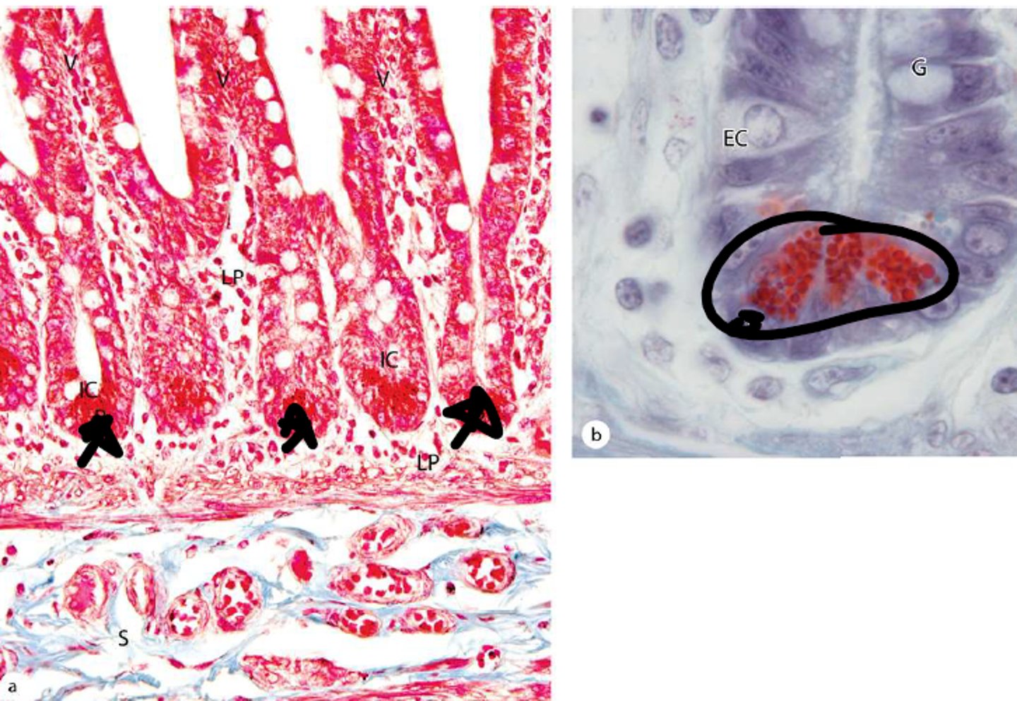
Intestinal Crypts
-formed by epithelia of the villi that extend down into the lamina propria
-stem and paneth cells located here
Enterocytes
simple columnar; absorptive cells
Goblet
secrete mucins; lubricate (protection)
Stem Cells
mitotic; replenish epithelial cells further up the villi (right image)
Paneth Cells
eosinophilic; secrete enzymes and defensins to protect against microbes; provide innate immunity
Brunner's Glands
-in submucosa
-mucigen filled clear cytoplasm and basally located nucleus
-secretes a thin, alkaline mucous to neutralize chyme
-produces lysozyme and epitdermal growth factor
Peyer's Patches
- location: mucosa, submucosa
- abundant
- lymphoid nodules
Peyer's Patches
ileum
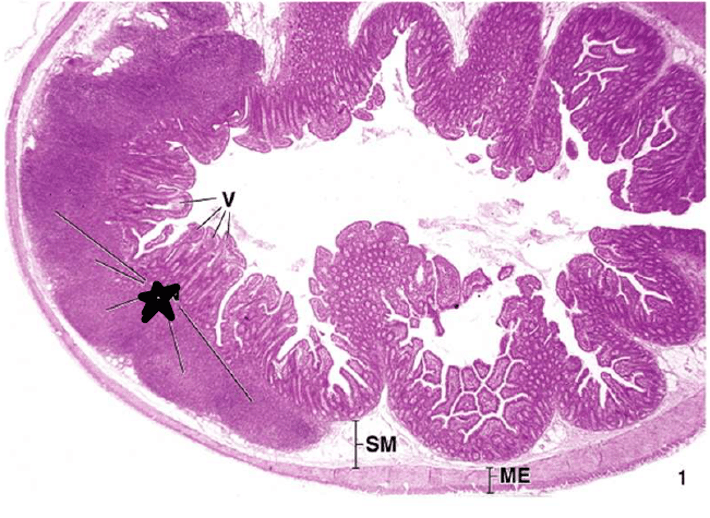
Dudoenum
Brunners glands are only present where?
Duodenum
Villous length is longest in the ____________________
Ileum
Villous length is shortest in the __________________
Plica Circulares
- absent in proximal duodenum and distal ileum
- pronounced in jejunum and proximal ileum
Ileum
Lymphoid tissue is more prominent where?
Ileum
there are more goblet cells in the _____________
Lymphatic Nidule
1
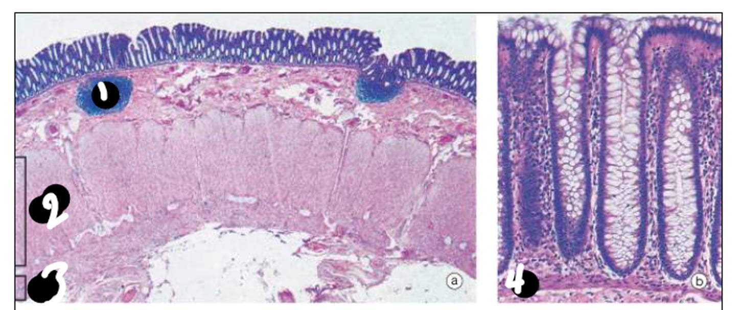
Circular Layer of the Muscularis Externa
2
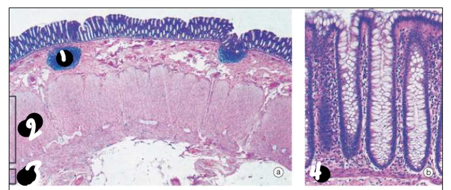
Longitudinal Layer of the Muscularis Externa
3
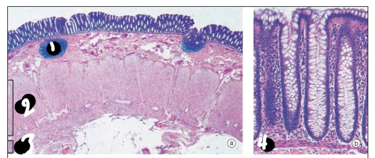
Muscularis Mucosa
4
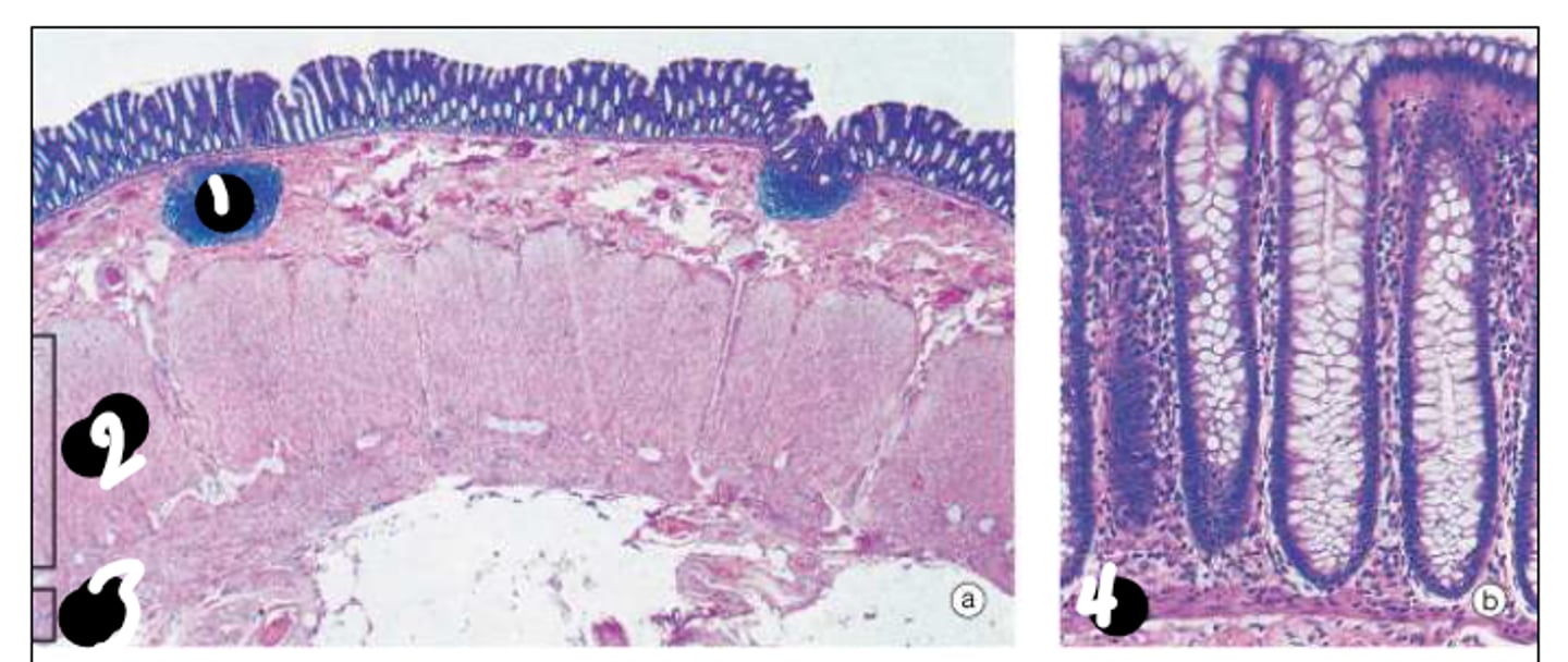
Stratified Squamous
type of epithelium in the anus
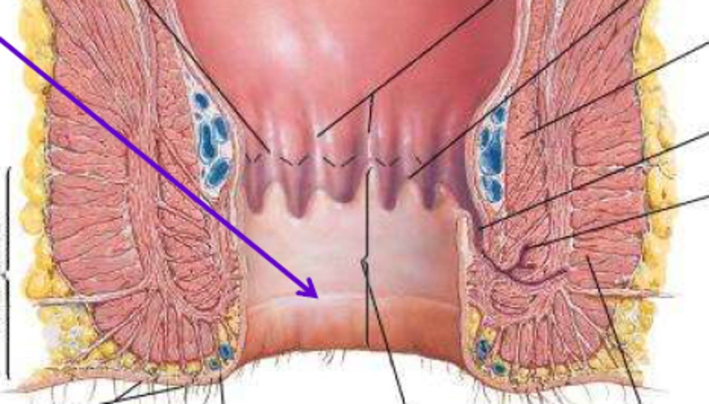
White Line of Hilton
is situated between the external and internal anal sphincters.
Vermiform Appendix
Features:
• cell types: enterocyte, goblet, enteroendocrine, stem cells, Paneth cells
• nodular lymphoids in lamina propria and submucosa; lymphoid follicles
Gastroesophageal Junction
regional change from stratified squamous to gastric secretory
Gastroduodenal Junction
regional change from gastric secretory to intestinal absorptive/villous
Ileocecal Junction
regional change from intestinal absorptive/villous to colorectal absorptive/protective
Rectoanal Junction
regional change to colorectal absorptive/protective to stratified squamous
En progreso (7)
Comenzaste a estudiar estos términos. ¡Sigue así!