Unit 5: Body Systems
 Call Kai
 Learn
Learn Practice Test
 Spaced Repetition
 Match
Match Flashcards
 Knowt Play
1/44

Earn XP
Description and Tags
+ Chemical Signalling // ERQ Practices: Pathways – Muscle contraction, Cell signalling, Depolarisation, Respiration, Photosynthesis
Name | Mastery | Learn | Test | Matching | Spaced | Call with Kai | Chat |
|---|
No analytics yet
Send a link to your students to track their progress
45 Terms
Receptors as Proteins with Binding Sites for Specific Signalling Chemicals
Ligand = Signalling chemical; Bind reversibly to specific receptors on / in target cells → Specific cellular response via signal transduction pathway
Binding of ligand to receptor
Conformational change in receptor protein
Activation of downstream signalling molecules
Internalisation of receptor-ligand complex
Cell Signaling by Bacteria in Quorum Sensing
Example of bioluminescence in marine bacterium Vibrio fischeri
When cells aggregate, inducer accumulates in confined environment → Concentrations of autoinducers increase → Luminescent system of ALL bacteria is activated → Emit light through bioluminescence
V. fischeri form symbiotic relationship with Hawaiian bobtail squid
When squid hatches, capture V. fischeri from environment and house in specialised light organ
Bacteria produce autoinducers which accumulate to a certain point
Once threshold is reached, autoinducers enter cell and bind to LuxR protein → Activates lux operon → Production of luminescent protein (luciferase) → Emission of light
Light allows squid to match surrounding moonlight / Camouflage itself from predators by illuminating underside
Squid resets bacterial population by expelling 90% every morning; Remaining bacteria multiply / reach the quorum at night to produce light → Prevents overgrowth + Reduce risk to immune system
Functional Categories of Signalling Chemicals in Animals & Differences (HINT: 4 types)
Hormones
Secreted by endocrine glans
Travel through bloodstream to reach target cells across body
Attaches only to specific receptor; Only specific cells respond
Takes time for secretion
Prolonged effect
Neurotransmitters
Released from one cell; When an electrical impulse reaches presynaptic nerve
Affect nearby target cell
Occurs at the synapse
Travels across the synaptic cleft to reach receptors on postsynaptic membrane
Results in an electrical impulse at the postsynaptic nerve cell
Cytokines
Glycoproteins
Functions include cell to cell messenger, enhance cell function, inflammation, cell proliferation, embryo development
Binds to cell membrane receptor; Triggers signals inside cells to activate proteins in cytoplasm → Sets of cascade reaction
Calcium Ions
Signalling molecules / second messengers
Participate in signal transduction pathways
Regulate cellular functions including nerve transmission, muscle contraction, hormone secretion, enzyme activity, and cell division
Released from intracellular stores
Enter cells via ion channels
Chemical Diversity of Hormones and Neurotransmitters
Reasons for wide range of chemical substances as signalling chemicals
Diverse functions (Distance and duration)
Ability to bind to differently-located receptors / Interact with chemically-specific receptors
Different modes of action on target cells
Target various cells efficiently
Hormones = Amines, proteins, steroids chemical groups; For long-distance
Amines, proteins are hydrophilic → Cannot cross cell membrane; Must bind to receptors on surface
Steroids are hydrophobic → Easily cross cell membrane; Bind to intracellular receptors
Neurotransmitters = Amino acids, peptides, amines, nitrous oxide; For localised (across synaptic gap, ~20 mm)
Most are hydrophilic → Extracellular receptors
Localised and Distant Effects of Signalling Molecules (& Contrasts)
Contrast between hormones transported by blood system and neurotransmitters that diffuse across synaptic gap
Neurotransmitters = Across synapses; Affect neighbouring cells
Oxytocin = E.g. Oxytocin affects reproductive system, social behaviour, stress response, etc.
Transmembrane Receptors in Plasma Membrane & Intracellular Receptors in Cytoplasm / Nucleus (& Difference)
Distribution of hydrophilic or hydrophobic amino acids in receptor
TRANSMEMBRANE RECEPTORS:
Consist cytoplasmic and extracellular domains (hydrophobic amino acids ~ IN cell / hydrophilic amino acids ~ OUTSIDE cell)
For signal transduction
Converts external signal into internal action; Ligand binding → Undergo conformational change → Initiate cascade → Activation of enzymes / Changes in gene expression / Change in cellular metabolism
Types include…
Chemically gated ion channel = Multi-pass protein w/ central pore → Possess gates
Enzymatic = Single-pass protein → Binds ligands extracellularly + Activates enzymes intracellularly
G protein coupled receptors (GPCRs) = Multi-pass protein w/ intracellular binding site for G-protein → Bind with ligands → Activate G protein
INTRACELLULAR RECEPTORS:
Have hydrophobic regions to interact with lipid-soluble ligands
Act as transcription factors → Affect gene expression → Long-term change in cell’s function (E.g. growth, differentiation, metabolic regulation, etc.)
Whether signalling chemical penetrates the cell / remains outside based on size and polarity
Hydrophilic signalling chemicals bind to transmembrane receptors
Hydrophobic signalling chemicals diffuse through membrane + bind to intracellular receptors
Initiation of Signal Transduction Pathway
Binding of ligand to receptor sets off a sequence of responses within the cell
Reception
Target cell detects ligand which binds to transmembrane receptor
Transduction
Signal is converted into new form that allows for response
Response
Transduced signal triggers cellular activity e.g. enzyme activity, gene expression changes, cytoskeletal changes
Transmembrane Receptors for Neurotransmitters & Changes in Membrane Potential
Ligand binding causes ion channel to open → Positively-charged ions (Na+, K+) diffuse into cell → Generate action potential
Acetylcholine receptor = Found at neuromuscular junctions & neuron-neuron synapses
Chemically gated ion channel; Opens only when acetylcholine binds to it → Diffusion of ions (Na+ and Ca2+ into cell and K+ exit cell, along concentration gradient)
Results in depolarisation of membrane → Muscle contraction → Repolarization & return to resting potential
Transmembrane Receptors that Activate G-Proteins
How GPCRs convey signal into cells
Ligand binds
Conformational change in receptor
Activates associated G-protein
Exchange of GDP (bound to G-protein) for GTP (guanosine di- / triphosphate)
α subunit + GTP disassociates from the β and γ subunit of G protein
Triggers cellular response; Interacts with other proteins / enzymes in cell; Transmit signal from activated receptor to downstream effector molecules
—
GTP is hydrolysed to GDP and inorganic phosphate group / Ligand unattached from receptor
α subunit + GTP comes back together with receptor & β and γ σ subunits
Many GPCRs are in humans
Mechanisms of Action of Epinephrine Receptors
Roles of G protein and cAMP as second messenger
Epinephrine receptors = GPCRs; Use cAMP to relay signal inside cell → Fight-or-flight response
Increased heart and breathing rate, dilation of airways, breakdown of glycogen to glucose, increased oxygen and glucose delivery to muscle cells, vasoconstriction of blood vessels
After G-protein activation…
α-subunit + GTP binds with adenylate cyclase
Converts ATP to cAMP
Activates several protein kinase A (PKA)
PKA phosphorylates enzymes and proteins e.g. phosphorylase in liver to break glycogen into glucose
Inactivation occurs
Adrenaline terminates to prevent overstimulation + return to resting state in preparation for new signals and responses by…
Degradation of cAMP
Deactivation of G proteins
Receptor endocytosis / desensitisation
Transmembrane Receptors with Tyrosine Kinase Activity
Kinase = Proteins that phosphorylates other substances using ATP
RTKs = Consist of three domains, single-pass protein; Composed of two α subunits (extracellular) and two β subunits (transmembrane) Occur in pairs
Can activate MULTIPLE signal transduction pathways simultaneously
Initial signal becomes amplified
Insulin binds to extracellular domain (α subunit)
Receptors dimerize
Autophosphorylate tyrosine residues on β subunits on intracellular domain
Activates kinase
Phosphorylation cascade occurs
—
Mobilisation of glucose transporter vesicles containing GLUT4
GLUT4 = Facilitates glucose uptake into cells
Vesicles merge with plasma membrane → Increased active glucose cotransporters → More glucose enters cell from bloodstream → Lowers blood glucose levels → Provides energy for cell; Prevent hyperglycaemia
Insulin resistance = Cells do not respond effectively to insulin → Reduced glucose intake → Development of Type II diabetes
Intracellular Receptors that Affect Gene Expression
Steroid hormones: Oestradiol, progesterone, and testosterone
Lipid soluble, steroid hormones pass through membrane and bind to intracellular receptors
Ligands bind to site on receptor → Activates receptor → Binds to specific DNA sequence → Promote gene transcription
Upon binding, hormone-receptor complex undergoes conformational change → Activation
Translocation to cell nucleus + Binds to hormone response elements (HRE)
Promote / suppress transcription of target genes
Effects of Hormones Oestradiol and Progesterone on Target Cells
For estradiol cells: Cells in hypothalamus secrete GnRH
Oestradiol binds to oestrogen receptors within cells in hypothalamus → Secretes GnRH
Stimulates release of LH & FSH from pituitary gland
Stimulate ovaries to produce eggs + Production of estradiol from ovaries
For progesterone cells: Cells in endometrium
Progesterone binds to progesterone receptors in endometrium cells
Promotes gene transcription (nutrient storage and angiogenesis)
Prepares endometrium for potential pregnancy (Induces thickening + Formation of additional blood vessels within)
During pregnancy, maintains integrity and functionality of endometrium; Otherwise, secretion decreases, uterus lining sheds
Regulation of Cell Signalling Pathways by Positive and Negative Feedback
To regulate hormone levels + Maintain homeostasis
Difference between two forms of regulation + Brief outline of an example each
Positive feedback = Amplifies response; E.g. Release of oxytocin during childbirth
Baby moves across cervix, activating stretch receptors in uterus → Brain signals oxytocin release → Oxytocin causes uterine muscle to contract → Contractions strengthen until baby is delivered
Negative feedback = Diminishes response; E.g. Insulin secretion
During high blood glucose levels, β cells are stimulated to secrete insulin → Prompts cells to absorb glucose / Convert glucose into glycogen or fat in liver → Reduce blood sugar → Prevent hyperglycaemia
Adaptations for Movement
Universal feature of living organisms; Range of organisms (Motile and sessile)
Motile = Organisms that have adaptations allowing movement within habitat, e.g. Three-toed sloth
Three long toes → Hanging; Movement on ground is almost impossible
Sessile = Cannot move from place to place but can alter body in response to environmental stimuli e.g. Venus flytrap
Pair of leaves with short and sturdy trigger hairs; When triggered, leaves close + Enzyme secretion → Trap and digest insects
(When one hair is triggered, ~20 seconds wait for a second hair to be triggered as confirmation for “bug”)
Sliding Filament Theory
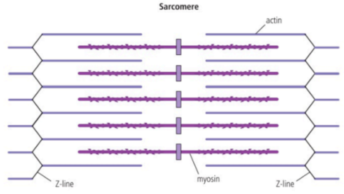
Sarcomere contracts by sliding of actin and myosin filaments
Muscle → Bundle of muscle fibres → Single muscle fibre → Myofibrils → Sarcomere
Myofibrils = Composed of repeating sarcomeres units; Many of these make up one muscle fiber
Sarcomeres = Long fibrous proteins that are attached to each other end to end; Synchronised movement (One moves, all move) → Shortens muscle fibre / entire muscle
Striations of skeletal muscle = Alternating fibers of myosin and actin
“I band” = Thin actin filaments = Light
“A band” = Thick myosin filaments + actin filaments on outer edge = Dark
Actin = Round proteins linked in chain to form filament
Myosin = Long thin protein + Movable head; Linked together in bundles to form filament (Multiple heads)
In sarcomere, myosin remains stationary while the actin on both sides move towards the centre using movable heads on myosin
When sarcomeres contract, the actin filaments slide over the myosin fibers, resulting in sarcomere shortening
Due to overlap, muscle appears lighter when contract
Describe the Process of Muscle Contraction (ERQ Practice) (7 marks)
Action potential in motor neuron triggers release of Ca2+ ions from sarcoplasmic reticulum
Calcium ions bind to troponin on actin and cause tropomyosin to move → Expose bindings sites for myosin heads
Actin filaments and myosin heads form a cross-bridge
ATP hydrolysis breaks cross bridge and myosin heads to change orientation into a high energy configuration
Myosin heads bind to actin filament before returning to original conformation
Repositioning of myosin head moves the actin filaments as well towards the center
Sliding of actin along myosin shortens the sarcomere, causing muscle contractions
Role of Titin
Titin = Large protein (~27,000 AA)
Helps sarcomeres to recoil after stretching (Reset to original position; During contraction, a spring-like tension is present and released when relaxed)
Hold myosin fibers in place
Prevents muscle from overstretching
Role of Antagonistic Muscles in Protein Relaxation
Needed because muscle tissue can only exert force when it contracts; If one bone is moved by one muscle, another muscle is required for the opposite movement
Contracting = Agonist
Relaxing = Antagonist
Structure and Function of Motor Units in Skeletal Muscle
Motor Unit = A motor neuron are joined to a set of muscle fibres; Strength of contraction ~ Nerve impulse to # of motor units (Motor neuron to muscle fiber ratios range from 1:10 to 1:200)
Consists of…
Motor neuron = On one end is the cell body at brain stem or spinal cord; Motor end plate (nerve ending) is connected to muscle cell (at neuromuscular junction)
Connect to effectors; Carry response to stimulus
Controls set of muscle fibers
Neuromuscular junctions = Type of synapse particularly at muscle tissue to simulate contraction
Muscle fibres
Skeletal Framework
Skeletons as anchorage for muscles and as levers
In body…
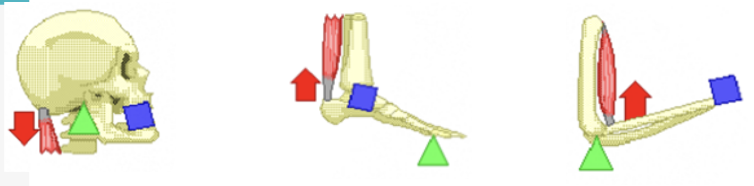
Bone = Lever / Joint = Fulcrum / Muscle contraction provides effort force on bone → Moves weight of body part (Load)
First-class lever = Contraction of neck muscle pulls on skull → Face rise
Second-class lever = Contraction of calf muscle causes ball of foot to pivot → Foot rise
Third-class lever = Contraction of bicep muscle acts on elbow joint → Forearm rises to lift object
Arthropods have exoskeletons; Vertebrates have endoskeletons
Synovial Joint + Example
Synovial joint = Where two bones need to move against each other; Allows wide range of motions
Ball and socket joints (Shoulder and hip) = Can move bones along several axes
Abduction (Out) and adduction (In)
Flexion (Backward; bending) and extension (Forward; straightening)
Rotation
Hinge joints (Ankle, elbow, knee) = Can only move along one axis
Flexion and extension
Roles of… (in Hip Joint)
Bone = Constituents of joint (E.g. Pelvis and femur in ball-and-socket hip joint)
Head of femur forms a “ball” that fits within the socket of the pelvis
Cartilage = Smooth protective connective tissue; Lines both pelvis and femur at hip joint to avoid bone on bone contact
Synovial fluid = Lubricating fluid; Reduces friction in hip joint
Ligaments = Tough connective tissue; Holds bones of hip joint in place while allowing range of motion
Muscles = Tissues that contract and relax; Controls movement of hip joint
Tendons = Connective tissue; Connects muscle (of hip joint) to bone
Range of Motion ~ Goniometer
Range of motion = Distance and direction joint can move; Measured in degrees
Used to document change in joint movement after surgery, etc.
Example of Antagonistic Muscles
Antagonistic muscle action → Facilitates internal body movements
Different orientations of muscle fibers in the internal and external layers of intercostal muscles mean that they move the ribcage in opposite directions; When one of the layers contract, it stretches the other, storing potential energy in the sarcomere protein titin.
External intercostal muscles contract → Pulls rib cage up and out → Inspiration (Thoracic cavity increase)
Internal intercostal muscles contract → Pulls rib cage down and in → Expiration (Thoracic cavity decrease)
Contraction of one stretches the other → Stores PE in titin of other muscle fibers
Reasons for Locomotion + Examples (FESM)
Foraging for food
E.g. Honey bees; Fly to collect nectar and pollenEscaping from danger
E.g. Flying fish; Extend pectoral fins → Glide in water + Swim fast → Evade predatorSearching for a mate
E.g. Loggerhead sea turtle; M & F swim back to “birth beach” to lay eggsMigration
E.g. Arctic tern; From Arctic breeding grounds → Antarctic region & back for food
Adaptations for Swimming in Marine Animals
Streamlining → Allow dolphin to move through viscous water easily & fast
Adaptions of limbs to form flippers → Steering
Adaptation of tail to form a fluke → Up and down motion for propulsion
Changes to airways into blowhole at the dorsal surface of head → Allow periodic breathing between dives + Avoiding body leaving the water
Pathogen
Broad range of organisms that cause infectious diseases; Typically reserved for:
Viruses (HIV, Measles, Smallpox, Influenza)
Bacteria (Salmonella, Cholera, Conjunctivitis)
Fungi (Athlete’s Foot)
Protists (Malaria - Plasmodium)
(No archaea !!)
Skin and Mucous Membranes as Primary Defense
Skin = Physical & chemical barrier to pathogens; 2 main layers
Underneath Layer
Sweat + Oil glands, Capillaries, Sensory neurones, Dermal cells
Top Layer
Mainly dead cells; New cells move up to replace the dead cells
CHEMICAL DEFENSES
Secrete antimicrobial peptides → Destroy pathogens
Sebaceous glands produce sebum → Maintains skin moisture + Lower pH → Inhibit bacterial growth
Lysozymes in sweat → Degrades bacterial cell walls
Non-harmful skin microbes inhibit pathogens by competitive exclusion
PHYSICAL DEFENSES
Continuous → Hard to find opening for pathogens to enter
Many layers + tough
Dry environment
Mucous membrane = Sticky mucus produce to trap pathogen; Produced by goblet cells
Cilia → Helps move pathogens up & out of respiratory tract
Found in trachea, nasal passages, vagina, urethra
Sealing of Cuts in Skin by Blood Clotting
Pathogens can enter via cuts, abrasions, or other breaches → Evade immune system + Establish infections
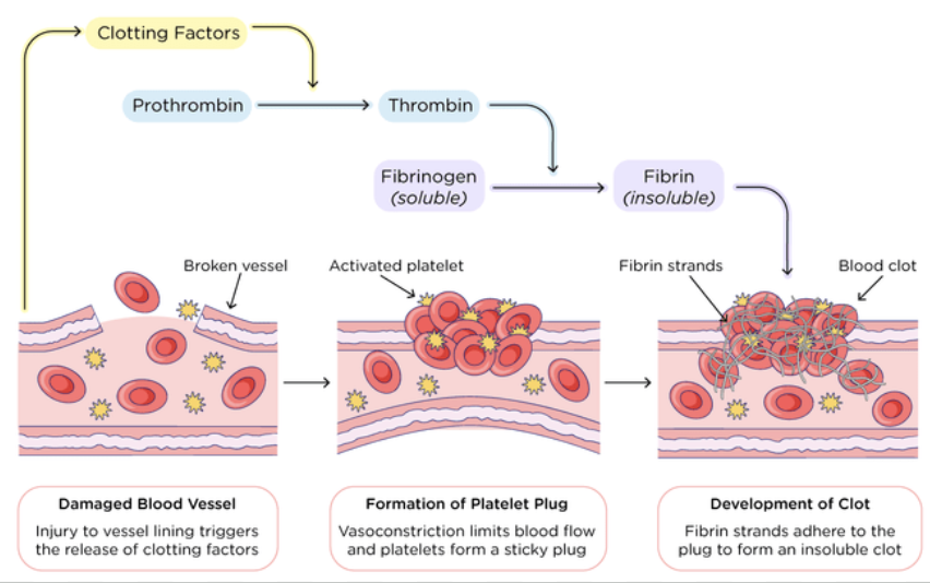
Release of clotting factors from platelets causes a cascade of reactions
⇒ Rapid conversion of fibrinogen → fibrin by thrombin
⇒ Trapping of erythrocytes to form clot
Damaged blood vessel cells release clotting factors → Stimulate platelets to stick to damaged area → Forms platelet plug + Cause prothrombin to change into active form → Thrombin
Thrombin changes soluble fibrinogen → Insoluble fibrin protein
Fibrin fibres adhere to platelet plug + Form mesh which captures blood cells and platelets → Create a clot across wound site
In presence of air, clot dried to form scab → Shields healing tissues underneath + Stop blood leaking and entry of pathogen
Note: Fibrinogen and prothrombin are present in blood plasma at all times
Innate VS Adaptive Immune System
Innate system (Up to macrophage)
Responds to broad categories of pathogens
Recognises “self” and “non-self” (e.g. pathogens, pollen, and dust) via antigens (lipoproteins)
Does not change during organism’s life; No memory
Rapid
Adaptive system (Starting from T-cells)
Responds in specific way to particular pathogen
Slower to activate
Builds up memory of pathogens encountered → Immune response becomes more effective (with age, too)
Memory cells = Type of white blood cell formed when first exposed to a pathogen; When exposed to pathogen a second time, memory cells are activated quickly
Vaccinations work by training immune system to recognise pathogens
Infection Control by Phagocytes
Amoeboid movement from blood to infection site
Phagocyte = Type of white blood cell (leukocyte) that can move with amoeboid movement, i.e.
Squeeze out of capillaries
Leave bloodstream to move to body tissues
Phagocytes recognise pathogens → Engulf them via endocytosis → Digest them using enzymes from lysosomes
Phagocytosis = Engulfing of molecules with non-self antigens
Foreign particles are placed inside vesicle + Digested by enzymes in lysosome
Lymphocytes
Lymphocytes = Cells in adaptive immune system; Cooperate to produce antibodies
Two types → B-lymphocyte (B-cell) & T-lymphocyte (T-cell)
B cells = Each individual has a large number of these cells that each make a specific type of antibody
Circulate in blood & contained in lymph nodes of the lymphatic system
Antibodies = Y-shaped proteins with a variable region at the tip of the ends acting as a binding site for a specific antigen
NOTE: Some pathogens have multiple antigens and require multiple antibodies for them to be “disabled”
Antigens
Antigens = Recognition molecules; Trigger antibody production
Most antigens are glycoproteins / other proteins usually located on the outer surface of pathogen
In the case of the surface antigens of erythrocyte…
If the blood is transfused to an incompatible receiver / different blood group, antigens may stimulate antibodies production → Clumping / Agglutination
Activation of B-lymphocytes by Helper T-lymphocytes
There are many B-cells and Helper T-cells but each are antigen specific
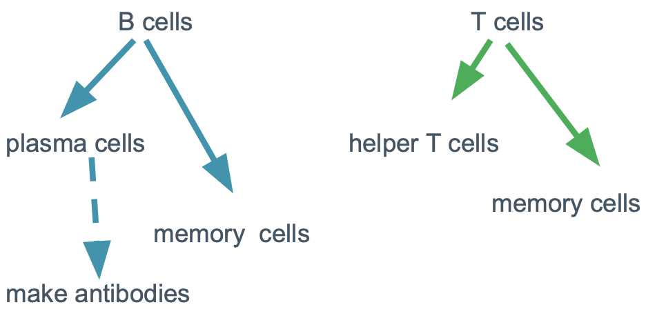
B-cells = Produce antibodies + Become memory cell ONLY when activated
Activation = Requires both…
Direct interaction with the specific antigen
Contact with a helper T-cell that has been activated by the same type of antigen
Multiplication of Activated B-cells (Plasma B-cells)
Multiplication of B-lymphocyte by mitosis → Form clones of antibody-secreting plasma cells
Relatively small number of B-cells respond to a specific antigen
Therefore, to produce enough antibodies, activated B-cells first rapidly divide by mitosis to produce lots of plasma B-cells
Plasma B-cells = Capable of producing same type of antibody; Have…
Large amount of cytoplasm + rough ER, ribosomes, and Golgi → Produce large amounts of antibody proteins
Immune Response (FULL)
Macrophage engulfs pathogen and presents its antigen
Helper T-cell imprints pathogen antigen(s)
~~~~~~~~~~~~
B-cell has a random antibody that binds to the pathogen antigen BY CHANCE
B-cell also engulfs pathogen and presents its antigen on the surface
~~~~~~~~~~~~
Helper T-cell with imprint binds to the B-cell with antigen in order to activate it
B-cell differentiates into plasma B-cells and divide
Plasma B-cell produces lots of antibodies to bind to pathogen and neutralise it by marking them for destruction
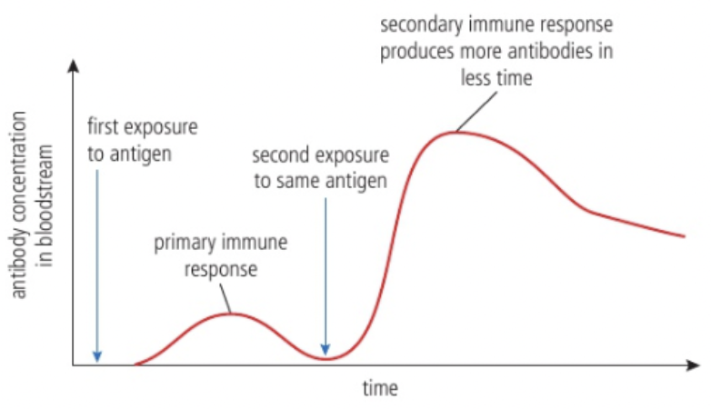
Immunity (Memory Cells) & Primary / Secondary Immune Response
Immunity = Consequence of retaining memory cells; Ability to eliminate an infectious disease from body
Due to long-term survival of lymphocytes (memory cells) capable of making specific antibodies needed to fight the infection
Memory (B/T) cells = Formed after initial infection / vaccination; Remain in body long-term
Provide rapid and robust response to subsequent exposures to same pathogen → Secondary immune response
Primary immune response = Takes time (B/T cells must be activated)
Secondary immune response = Faster + Produce more antibodies than first exposure
Transmission of HIV in Body Fluids
HIV = Human immunodeficiency virus; Gradually attacks immune system
Leads to AIDS
Transmission must involve body fluids, e.g.
Blood, semen, rectal fluids, vaginal fluids, breastmilk
Mechanisms of HIV transmission include…
Unprotected sex, birth, breastfeeding, blood transfusion, contaminated needles, (rarely) across placenta
HIV positive mothers can reduce the risk by…
Taking antiretroviral medications during pregnancy
Avoid breastfeeding
Infection of Lymphocytes by HIV with AIDS as a Consequence
Only certain types of lymphocytes are infected and killed
Reduction in these lymphocytes limits the ability to produce antibodies and fight opportunistic infections
HIV infect helper T-cells → Cannot activate B-cells → Unable to produce antibodies and fight off other infections → AIDS (Acquired immune deficiency syndrome)
HIV is monitored by measuring T cell count and viral load
Antiretroviral therapy (ART) can be used to control HIV → Allow immune system to recover
Antibiotics
Antibiotics = Chemicals that block processes occurring in bacteria BUT not in eukaryotic cells
Stop chemical pathways unique to prokaryotes → Eukaryotic cells are unaffected
Affects…
Cell wall and membranes
Protein synthesis
DNA/RNA synthesis
Antibiotics fail to control infection with viruses because…
Viruses have no metabolism; Cannot be affected
Evolution of Resistance to Several Antibiotics in Strains of Pathogenic Bacteria
Careful use of antibiotics is necessary to slow the emergence of multi-resistant bacteria
Antibiotic resistance occurs when bacteria mutate and no longer respond to drugs; Accelerated by overuse and misuse of antibiotics
Can be developed via mutations or horizontal gene transfer (Resistance spreads between bacteria of same and different species)
E.g. MRSA = Superbug
Threat to public health; Lead to longer illness, more hospital stays, increased mortality
Zoonoses
Zoonoses = Infectious diseases that can be naturally transferred from vertebrate animals to humans
Highly prevalent; ~60% emerging infectious diseases globally are zoonoses → High economic burden (Healthcare costs + Loss of productivity)
Examples:
Tuberculosis
Primarily human disease, can be transmitted through infected animals, especially cattle
Transmitted via inhalation of aerosols
Rabies
Viral disease spread through the bite of an infected animal
Japanese encephalitis
Mosquito-borne viral infection
Reservoirs of virus = Pigs & wild birds
COVID-19
Caused by SARS-CoV-2 virus
Reservoirs of virus = Bats
Spread through human via intermediate host at a wildlife market
Primarily spreads through respiratory particles released by infected person
Vaccines and Immunisation
Vaccines contain antigens, or nucleic acids with sequences that code for antigens → Stimulate the development of immunity to a specific pathogen without causing disease
Vaccines introduce antigens / genetic instructions to produce antigens into body, which simulates an infection → Triggers immune system to produce T-cells and antibodies + Memory cells which recognise and fight pathogen in future exposure
Live vaccines use a weakened form of the pathogen
Inactivated vaccines contain the “killed” version of the pathogen
mRNA vaccines use mRNA to instruct cells to produce a protein that triggers an immune response
Immunisation = Process of developing immunity without disease symptoms
Herd Immunity and Prevention of Epidemics
Members of a population are interdependent in building herd immunity
Herd immunity = Occurs when large portion of community become immune to disease, preventing spread from person to person
Protects those who cannot be vaccinated e.g. immunocompromised patients and newborns
If a sufficient % of a population is immune too a disease, transmission is greatly impeded
For highly contagious diseases, e.g. measles, ≥95% of population must be immune to stop spread
Evaluation of Data related to COVID 18 (% Change VS Difference)
Percentage change = Two values over time
Difference/Original * 100
Percentage difference = Two values at the same time
Difference/(Average of two values)*100