Lecture 13 - Fractionation and the Dose Rate Effect
1/38
Earn XP
Description and Tags
ONCOL 335 - Radiobiology. University of Alberta
Name | Mastery | Learn | Test | Matching | Spaced |
|---|
No study sessions yet.
39 Terms
Potentially Lethal Damage definition
damage that could cause death, but is modified by post-irradiation conditions
when is potentially lethal damage repaired?
the cellular damage is repaired during the interval between treatment and assay, especially under suboptimal growth conditions
how is trypsin used in experiments measuring lethal damage repair?
Trypsin is used to digest proteins and detach cells, allowing for the assessment of cellular repair mechanisms and the measurement of lethal damage.
the earlier we trypsinize, the more the trypsin stresses the cell, thus leading to potential lethal damage
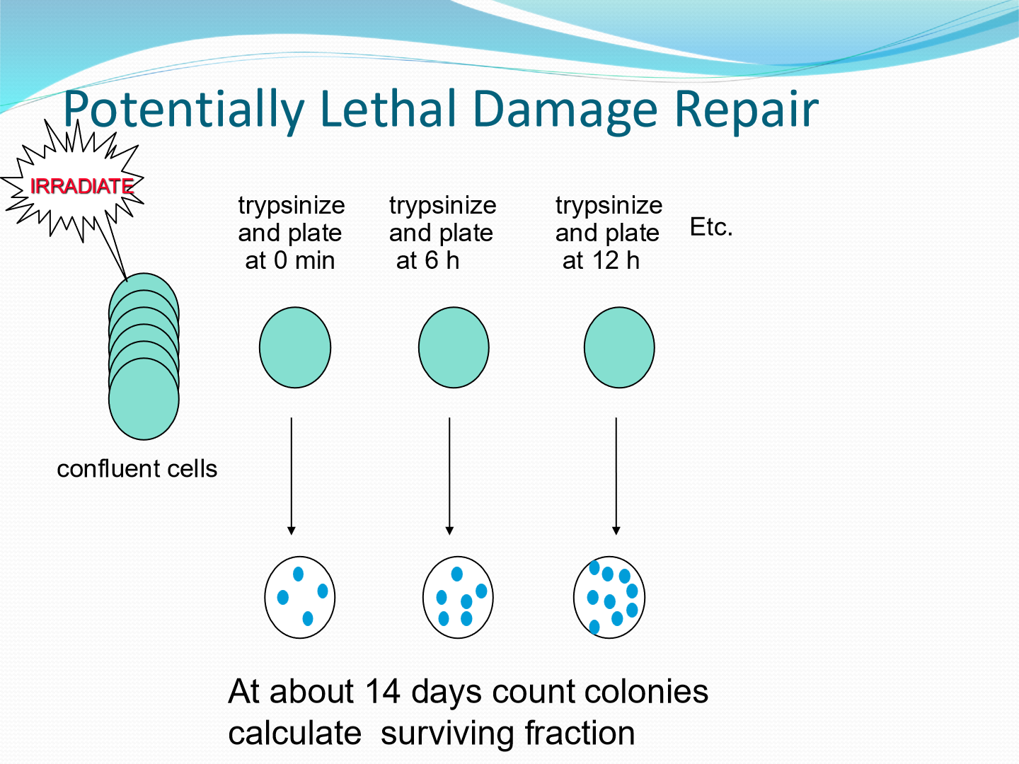
What is done after the trypsin experiment?
Cells are assessed for survival and repair efficiency. results are recorded on a graph
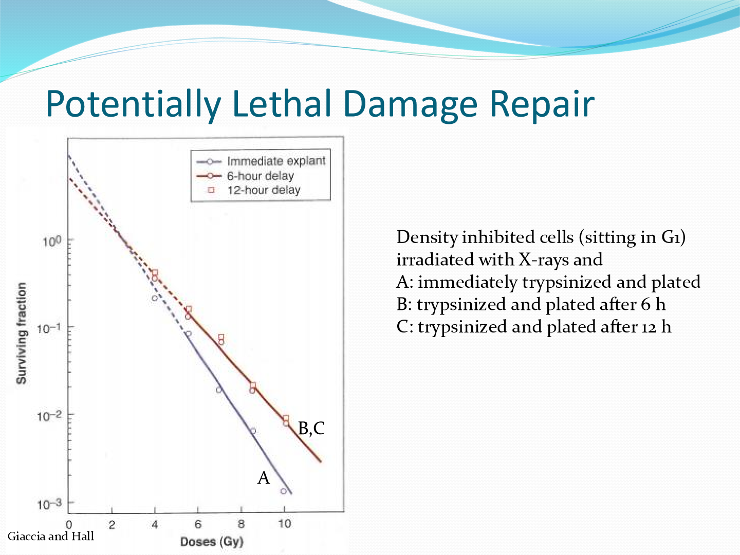
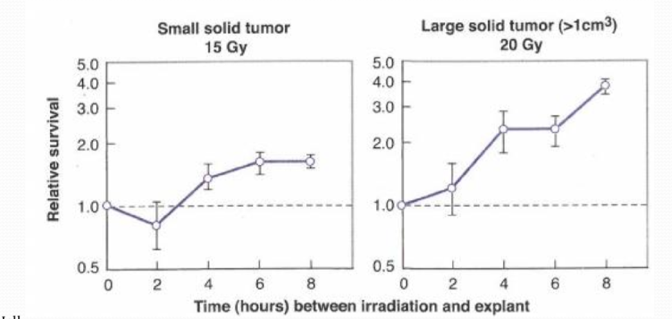
what is the conclusion of this lethal damage repair experiment in vivo?
the more time that is passed for PLD repair to occur, the more cells survive.
this test was done wiht mouse fibrosarcomas irradiated in situ
what is sublethal damage?
nonlethal cellular damage that can be repaired or can become lethal with further dose
increase in cell survival that is observed if a give radiation dose is split into two fractions by a time interval
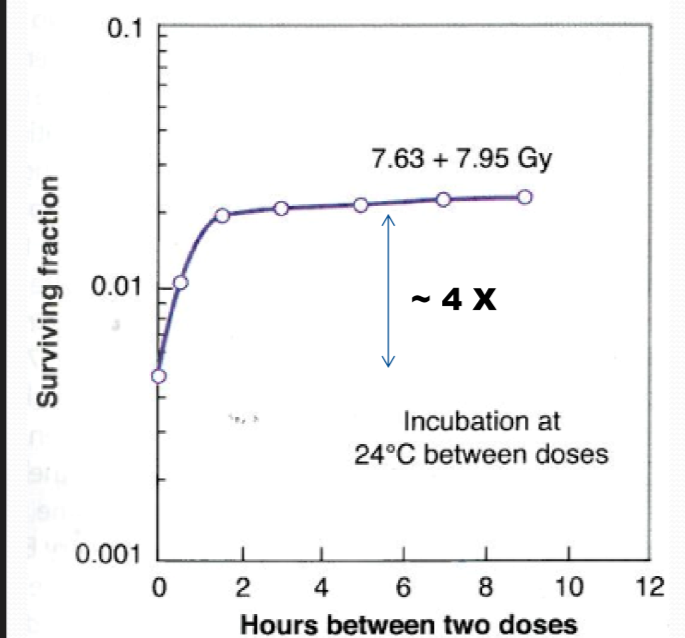
what does this in vitro graph say?
It illustrates the relationship between time of dose fractionation and cell survival, showing improved survival rates with increased time intervals.
the more hours you give in between fractions, the sublethal damage has time to repair
as shown by the flat curve

why is there a dip in cell survival at 6 hours between doses in vivo
at this time point, the cell has gone through the cell cycle so now it is in a sensitive phase of the cell cycle as compared to a radioresistant phase
what is the most radiosensitive phase?
G2/M
what is the most radioresistant phase?
late S phase
why do the dose respose curves of CHO and HeLa cells look different
CHO cells have a shorter G1 phase, resulting in less time in repair damage in this phase
therefore G1 in CHO cells is a little more sensitive


How do cells in sublethal damage experiments differ in vivo as compared to in vitro
In vivo cells experience a more complex microenvironment, including interactions with other cell types and extracellular matrix, which can influence their response to sublethal damage compared to the controlled conditions of in vitro experiments.
Cells in vivo will have a different oxygen concentrations, different metabolic levels, etc
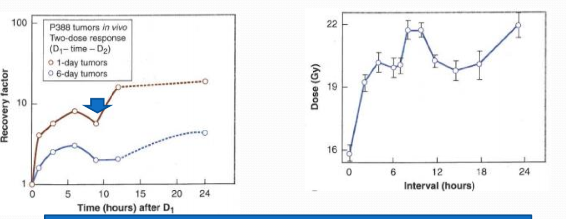
why is the dip smaller in vivo than in vitro?
because in vivo cells have slower cycling as they have a more complex microenvironment
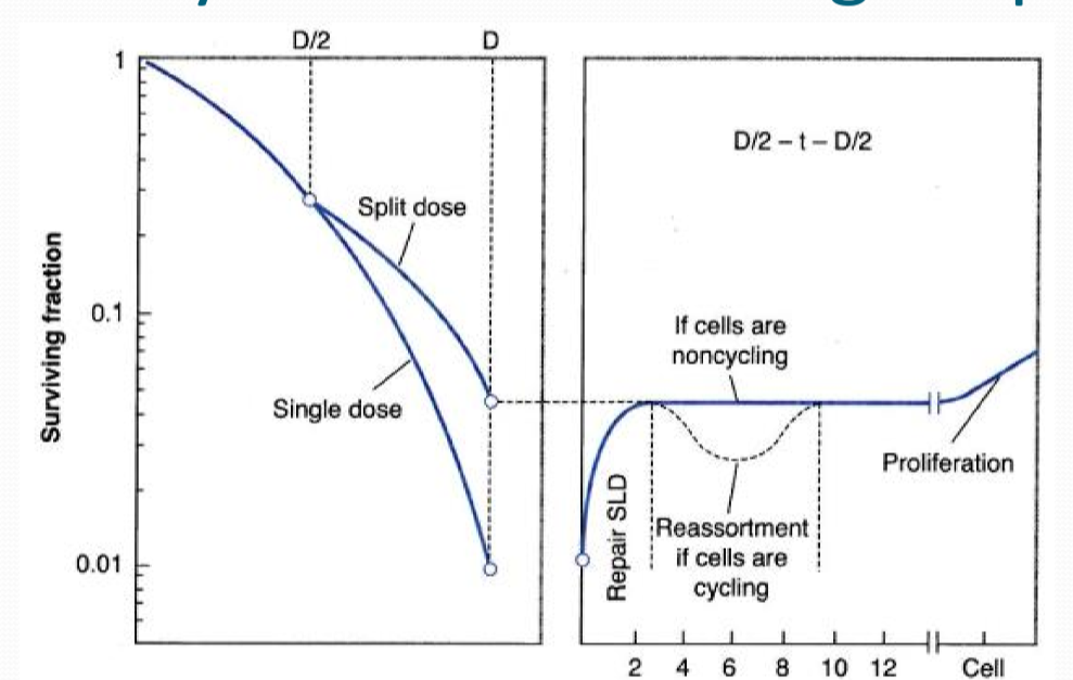
why do we get a sparing effect if we split dose?
we get a repair shoulder

why do we get a dip in the graph on the right
the dip occurs as the cells cycle into a more sensitive phase

why do we get an increase in the graph on the right
if long enough time is waited between doses, the cells will reactivate and start proliferating again
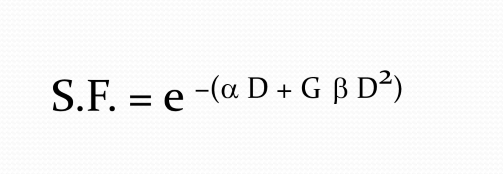
How does the Lea and Catchside equation differ from the LQNT model?
has extra factor G to take into account the repair pulses in cells
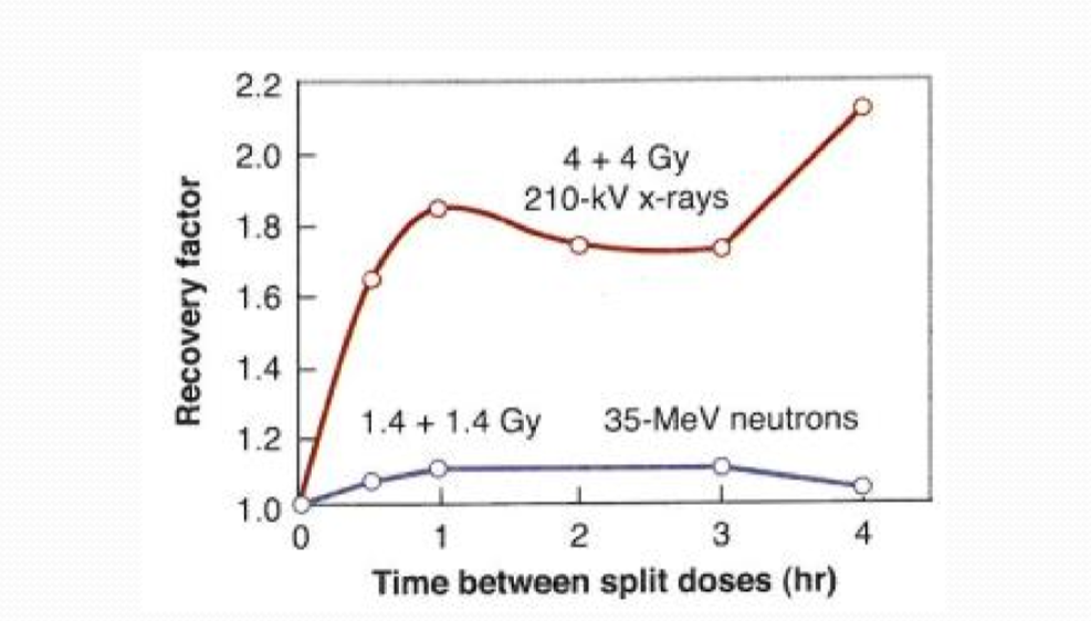
What effect dose LET have on recovery factor in split-dose experiments?
Higher LET decreases recovery factor as they produce more sublethal damage, and less repairable damage
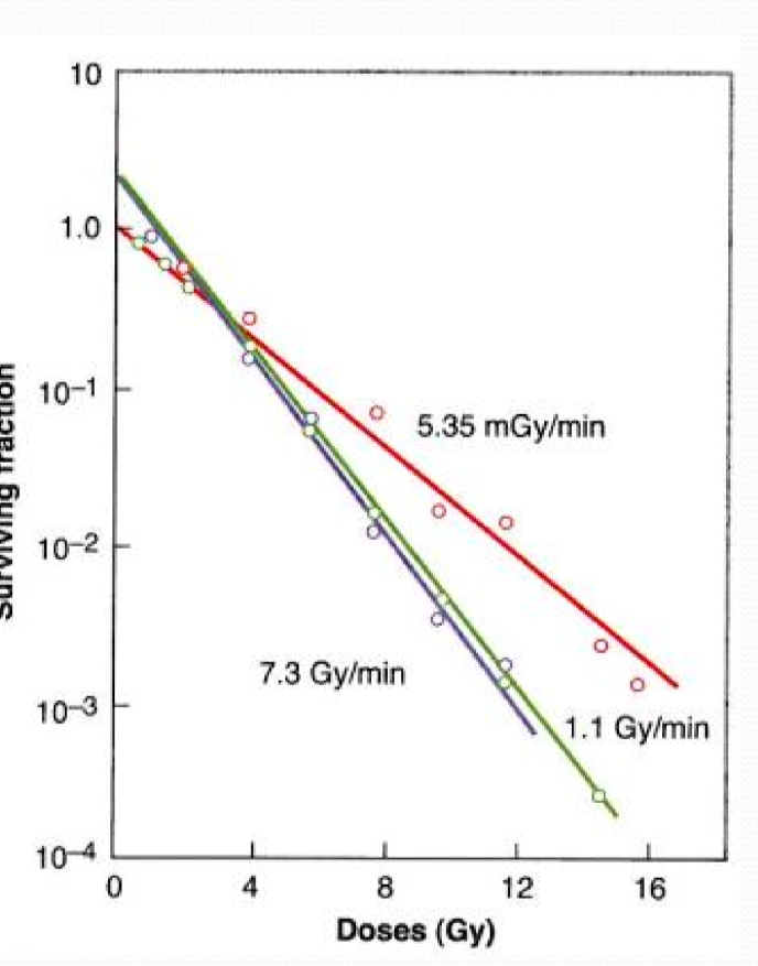
what is the dose rate effect?
an increase in isoeffective radiation dose with decreasing radiation dose rate
give dose continously over a longer time, and over this time repair can then begin
This phenomenon indicates that lower dose rates allow for cellular repair mechanisms to function more effectively, leading to a higher overall tolerance to radiation.
what phenomena play a role in the dose rate effect (hint: the 4Rs)
repopulation
reoxygenation
reassortment
recovery
the same factors that play a role in fractionation play a role in dose rate as well!
what two phenomena are present at lower dose rates (taking longer to give 2 Gy)?
repopulation and reoxygenation
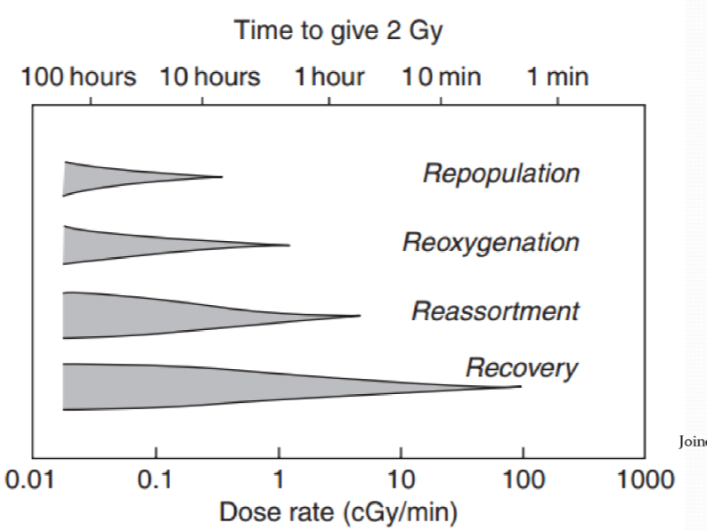

what two phenomena are present at higher dose rates (taking less time to give 2 Gy)
reassortment and recovery

when does repair for DNA damage begin?
immediately after DNA damage, ATM is at damage site within seconds

what happens with increased fractions?
the shoulder is repeated with each fraction
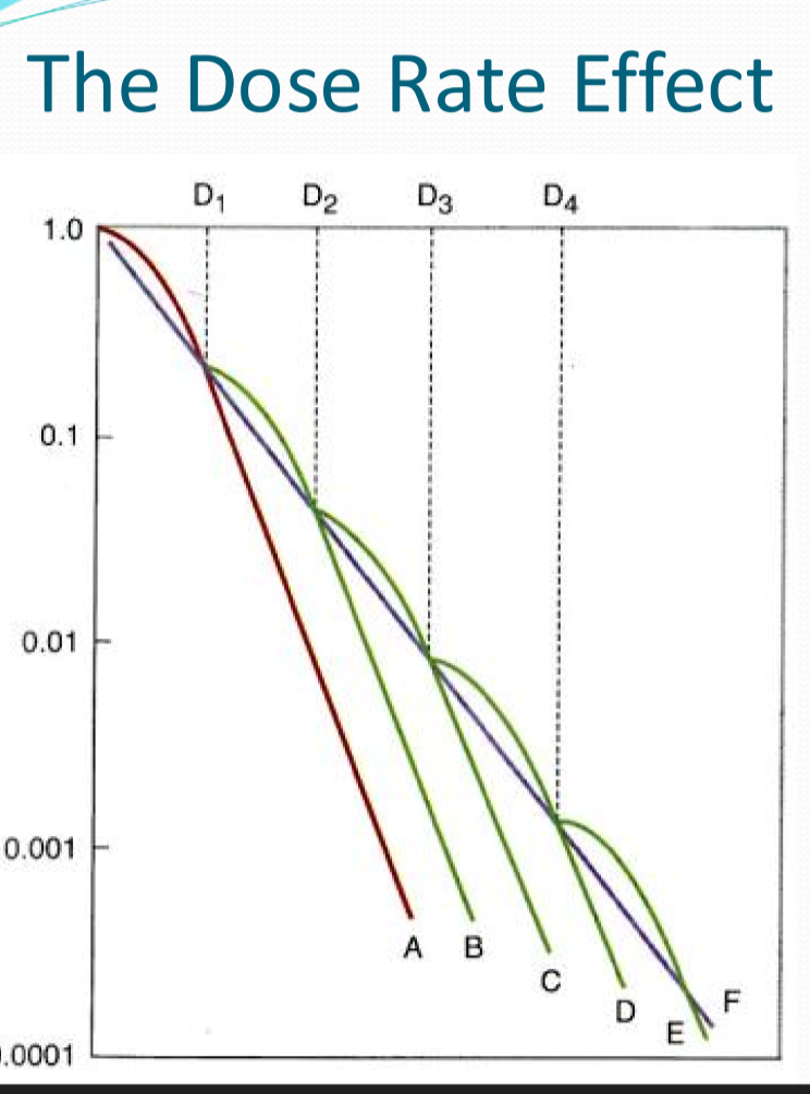
what happens with continous low dose irradiation?
akin to infinetly small fractions, would give a survival curve with no shoulder
What doses give an increased slope of a dose survival curve?
high dose rates and really small dose rates!
explain what happens at each dose rate
at high dose rates: a substantial dose rate effect is evident
medium dose rate: still a little dose rate effect, slope is smaller
small dose rate: dose rate affect increases again!!!
smallest dose rate: cells are too slowly killed off, proliferation counteracts the effect
what is the dose rate effect due to ?
repair of sublethal damage
what type of therapy is the dose rate effect an important consideration?
brachytherapy
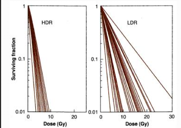
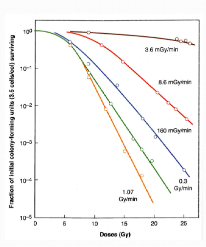
these are dose survival curves at high and low dose rates for 40 different cell lines of human origin. why are the curves not overlapping?
different radiosensitivities

these are dose survival curves at high and low dose rates for 40 different cell lines of human origin. why do the curves fan out at low dose rate?
different repair times
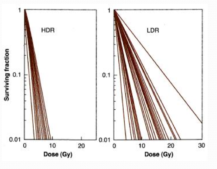
HDR curve phenomena is due to ______, LDR curve phenomena is due to ______
HDR: intrinsic radiosensitivity
LDR: different repair times
at a lower dose rate, what do you need to cause the same amount of damage as high dose rates?
you need a larger total dose to give the same effect has high dose rates
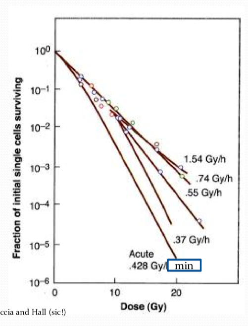
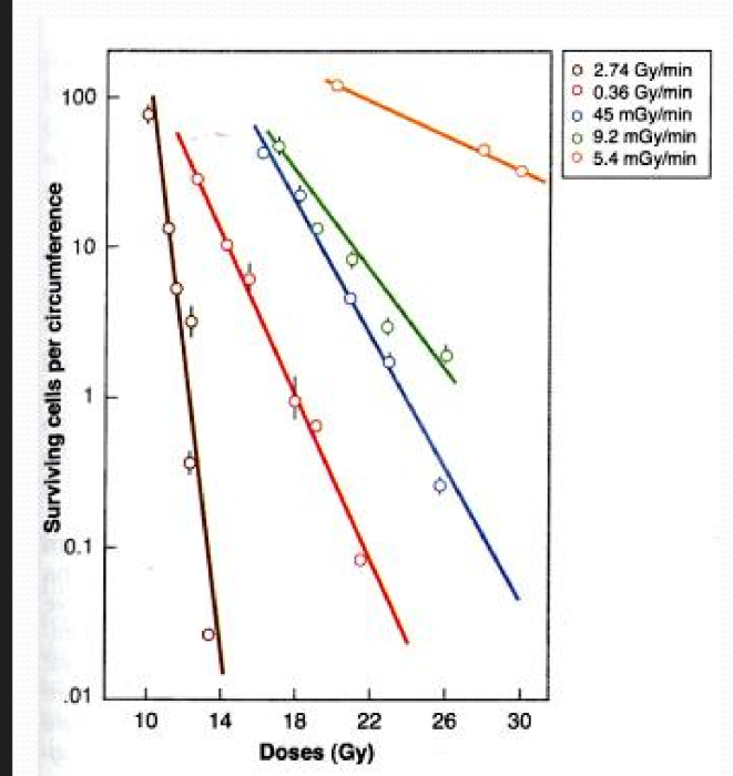
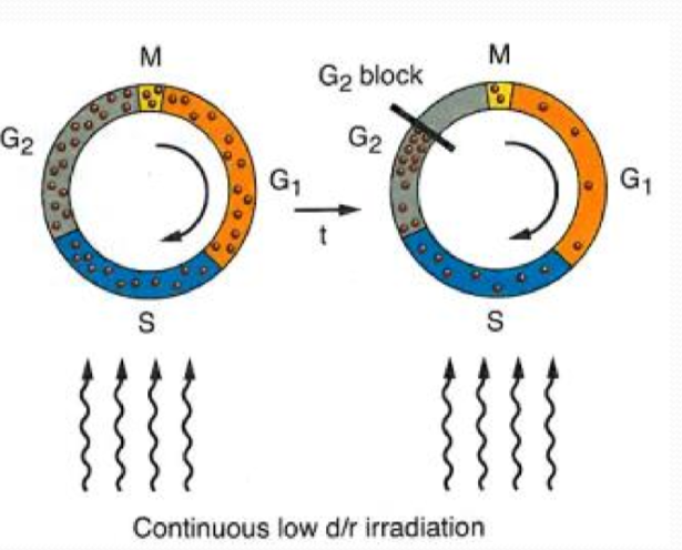
what is the Inverse Dose Rate Effect?
when the dose rate is lowered below a certain level, the amount of cell killing INCREASES
at what dose rate does the inverse dose rate effect kick in?
1.54 Gy/hr
What are the two explanations of the inverse dose rate effect?
Dose Rate Sensitivity
Joiner Effect
Dose rate sensitivity explanation
at high dose rates, cells are frozen in the phase of the cell cycle
but in low dose rates, the cells continue through the cell cycle during irradiation, making them more sensitive
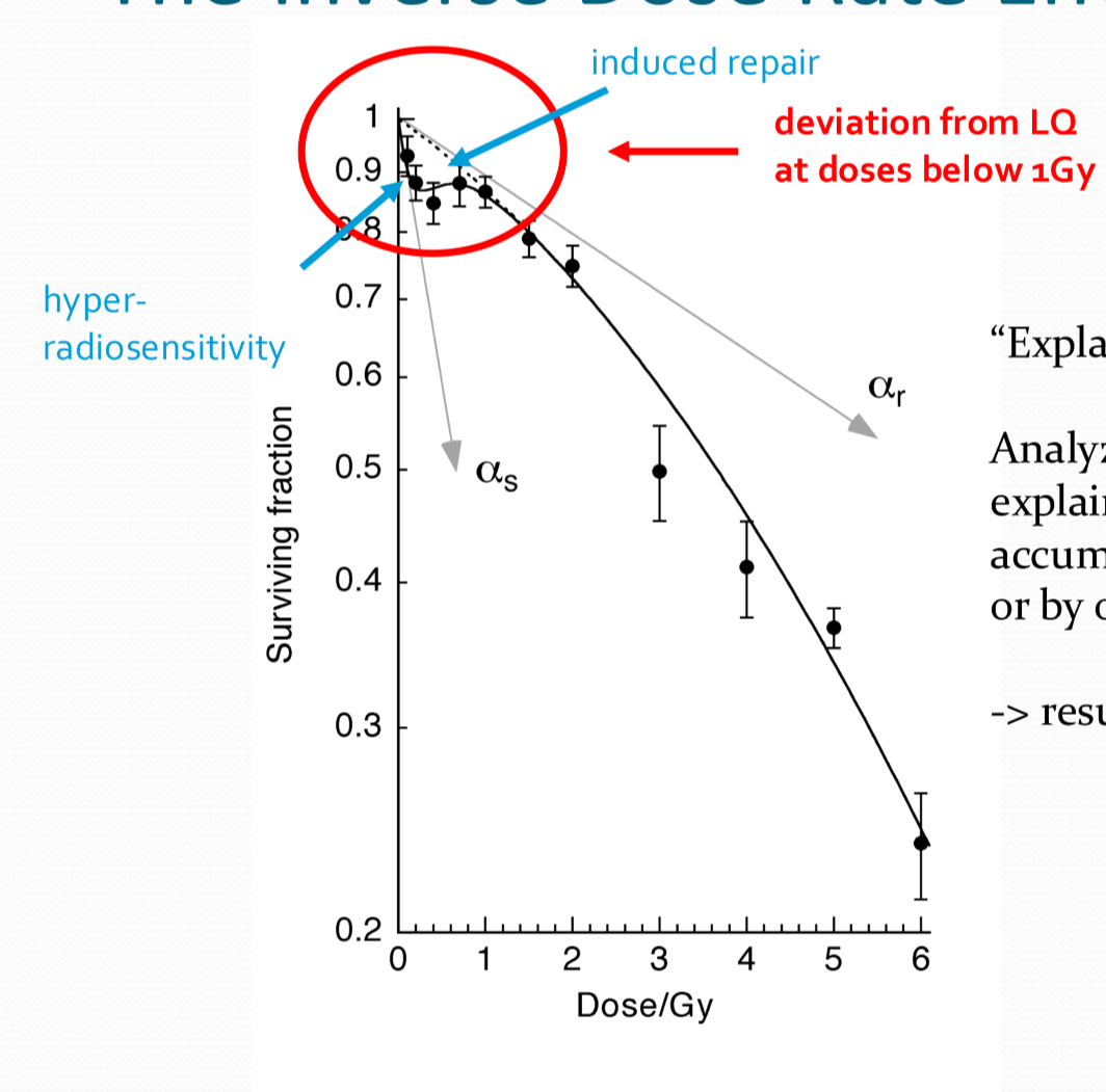
Joiner Effect explanation
at very low doses, cells have a peak of hyperradiosensitivity that deviates away from the LQ model as the DNA damage repair mechanism are not fully activated
with super low dose rates, the cell is stuck in this peak of hyperradiosensitivity so the cell is affected more by damage
Dose rate effect in a nutshell
High dose rate = high death
Medium dose rate = lower death rate
Low dose rate = high cell death
Even lower dose rate = proliferation
