Case 6: Take My Breath Away
 Call Kai
 Learn
Learn Practice Test
 Spaced Repetition
 Match
Match Flashcards
 Knowt Play
1/31

There's no tags or description
Looks like no tags are added yet.
Name | Mastery | Learn | Test | Matching | Spaced | Call with Kai | Chat |
|---|
No analytics yet
Send a link to your students to track their progress
32 Terms
True
The best first question you can ask yourself when you are evaluating the pleural space is, "Is there something in the pleural space?".
TRUE OR FALSE
True
If there is something in the pleural space, your second best question to ask is, "What opacity is making the potential pleural space an actual space?".
TRUE OR FALSE
a. normal
Identify the opacity in the pleural space:
a. normal
b. air
c. soft tissue
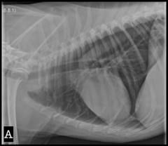
c. soft tissue (fluid)
Identify the opacity in the pleural space:
a. normal
b. air
c. soft tissue
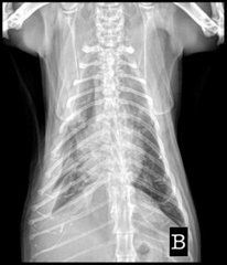
b. air
Identify the opacity in the pleural space:
a. normal
b. air
c. soft tissue
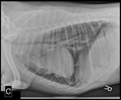
f. All Of The Above
What are some possible causes for pleural effusion in cats?
a. Congestive Heart Failure
b. Neoplasia
c. Pyothorax
d. Idiopathic
e. Chylothorax
f. All Of The Above
a. lungs
b. fluid in pleural space
Identify the labeled structures:
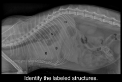
1. extra thoracic structures
2. pleural space
3. pulmonary parenchyma
4. mediastinum (cranial, middle, and caudal)
Thoracic radiographs can be difficult to interpret. What 4 "chunks" does she break the thorax down into the make evaluation easier?
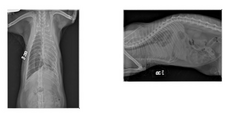
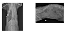
A feline right lateral thoracic projection and a VD projection are available for view
Describe the study you are seeing here
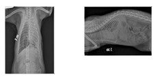
Patient positioning is adequate for interpretation. Study technique is adequate for interpretation.
Describe patient positioning and technique
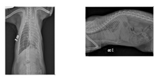
On initial lateral projection there is marked inc. soft tissue opacity within pleural space causing marked retraction of lung lobes from thoracic wall. Lung lobe margins are mildly rounded in contour. Cardiac silhouette not visualized, and marked increased soft tissue opacity is superimposed over cranial mediastinum. There is ill defined increased soft tissue opacity visible within partially aerated lungs within caudodorsal thorax. On VD projection there is large amount of free gas and minimal inc. soft tissue opacity within right hemithorax, with marked retraction of R. lung lobes from thoracic wall. Moderate-marked inc. soft tissue opacity present within L. hemithorax, most severe cranially and caudally, again with retraction and rounding of lung lobes. Cardiac silhouette is not identified, but there is mild shift of the carina to the left. Included portions of cranial abdomen are within normal limits.
Describe your findings
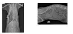
Severe pleural effusion, partially resolved after right sided thoracocentesis, with marked iatrogenic right sided pneumothorax and moderate left sided pleural effusion post-centesis.
Describe Conclusions: Give your radiographic diagnosis and list possible differential diagnoses for all your findings
None indicated as thoracocentesis was already performed.
Describe Next Steps: Are there any additional imaging suggestions that would help clarify your findings? Are there critical next steps to take from a stabilization/management/treatment plan standpoint?
note: this patient has already had thoracocentesis and radiographs done
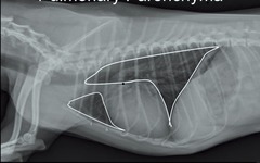
Pulmonary Parenchyma
What is hilighted here?
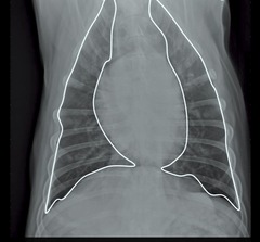
Pulmonary Parenchyma
What is this?
Mediastinum: cranial, middle (cardiac silhouette), and caudal respsectively
What do these circles represent?
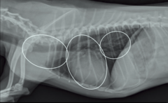
Pleural space
What is this?
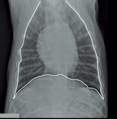
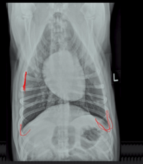
Normal = lungs expand all the way to the body wall and there are normal sharp points
Are these normal or abnormal radiographs?
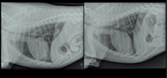
Normal = lungs expand all the way to the body wall and there are normal sharp points
Are these normal or abnormal radiographs?
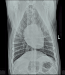
Costophrenic angle
What do those little "u's" represent at the lower right and left hand corners of the screen in this normal thorax?
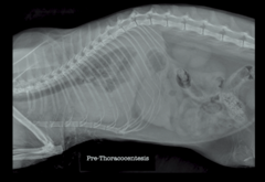
Soft tissue opacity = fluid = differentials include chylothorax, hemothorax, any kind of fluid in the thoracic cavity
Lungs are very small - TON of space between lungs and body wall
This is the cat BEFORE thoracocetesis. If there is something between the lung and thoracic wall, what is it? What opacity?
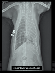
Thoracocentesis done on RIGHT side
This is the VD of the cat AFTER thoracocentesis. What side was the thoracocentesis done on?
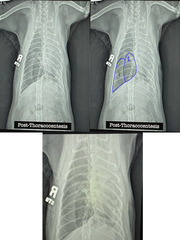
1: air opacity = came from introducing air into the lung into the pleural space = iatrogenic pneumothorax from thoracocentesis
2: lung opacity = normal = can vizualize airways and vessels = atelectic lung that is collapsed but there is still vasculature
This is the all same image. Tell me the difference between the opacity in 1's circle versus 2's circle.
NORMAL = Clear cardiac silhouette margin with soft tissue opacity. There is fat opacity in the pericardial sac (more visible at the apex of the heart in this image). There is air opacity in the lung.
Is this a normal or abnormal thorax? Describe what you're looking at in terms of opacity.
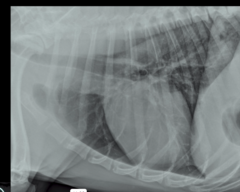
TRUE: you cannot see blood in the heart distinct from chambers in the heart because it is all "soft tissue opacity"
TRUE OR FALSE: You cannot discern fluid within the heart from chambers in the heart.
1. gives the cat room for the lungs to expand = eliminates trouble breathing
2. allows you to sample the fluid and determine what it is
What are the 2 main indications for doing thoracocentesis in a cat that has fluid in the thorax and is in respiratory distress?
Pleural space
The Parietal pleura is the side that is wrapped around the lungs
Visceral pleura lines the inside of the thoracic cavity/body wall
Between the two of them is a "potential space". What is it?
False
Pleural effusion in cats is usually unilateral.
TRUE OR FALSE
False = the most common cause is CONGESTIVE HEART FAILURE
Neoplasia is the most common cause of pleural effusion in cats.
TRUE OR FALSE
False
Most cats with pleural effusion have a good prognosis.
TRUE OR FALSE
True
Ultrasound is very helpful for evaluating whether pleural effusion is present in the thoracic cavity of cats.
TRUE OR FALSE
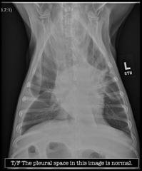
False
TRUE OR FALSE: The pleural space in this image is normal.