CNRN Certification Study - terms/symptoms/anatomy
 Call Kai
 Learn
Learn Practice Test
 Spaced Repetition
 Match
Match Flashcards
 Knowt Play
1/71

Earn XP
Description and Tags
Vocabulary flashcards covering core neuroanatomy, neurophysiology formulas, neurotrauma, spinal cord emergencies, stroke syndromes, neuro-oncology, neuromuscular disorders, and bedside patient management based on the 2026 CNRN exam blueprint.
Name | Mastery | Learn | Test | Matching | Spaced | Call with Kai | Chat |
|---|
No analytics yet
Send a link to your students to track their progress
72 Terms
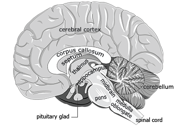
Frontal Lobe
Brain region responsible for executive function, judgment, personality, motor planning, and contains Broca speech area in the dominant hemisphere.
Parietal Lobe
Brain region responsible for somatosensory processing, spatial awareness, and integration of sensory input.
Temporal Lobe
Brain region responsible for memory, auditory processing, Wernicke speech area, and frequently serves as a seizure focus.
Occipital Lobe
Brain region responsible for vision and visual interpretation.
Cerebellum
Brain structure responsible for coordination, balance, motor learning, and smooth movement.
Brainstem
Brain region containing cranial nerves, the reticular activating system, and respiratory/cardiovascular centers.
Basal Ganglia
Deep brain structures involved in movement regulation and muscle tone.
Thalamus
Brain structure serving as a sensory relay network and facilitating consciousness.
Cerebral Perfusion Pressure (CPP)
Neurologic measurement defined by CPP=MAP−ICP; cerebral perfusion falls when ICP rises or MAP drops.
Mean Arterial Pressure (MAP)
Estimate of average driving pressure for organ perfusion, calculated as MAP≈3SBP+2×DBP.
Monro-Kellie Doctrine
Physiological principle stating that skull volume equals brain plus blood plus CSF (Skull Volume=Brain+Blood+CSF), meaning an increase in one component must be offset or ICP rises.
Cushing Response
Late sign of increased ICP and impending brain herniation characterized by hypertension with a widened pulse pressure, bradycardia, and irregular respirations.
Epidural Hematoma
Arterial bleeding often associated with a temporal skull fracture, classically presenting with a lucid interval followed by rapid neurologic deterioration.
Subdural Hematoma
Venous bleeding between the dura and arachnoid layers, more common in older adults or patients taking anticoagulants.
Diffuse Axonal Injury
Traumatic brain injury involving shearing of axons resulting in coma out of proportion to CT findings.
Basilar Skull Fracture
Fracture at the base of the skull characterized by clinical signs such as CSF rhinorrhea, CSF otorrhea, periorbital ecchymosis, Battle sign, and hemotympanum.
Central Cord Syndrome
Spinal cord injury pattern causing upper extremity weakness greater than lower extremity weakness, often following cervical hyperextension.
Anterior Cord Syndrome
Spinal cord injury pattern resulting in motor loss and pain/temperature loss below the lesion with preserved dorsal column function.
Brown-Séquard Syndrome
Incomplete spinal cord injury characterized by ipsilateral motor and proprioception loss with contralateral pain and temperature loss.
Autonomic Dysreflexia
Potentially life-threatening condition occurring in spinal cord lesions at or above T6, triggered by noxious stimuli like bladder distention, causing severe hypertension, pounding headache, flushing/sweating above the lesion, and bradycardia.
Spinal Shock
Transient state of acute flaccid paralysis, areflexia, and loss of autonomic function below the lesion following spinal cord injury.
Neurogenic Shock
Hemodynamic instability caused by loss of sympathetic tone following high spinal cord injury, presenting with hypotension, bradycardia, and warm, dry skin.
Left MCA Stroke Syndrome
Vascular stroke presentation marked by right face and arm weakness, aphasia, right visual field cut, and left gaze preference.
Right MCA Stroke Syndrome
Vascular stroke presentation marked by left spatial neglect, left weakness, impulsivity, anosognosia, and left visual field cut.
ACA Stroke Syndrome
Vascular stroke presentation characterized by leg weakness greater than arm weakness, abulia, urinary incontinence, and personality changes.
Myasthenia Gravis
Autoimmune neuromuscular junction disorder characterized by fatigable muscle weakness, ptosis, diplopia, dysphagia, and respiratory muscle weakness.
Guillain-Barré Syndrome
Post-infectious polyneuropathy presenting with ascending muscle weakness, areflexia, paresthesias, and risk of autonomic instability and respiratory failure.
Multiple Sclerosis
Demyelinating central nervous system disease characterized by relapsing neurologic deficits, optic neuritis, fatigue, weakness, and heat sensitivity.
Amyotrophic Lateral Sclerosis (ALS)
Progressive neurodegenerative disease involving upper and lower motor neurons, causing muscle weakness and respiratory failure while cognition is often preserved.
Wernicke Encephalopathy
Acute metabolic disorder caused by thiamine deficiency, characterized by the clinical triad of confusion, ophthalmoplegia, and ataxia.
Posterior Reversible Encephalopathy Syndrome (PRES)
Neurologic condition triggered by severe hypertension, eclampsia, renal disease, or immunosuppression, presenting with headache, visual symptoms, and seizures.
Normal Pressure Hydrocephalus
Neurologic condition classically characterized by the clinical triad of gait disturbance, cognitive decline, and urinary incontinence.
Parkinson Disease
Progressive neurodegenerative movement disorder characterized by bradykinesia, rigidity, resting tremor, and postural instability.
External Ventricular Drain (EVD)
Invasive device used for CSF drainage and ICP monitoring, requiring strict reference leveling, correct drainage height, sterile handling, and clamping during positioning or transport per protocol.
symptoms of a stroke in the frontal lobe
disinhibition, poor judgement, contralateral weakness, expressive aphasia, impaired initiation.
symptoms of a stroke in the parietal lobe
contralateral sensory loss, neglect, difficulty with spatial awareness, and difficulty with language comprehension.
symptoms of a stroke in the temporal lobe
receptive aphasia, memory disturbance, auditory hallucinations, complex partial seizures
symptoms of stroke in the occipital lobe
homonymous hemianopia, cortical blindness, visual agnosia
symptoms of stroke in the cerebellum
ataxia, dysmetria, intention tremor, coordination issues (wide based gait)
symptoms of stroke in the brainstem
altered consciousness, pupillary changes, dysphagia, abnormal respirations, crossed findings
symptoms of a stroke in the basal ganglia
bradykinesia, rigidity, tremors, difficulty with movement initiation
symptoms of a stroke in the thalamus
sensory loss, altered pain perception, motor disturbances, thalamic pain syndrome
symptoms of a stroke in the spinal cord
sensory changes, bowel/bladder dysfunction
subtle changes in LOC is often the earliest sign of ___ decline.
neuroligc
unilateral fixed/dilated pupils can signal CN __ compression and herniation
cranial nerve III
Neuro nursing considerations for Noncontrast CT head
frequent first line acute neuro imaging, check transport safety, airway monitoring and contrast status if ordered with CTA/CTP.
neuro nursing implications for CTA/MRA
assess contrast allergy/renal policy. monitor access site if invasive angiography.
what pain is described as “tingling, shooting, burning”
neuropathic pain
which of the benign headaches is more common in the male population?
cluster headaches. A type of primary headache disorder characterized by severe, unilateral pain, often accompanied by autonomic symptoms.
menieres disease is a neurogological disorder affecting the ___ nerve, also known as cranial nerve VIII
acoustic
is Alzheimers disease a cortical or subcortical dementia?
Alzheimer's disease is considered a cortical dementia, primarily affecting the cerebral cortex and leading to cognitive decline and memory loss.
Cortical dementia, such as AD, involves the degeneration of the cortical area of the brain and affects memory. This is demonstrated by severe significant neuronal loss and brain atrophy.
An example of subcortical dementia is
parkinsons disease
what is an example of a subcorbital dementia is ______
parkinsons disease- where the subcortical area of the brain controls motor skills.
one of the most common forms of parkinsonism is ______ induced.
medication
what is the neurological disorder that is a result of an imbalance between the production and reabsorption of cerebrospinal fluid (CS)?
hydrocephalus, characterized by increased intracranial pressure and enlargement of the ventricles.
an obstruction of CSF between the third and fourth ventricle is classified as which type of hydrocephalus: Communicating or non-communicating?
Noncommunicating hydrocephalus is the obstruction to flow of CSF and is often caused by brain tumors, cysts, scarring, or infections. The obstruction commonly occurs in the ventricles, but can occur anywhere in the ventricular/CSF system.
what is the most common cause of communicating hydrocephalus?
decreased reabsorption
forms of congenital hydrocephalus
chiari malformations
spina bifida
aqueductal stenosis
causes of acquired hydrocephalus
meningitis
hemorrhage
tumors
encephalitis
TBI
inflammation of bones and cartilage of joint
osteoarthritis
symptomatic, degenerative changes of osteoarthritis, which include degenerative disc disease and spinal stenosis
spondylosis
myelopathy
involves compression of the spinal cord and tends to be the result of central stenosis
involves compression of nerve roots and is associated with lateral recesses and foraminal stenosis
radiculopathy
disc protrusion into the spinal canal can cause what type of stenosis?
central stenosis
disc protrusion into the spinal canal can cause what type of stenosis?
central stenosis
Disc protrusion can narrow the central cord canal, causing a central stenosis . Hypertrophy of the facet joints can result in compression of nerve tracts. Osteophytes can impinge either laterally or centrally, causing cord or nerve involvement.
Which portion of the spinal cord do the sensory impulses enter from the peripheral nerves?
Dorsal horn
The dorsal horn receives the sensory impulses from the peripheral nerves, and the ventral horn contains the cells that send out the motor impulses. In neuropathic pain, sympathetic nerve fibers sprout in the dorsal horn. This is called deafferentation of pain. It results in increased nociceptive neurons and responsiveness in the CNS.
what portion of the inner ear is involved in the symptoms of menieres disease
the labyrinth
the symptoms of menieres disease are associated with a change in fluid volume within a portion of the inner ear the labyrinth of the inner ear is responsible for maintaining balance and hearing. Changes in fluid volume, particularly in the endolymph, can lead to the characteristic symptoms such as vertigo, tinnitus, and hearing loss.
What area of the brain is critical for memory?
hippocampus
What is produced in the brain that results in the main pathological change in AD?
myloid plaque and neurofibrillary fibers are produced in the brain and can be found primarily on autopsy. The symptoms seen in AD are the result of the death of many neurons in the hippocampus and cerebral cortex.The primary pathological change in Alzheimer's disease is the accumulation of amyloid plaques and neurofibrillary tangles in the brain, leading to neuron death.
Definitive diagnosis of AD is on autopsy.
What is the neurotransmitter responsible for inhibition of muscle tone?
Dopamine
The neurotransmitter responsible for inhibiting muscle tone is dopamine. The neurons in the substantia nigra are responsible for producing and projecting dopamine to the striatum in the basal ganglia. The excitatory neurotransmitter (in opposition) is acetylcholine. In PD, the nigra–striatal pathway degenerates, resulting in an imbalance between dopamine and acetylcholine. Acetylcholine then has a greater effect, resulting in increased muscle tone.
Where does the CSF get reabsorbed in the brain?
arachnoid villi