Cardiac #2: Congenital Heart Defects
1/27
There's no tags or description
Looks like no tags are added yet.
Name | Mastery | Learn | Test | Matching | Spaced |
|---|
No study sessions yet.
28 Terms
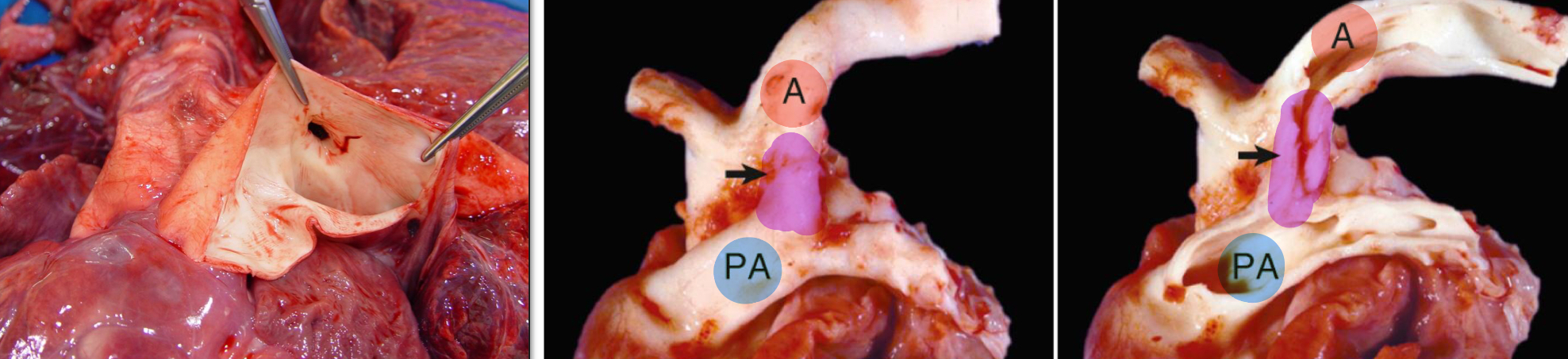
What is a Patent Ductus Arteriosus (PDA)?
The ductus arteriosus is a connection between the pulmonary artery and the aorta in the fetus
When it doesn’t close after birth, it becomes a PDA
A PDA is most common in what species? In what sex?
Dogs
Females
What are some Sequelae that a PDA could cause?
Continuous murmur
Bounding Femoral Pulse
Left congestive heart failure
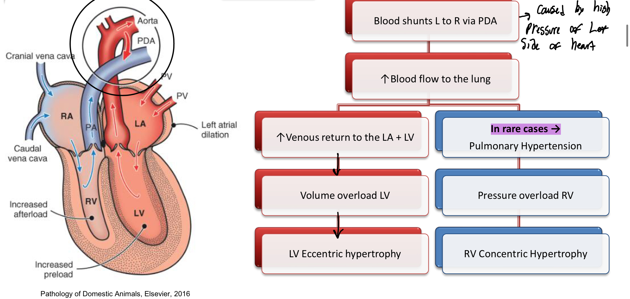
What are the Hemodynamics of PDA?
Blood shunts from L to R via the PDA
Inc of blood flow to the lungs
Inc of Venous blood to the LA+LV
Volume overload of LV
LV hypertrophy
Rare CASES: Pulmonary Hypertension (Inc B.P of arteries in the lungs)
Pressure overload of RV
RV COncentric hypertrophy
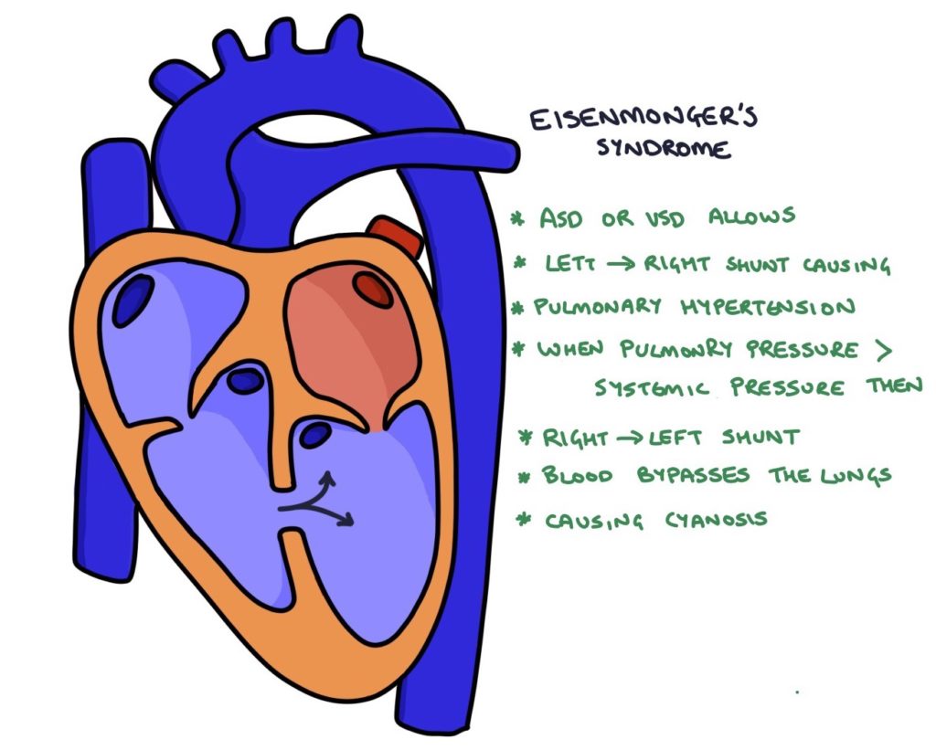
What is Eisenmenger Syndrome? What are the C.S associated with this syndrome?
Pressure of the RV becomes higher than the LV causing the shunt to reverse
C.S
Are caused by unoxygenated blood being pumped into the LV and then around the body (bc pressure in RV>LV)
Cyanosis
Lethargy
Exercise intolerance
Collapse
Erythrocytosis (Higher # of RBCs)
Image is wrong, just using it to associate
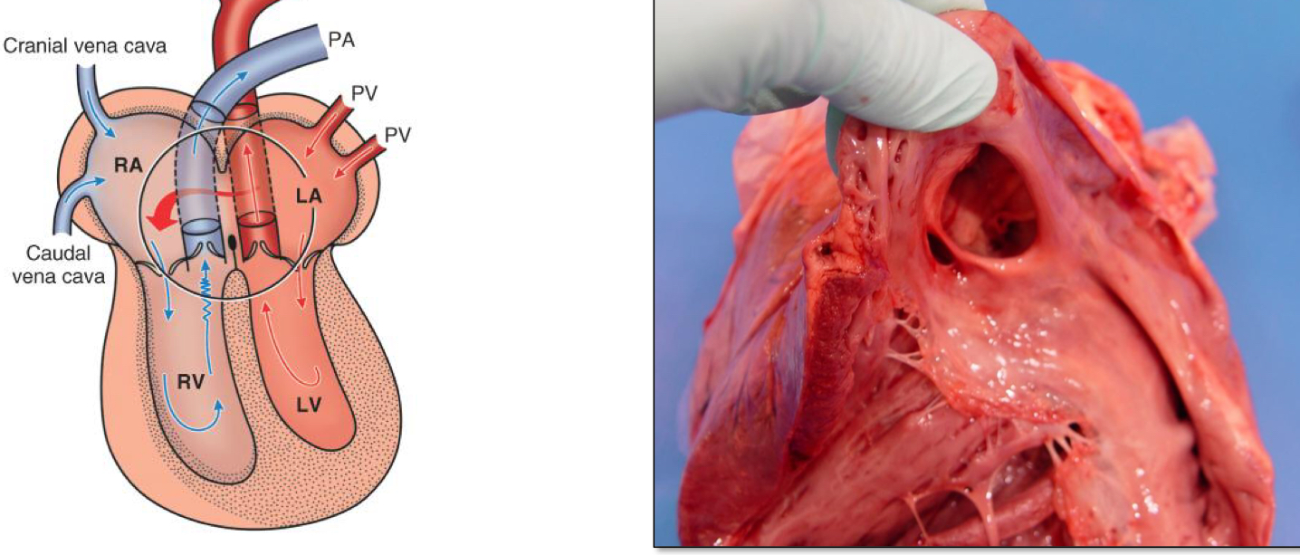
What are the hemodynamics of an Atrial Septal Defect (if the defect is large enough)?
Blood Shunts from LA to RA
Inc of Blood volume in the right ventricle
Volume overload of RV
RV essentric hypertrophy
Volume overload of a ventricle causes _________ hypertrophy
Eccentric
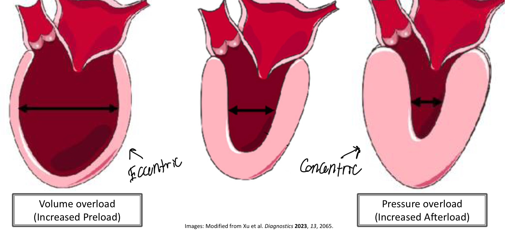
Pressure overload of a ventricle causes ________ hypertrophy
Concentric

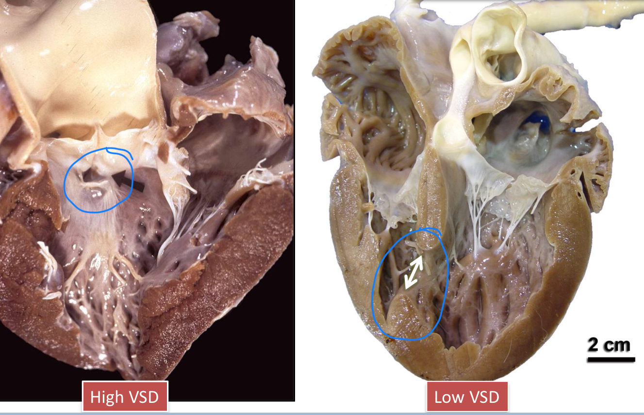
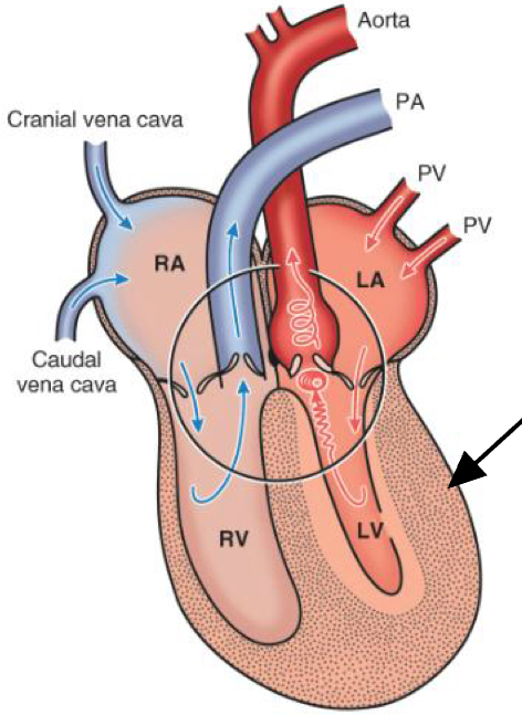
What is a ventricular septal defect (VSD)? What are the 2 variants?
A VSD is when the wall/septum that separates the ventricles does not form properly, resulting in communication between the LV and RV
High VSD
Defect in the upper membranous portion of the interventricular septum
Low VSD
Defect in the lower muscular portion of the interventricular septum
What are the Hemodynamics of a VSD?
Blood shunts from LV to RV (high to low pressure)
Inc of Blood Volume in the RV
Equalization of pressure between RV and LV (RV=LV)
LV and RV hypertrophy
What is Valvular Dysplasia?
Abnormal shape/appearance of the valves of the heart
Short, thick, misshapen
Rolled edges
Absence of chordae tendineae
Fusions of leaflerts or chordae to the ventricular wall
Thick or atrophic papillary muscle
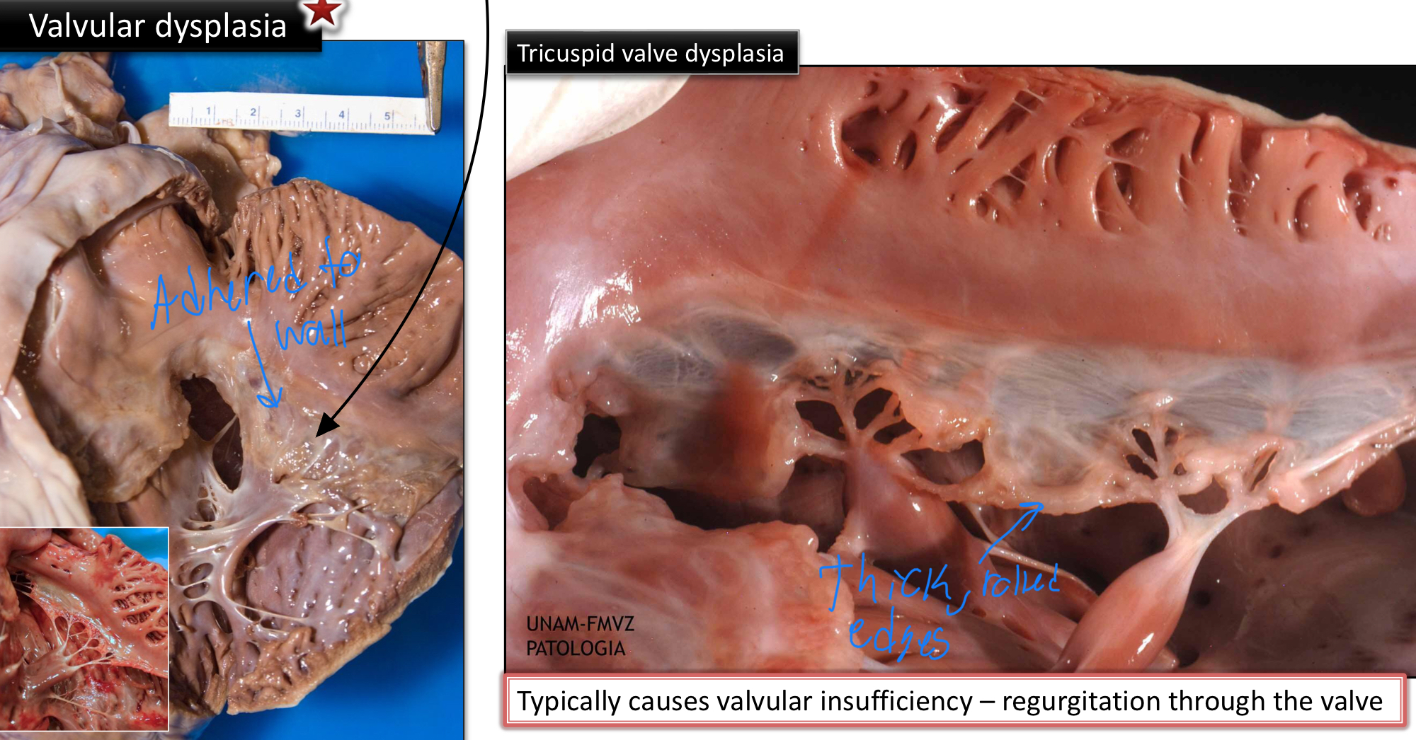
Valvular dysplasia may cause _____ or _____ of the valve
Insufficiency
Valvular insufficiency is when the valve doesn’t close and blood can move “backward“
Stenosis
A heart condition where a heart valve narrows, preventing it from opening fully and restricting blood flow
What are the Hemodynamics of Valvular Dysplasia?
Dysplastic valve is insufficient (blood can flow backwards)
Regurgitant blood flow during systole (contraction of the heart)
Volume overload of the atrium and ventricle
Atrial dilation and Eccentric ventricular hypertrophy
What is Pulmonic Stenosis? What are the 3 types
Narrowing of the Pulmonary a. (carries blood from RV→Lungs for oxygenation)
The types are classified based off of the location of stenosis in regard to the valve
Valvular
Subvalvular
Supravalvular
What are the hemodynamics of a Pulmonic Stenosis?
Stenotic valve restricts outflow
Pressure overload of RV
RV concentric Hypertrophy
Right Heart Failure
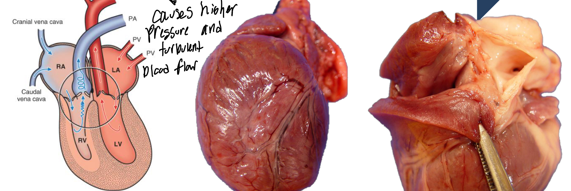
T/F: Pre-stenotic arterial dilation is often found in the artery, proximal to the stenosis
False, it is a Post-stenotic dilation that is often found distal to the stenosis
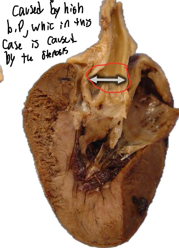
What is a Subaortic Stenosis (SAS)?
A narrowing of the aortic valve orifice caused by a fibromuscular membrane just below the aortic valve
T/F: A SAS is usually accompanied by a post-stenotic dilation in the aorta
True
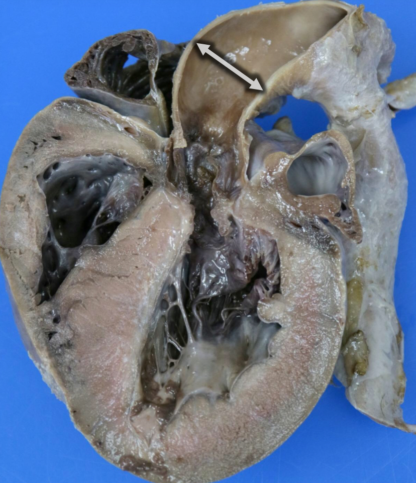
What is the Hemodynamics of a SAS?
Stenotic valve restricts outflow of blood from LV→Aorta
Pressure overload of LV
LV concentric hypertrophy
Arrhythmias and sudden cardiac death (if severe enough)
Congestive Left Heart Failure
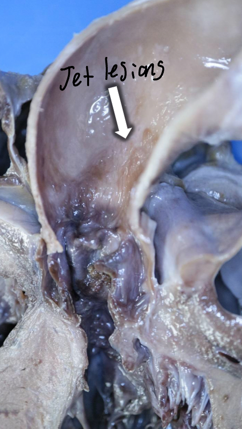
Along with Post-stenotic dilation, what other lesions may appear in the Aorta of a dog with SAS?
Jet Lesions
The result of turbulent blood flow
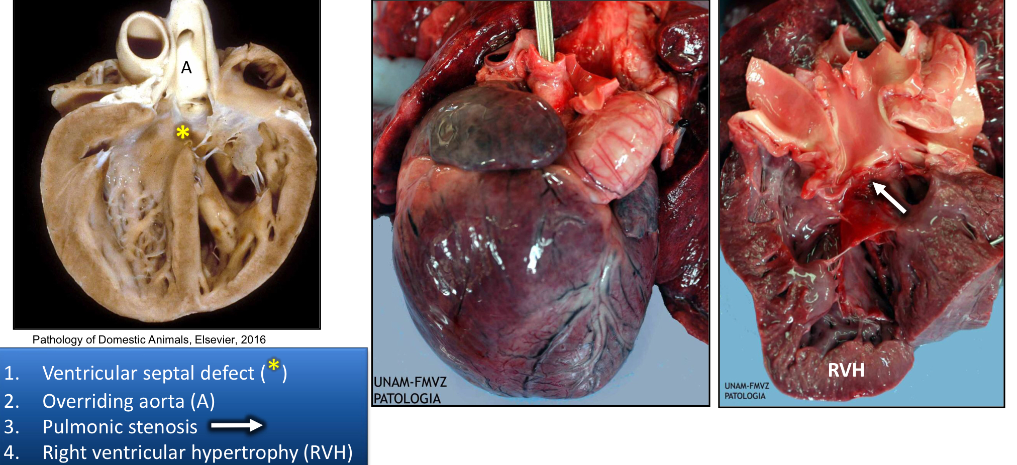
What are the 4 parts of Tetralogy of Fallot?
Ventricular Septal Defect
Overriding Aorta
Pulmonic Stenosis
Right ventricular Hypertrophy
What are the Hemodynamics of the Tetralogy of Fallot?
Pulmonic Stenosis valve restricts valve outflow
pressure overload of RV
RV concentric hypertrophy
R→L shunt of blood through Ventricular Septal defect (VSD)
Cyanosis
From un-oxygenated blood going from RV→LV→Rest of the body
What usually results in the death of animals with Tetralogy of Fallot?
Hypoxia or Hyperviscosity
Which Congenital Cardiovascular Defect(s) cause(s) L→R shunting of blood?
PDA
ASD
VSD
Which Congenital Cardiovascular Defect(s) cause(s) R→L shunting of blood?
Tetralogy of Fallot
Which Congenital Cardiovascular Defects cause volume overload?
Volume Overload=Eccentric Hypertrophy
PDA
ASD
VSD
Valvular Dysplaisa
Which Congenital Cardiovascular Defect(s) cause(s) pressure overload?
Aortic and Pulmonary Stenosis
Which Congenital Cardiovascular Defects cause cyanosis?
Tetralogy of Fallot