2D and M-Mode
 Call Kai
 Learn
Learn Practice Test
 Spaced Repetition
 Match
Match Flashcards
 Knowt Play
1/61

There's no tags or description
Looks like no tags are added yet.
Name | Mastery | Learn | Test | Matching | Spaced | Call with Kai | Chat |
|---|
No analytics yet
Send a link to your students to track their progress
62 Terms
Suboptimal
When angle isn’t perpendicular to the probe.
What should be assessed for Abnormalities?
Wall motion
Wall thickness
Chamber size
Valve motion
Valve thickness
Overall function
When does Systole occur?
Occurs just after the onset of the QRS to near the end of the T wave on the EKG.
What is the first part of Systole?
IVCT
When does Diastole occur?
Occurs near the end of the T wave to just after the onset of the QRS on the EKG.
What is the first part of Diastole?
IVRT
When the 2D image is off-axis, you should measure using 2D or M-Mode?
2D
End diastole:
at the onset of the QRS.
MV just snapped shut, or is about to, and LV is at biggest dimension.
End systole:
minimum internal dimension.
AOV just snapped shut, or is about to, and LV is at smallest dimension.
EF
The percentage of blood ejected each heart beat.
EF Formula
SV X 100 (normal 50 – 70 %)
EDV
Normal EF
50 -70% or 53 -73% (20+ years old)
(Teicholtz method):
Aquire LVIDd & LVIDs
LVIDd³ = LVIDV and LVIDs³ = LVISV
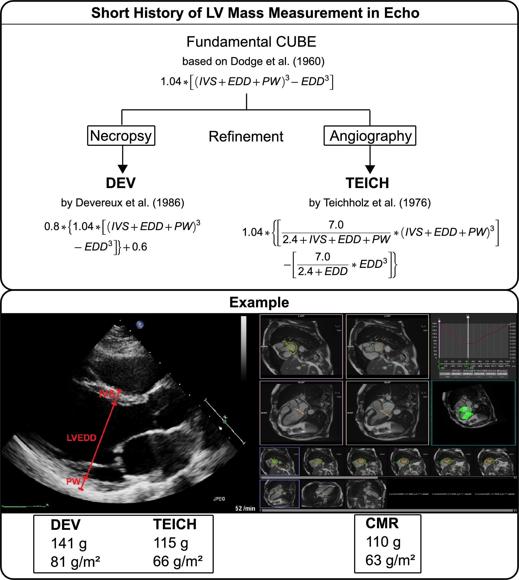
Measuring the ejection fraction (EF) by:
M-mode or 2D – Teicholtzs method:
Measurements in diastole and systole at one level.
Does not provide overall estimate of the EF.
Only the mid-basal wall segment is sampled.

Measuring the ejection fraction (EF) by:
2D echo – Simpson’s method (biplane).
The Best Method
Trace the chamber in systole and in diastole.
Provides an estimate of EF from the entire wall seen in the image plane. (2nd best method compared to 3DE.)
Measuring EF by 3DE Imaging
LV size, LV volume, and LVEF are best measured via 3D Echo.
Using AP4CH view with focus on the LV, optimize the image , maximize temporal and spatial resolution by using the smallest field of view possible.
Confirm good endocardial tissue definition.
Ask the patient to suspend respiration and activate the 3DE software.
Only available if the software is.
Strain Imaging
Strain echo has proven to add information about global function, regional function, and timing of myocardial contraction. (Reproducibility is limited however)
Ventricular strain is becoming more popular and is based off the premise of the deformation of the myocardium.
Different types exist. One of the most common is Longitudinal strain which is acquired by 2D Speckle tracking from the apical views.
One must acquire apical images, calculate the peak systolic strain of the LV segments, display the bullseye map, and calculate longitudinal strain indices.
Fractional Shortening
Percentage of LV size reductions from diastole to systole as measured from the basal segments via 2D or M-Mode linear measurements.
Basically: systolic thickening of the IVS
Normal FS
>20-30% (Usually 25%)
What doesn’t FS do?
Does not provide the best assessment of LVEF due to assumptions:
Normal LV geometry
Symmetrical Function
No WMA(Wall motion Abnormalities)
No dyssynchrony/ LBBB
Excellent Quality Images
Perfect Orientation
Body Surface Area (BSA)
Depends on height and weight of patient. It is usually a more accurate indicator of body mass as compared to actual body weight.
Normal BSA: 1.73 m²
BSA Formula
Mosteller Formula = (height [inches] x weight [pounds] / 3131) (1/2)
Stroke Volume
amount (volume) of blood ejected with each heart beat (contraction).
Stroke Volume Formula
EDV – ESV (normal 70-100ml)
Cardiac Output
Amount of blood ejected per minute.
CO Formula
SV X HR (heart rate) (normal 4-8 L/min)
Cardiac Index
Cardiac output/body surface area.
cardiac output adjusted for body surface area.
CI Formula
CO/BSA (normal 3 – 4 l/min2)
M-Mode
Y Axis
Represents depth (Time of Flight)
M-Mode
X Axis
Represents Time
The rapid frame rate makes M-mode more accurate for:
Timing of rapid cardiac motion.
Precise measurements of cardiac dimensions.
M-mode limitation:
In order to obtain accurate measurements, the scan line must be perpendicular to the structure being imaged.
A non-perpendicular orientation to the structure of interest will result in incorrect measurements.
Ice Pick View
Produces a one-dimensional image of structures within the heart (and chest) according to their distance (depth) from the transducer along the M-mode scan line or cursor.
RV Measurement
2.3 cm
IVS
1.2 cm
LVIDd
5.6 cm
LVIDs
Systolic dimensions are dependent on function.
LVPW
<1.2cm >1.2 = Hypertrophy - increased
muscle mass.
M-Mode Sweep
should be performed demonstrating continuity of the aortic valve and mitral valve (AML should be continuous with posterior wall of the AoR).

Is this a Good picture for LV?
No Because the MV is in the way

What can you do with this picture?
Measure the RVFW and the Pericardium

What’s wrong with this picture?
IVS and LVPW is Irregular
LV is too Big

What’s wrong with this?
LV is Big and there’s a little Hyperdynamic Motion

What’s wrong?
Walls are thickening and thinning at different times. This is called Paradoxical/Akinetic motion
Early Diastole
The leaflets separate widely
(E-point) – 70% of blood enters ventricle.
EPSS
E point septal separation --- (in the absence of mitral stenosis) Increased EPSS represents the dilation of the LV (i.e. aortic regurg., systolic dysfunction).
*If both leaflets are not on the M-mode - You are
Not perpendicular to both leaflets.
Mid diastole (Diastasis)
Period when pressure has equalized between the LA and LV
-Very little flow across the MV - Leaflets begin to move back toward each other
Atrial Contraction
Atrium contracts and sends the last 30% of blood into the ventricle (P-wave on the EKG).
What is Fluttering of the MV caused by?
Aortic regurgitation
Late ventricular diastole (atrial contraction):
The leaflets separate again to form the A point.
*”B” bump (“B” notch) on the slope of the A-C closure of the MV may indicate
Increased LVEDP
LVEDP Stands for?
Left ventricular end diastolic pressure.
Ventricular systole (atrial diastole)
Coaptation (leaflets tips touching) or closure of the MV leaflets in systole is seen as a thin line that moves slightly anterior (paralleling the motion of the posterior wall).
Mitral Valve Prolapse occurs in
Systole, because the leaflets sag in the LA

MVP jet in the middle of the leaflets is called?
Centric
Excursion:
The opening of valves or the leaflets of a valve
Cardiomyopathy (CMO):
A disease that diffusely affects the myocardium.
SAM
Systolic Anterior Motion of the Mitral Valve
MVP
When the anterior or posterior MV leaflets sink or sag into the LA during systole.
Types of MVP
Mid to Late systole
2mm below C-D Points
Holosystolic or Pansystolic
3mm below C-D point