ap bio cell membrane study guide notes
1/43
There's no tags or description
Looks like no tags are added yet.
Name | Mastery | Learn | Test | Matching | Spaced | Call with Kai |
|---|
No analytics yet
Send a link to your students to track their progress
44 Terms
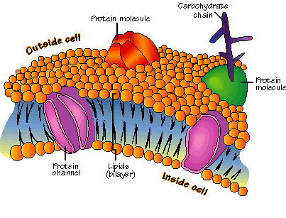
phospholipid bilayer
2 layers of phospholipids
Heads = hydrophilic (water-loving)
Tails = hydrophobic (water-fearing)
Makes the plasma membrane
Selectively permeable
Holds proteins, lipids, and carbs
fluid mosaic model
Membrane is fluid with mobile proteins embedded in a phospholipid bilayer
Phospholipids form a hydrophobic barrier that repels water and hydrophilic molecules
selective permeability of cell membrane
The membrane controls which molecules enter and exit
Phospholipid layer forms a hydrophobic barrier
Transport proteins regulate which molecules can pass through
simple diffusion
moves high to low concentration
no energy is required
possible bc small, nonpolar molecules can pass through phospholipid bilayer
transport of polar molecules
can’t pass through the bilayer by simple diffusion
polar or hydrophilic, so the membrane repels them
rely on transport molecules and proteins to enter/exit cell
integral proteins
Permanently embedded in the phospholipid bilayer
Have hydrophobic regions to stay within the membrane
Made by ribosomes on the rough ER
Structure includes α-helices (spiral) and β-barrels (pores)
Adapted to serve specific roles: pore, pump, receptor, or carrier
transmembrane proteins
Span the entire cell membrane, from extracellular to intracellular sides
Often integral proteins or closely affiliated with them
Function in transport, signaling, and cell adhesion
channel proteins
Proteins that allow materials to move into/out of the cell
Can function via passive or active transport
aquaporins
integral channel proteins that facilitate the rapid transport of water across a cell membrane
g-protein
Molecular switches: GTP = ON, GDP = OFF
Activated by GPCR
Change shape to start signaling inside the cell
GTPase turns them off
sodium potassium pump
Uses ATP to move 3 Na⁺ out and 2 K⁺ in
Maintains ion concentration gradients
Creates membrane potential needed for nerve signals and muscle contractions
glucose transponders
proteins that facilitate glucose across the membranes
anchor proteins
Attach proteins to the inner or outer membrane (often via lipid anchor)
Ensure proper positioning of membrane proteins
Help with signal transduction, cell-cell adhesion, membrane protection, and protein trafficking
Can be integral or peripheral proteins
peripheral proteins
Loosely attached; do not enter lipid bilayer
Extracellular: cell recognition/communication
Cytoplasmic: link cytoskeleton, enzymes, signaling
Functions: signaling, structure, enzymatic activity, vesicle transport
Easily detached
Carbohydrate Receptors
Bind carbs on cell surfaces (“sugar sensors”)
Functions:, cell recognition & communication, Immune recognition of pathogens, Pathogen entry , and Intracellular trafficking, Development & differentiation
Important for: immune response and tissue differentiation
ligands
Bind specific protein receptors
Can be proteins or non-proteins (e.g., hormones, neurotransmitters)
Induce conformational changes → control enzymatic activity, cell signaling, drug action
Examples: insulin, oxygen (erythrocytes), neurotransmitters, antibodies
endocytosis
the cell forms new vesicles from the plasma membrane
allows for the cell to take in macromolecules
exocytosis
bulk transport out of the cell through the membrane
phagocytosis
“cell eating,” brings in large particles or cells.
cell wraps a pseudopodia around a solid particle to bring into the cell
pinocytosis
“cell drinking,” brings in small droplets of extracellular fluid.
takes in small droplets of extracellular fluid within small vesicles
Receptor-Mediated Endocytosis
A specific process where ligand proteins bind to receptors on the cell surface.
Usually occurs in coated pits that pinch off into the cytoplasm.
Cell surface receptors
Binds specific extracellular molecules to transmit signals into the cell to trigger an intracellular response.
Cell surface identity markers
They’re usually glycoproteins that are on the outer membrane.
Help cells recognize each other (important for the immune system).
Allow the body to distinguish self from non-self.
enzyme
Carry out chemical reactions at the membrane surface.
-ase suffix
Cell adhesion proteins
proteins that help cells stick to each other or to the extracellular matrix.
Maintain tissue structure, cell positioning, and cell communication.
Attachments to the cytoskeleton
Anchor the cell membrane to the internal cytoskeleton fibers.
Help maintain cell shape, stability, and organization of membrane proteins.
Selective transport channel
Membrane proteins that allow only specific molecules or ions to pass through.
Anchoring Proteins (phosphatidylinositol )
Membrane proteins are attached to phosphatidylinositol (a lipid in the inner membrane).
Anchor the membrane to the cytoskeleton or extracellular structures.
Help position proteins and transmit signals inside the cell.
Anchors – receptors for signals
Membrane proteins that bind signaling molecules (like hormones or neurotransmitters)
Act as anchors by linking to the cytoskeleton or other membrane structures
Transmit signals from outside the cell to the inside.
Pores – B pleated sheets
Made of β-pleated sheets.
Create openings for molecules to move across the membrane.
Diffusion of ions through channels
Ions move through ion channels, which are protein tunnels in the membrane.
These channels let ions pass because they are hydrophilic, unlike the lipid bilayer.
facilitated diffusion
helps large, polar, or charged molecules move across membrane
Passive: no ATP required
Can saturate: transport rate maxes out when all carriers are occupied
Example application: adding phosphate to blood donations helps RBCs produce ATP and prolong shelf life
active transport
low to high concentration
Requires ATP energy
Transfer proteins are used to pump particles from low to high concentration.
Proton Pump
pumps H+ (hydrogen ions) across the membrane
Coupled Channels
one molecule moves down its gradient to help another move against its gradient
osmosis
diffusion of water through membrane
moves from high to low concentration
maintains cell homeostasis
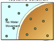
isotonic
when a solution is balanced
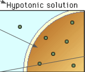
hypotonic
more water than solutes
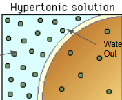
hypertonic
more solutes than water
concentration gradient
a boundary with varying levels of concentration
active transport
Inside of cell: negatively charged
outside: more positive → membrane potential
Ions move due to:
Chemical force: concentration gradient
Electrical force: voltage gradient attracts opposite charges
Drives ion movement (e.g., Na⁺, K⁺) across the membrane
electrogenic pump
Use ATP to move ions → create electrochemical gradient (concentration + charge)
Inside cell: negative → attracts positive ions, repels negative ions
Examples: Sodium-Potassium Pump, Calcium Pump, Proton Pump
cotransport
Indirect active transport: ATP pump moves one solute → drives movement of another solute against its gradient
Analogy: like water pumped uphill doing work as it flows back down
Generates: electrochemical gradient → source of cellular potential energy