Skeletal System
1/30
There's no tags or description
Looks like no tags are added yet.
Name | Mastery | Learn | Test | Matching | Spaced | Call with Kai | Chat |
|---|
No analytics yet
Send a link to your students to track their progress
31 Terms
Appositional Growth
Cartilage-forming cells in perichondrium secrete new matrix against external face of existing tissue
Interstitial Growth
Lacunae-bound chondrocytes divide and secret new matrix, expanding cartilage from within. Typically ends during adolescence, when skeleton stops growing.
Hyaline Cartilage
Most abdundant, support, flexibility, and resilience. Fine collagen fibers.
Elastic Cartilages
Only found in two locations (external ear, epiglottis). Stretchy, withstand repeated bending.
Fibrocartilage
Parallel rows of chondrocytes alternative with thick collagen fibers. Occur in areas subject to both pressure and stretch (menisci of knee, discs between vertebrae).
Function of Skeleton
Support, framework, protection, mineral storage, blood formation.A
Axial Skeleton
Skull, vertebral column, rib cage. Support and protect.
Appendicualr Skeleton
Upper, lower limbs, and girdles. Allow for movement and manipulation of environment.
Long bone
Most limb bones, longer than they are wide. Humerus, Tibia, Fibula, Femur, etc.
Short bone
Roughly cube shaped, wrist and ankle.
Flat bone
Thin, flattened, a bit curved. Sternum, scapulae, ribs, cranial bones.
Irregular bone
Doesn’t fit any previous category. Vertebrae, hip bones.
Endochondral Ossification Steps
All bones below base of skull form by endochondral ossification (besides clavicles).
Bone collar forms around disphysis of hyaline cartliage model. Consists of bone collar, primary ossification center.
Cartilage calcifies in center of diaphysis and developes cavities. Consists of deteriorating cartilage matrix.
Periosteal bud invades internal cavities, spongy bone forms. Blood vessel and spongy bone formation. Osteoprogenitor cells become osteoblasts, secrete osteoid around remaining calcified fragments of hyaline cartilage.
Diaphysis elongates, medullary cavity forms. Secondary ossification centers form in epiphyses.'
Epiphyses ossifies. Once ossification is complete, hyaline cartilage only remains in epiphyseal plates and articular cartilages.
Intramembranous Ossification
Forms cranial bones and clavicles. Most bones are flat bones.
Ossification centers develop in fibrous connective tissue membrane. Mesenchymal cells.
Osteoid is secreted and calcifies, trapped osteoblasts becomes osteocytes.
Immature spongy bone and periosteum form.
Compact bone replaces immature spongy bone, deep to periosteum. Red marrow developes.
Fracture repair
Hematoma forms. Blood vessels in bone and periosteum are torn, causing hemorrhaged blood clots and forms a hematoma.
Fibrocartilaginous callus forms
Bony callus forms (spongy bone, trabeculae)
Bone remodeling occurs (compact bone)
Comminuted Fracture
Three or more pieces
Spiral Fracture
Twisting forces applied to a bone, fractures.
Depressed Fracture
Broken bone portion pressed inward
Compression
Bone is crushed
Epiphyseal
Seperates from diaphysis along epiphyseal plate
Greenstick
Bone breaks incompletely, only one side of shaft breaks while other side bends.
Fibrous joints
Suture - Held together with short interconnecting fibers, only found in skull. No movement.
Syndesmosis - Joint held together by ligament. Little to no movement, can be found in distal ends of tibia and fibula.
Gomphoses - Peg-in-socket, tooth.S
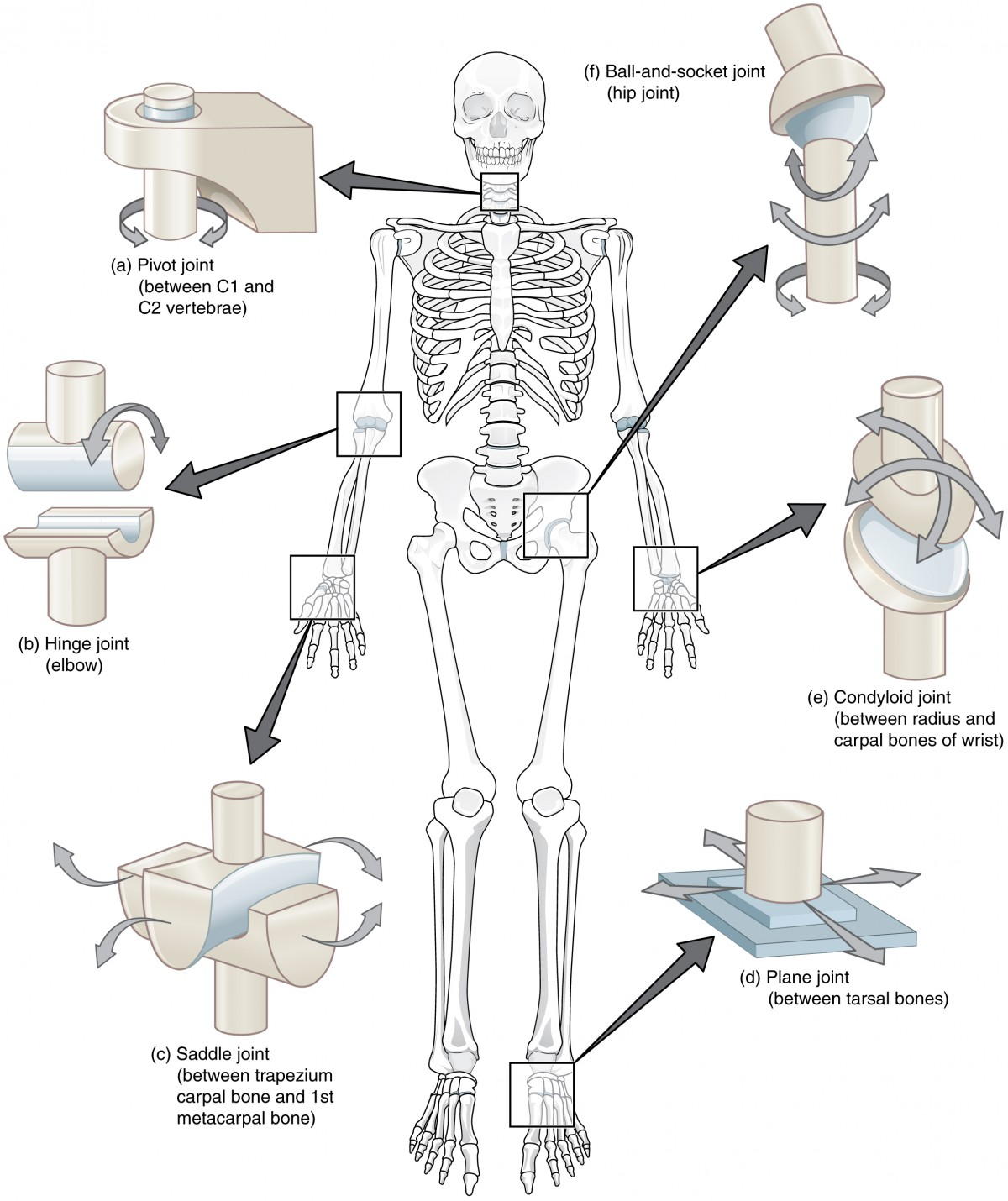
Synovial Joint
Bones seperated by fluid-containing joint cavity, most joints of the body. Has articular cartilage, joint cavity, articular capsule, synovial fluid, reinforcing ligaments, and nerves and blood vessels. Plane, hinge, pivot, condylar, saddle, ball-and-socket.
study
Flexion/Extension
decreasing/increasing angle between two bones in sagittal plane
Abduction/Adduction
Moving a limb away/towards body midline in frontal plane
Circumduction
Cone movement
Medial/lateral rotation
rotating toward/away from median plane (like twisting your leg from the hip inward or outward)
Inversion/eversion
Sole of foot twisting towards/away from medial
Elevation/depression
Up and down (movement of mandible elevation and depression, like chewing).
Opposite/reposition
Doing the “ok” symbol with your thumb and index finger, pinching them together is opposition.