Hardware QA-QC
1/122
There's no tags or description
Looks like no tags are added yet.
Name | Mastery | Learn | Test | Matching | Spaced |
|---|
No study sessions yet.
123 Terms
The location of the coils installed in the MRI scanner, from the inner point closest to the patient to the outer most edge
RF radiofrequency coils, gradient coils, shim coils, main magnet
Gradient amplitude
strength of the gradient.
gradient rise time and what its measured in
time it takes for the gradient to reach its full amplitude, measured in microseconds
10mT/m = ?G/cm
1
trapezoidal gradient pulse used in conventional pulse sequences
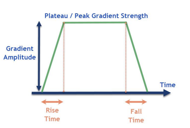
gradient slew rate
speed rate of ascent or descent of a gradient from zero to its maximum amplitude, either positive or negative; strength of the gradient over a specific distance
how to calculate gradient slew rate
amplitude divided by the rise time in msec. Measured in mT/m/msec or T/m/sec.
shorter rise time does what
the faster the gradients and therefore echo spacing
Gradients with a shorter echo spacing will have
better resolution capabilities and more available slices per TR period.
convert vendor spatial gradient map into manufacturer conditions chart
1 Tesla/meter = 100 Gauss / cm
what is gauss/cm important to know for
metallic implants
duty cycle
time the gradients are on during a TR period, the “gradient working time.
parameter changes that affect pulse duty cycle
Increased # slices
Employment of fat suppression pulses (SPAIR requires increased TR compared to SPIR)
Utilization of presaturation slabs/bands
Increased ETL
what causes noise in mr room
rapid, successive switching of the gradient coils
what gradient is used for sagittal to enable magnetic field around pt’s body
x gradient
what gradient is used for coronal to enable magnetic field around pt’s body
Y gradient
what gradient is used for axial to enable magnetic field around pt’s body supine
Z gradient
what happens when you apply two gradients at the same time
oblique slice is created
RF coils are located where in the magnet
closest to the patient
utilization of multiple, powerful RF pulses (Fast Spin Echo sequences) can lead to
increased SAR induction in a patient.
180° RF pulse is _____ the power of the initial 90° RF pulse
4 times
RF heating is more of a concern in what sequences and why
fast spin echo sequences due to the multiple echo train lengths
what are major parts of the RF system
Radiofrequency coils (transmitter and reciever)
Radiofrequency coils consist of
two electromagnetic coils, the transmitter and receiver coils generating and receiving electromagnetic fields
radiofrequency absorption measurement
Specific Absorption Rate (SAR) Watts per kilogram (W/kg
what type of energy is RF
low energy non ionizing
SAR is _____ to _____ for the resonant frequency
proportional, power of 2
what happens to SAR when you double field strength
increases 4 times
primary biological effect of RF fields
tissue heating
larger pt does what to SAR
increases
how to protect pt when larger in scanner from RF heating/burns
place manufacturer pads on them to not let skin touch
why should you be careful with blankets in exams with high SAR/increased FSE usage
need a means for heat to escape
SAR/body temp correlates to
the amt of thermal induction minus escaped heat
room temp should be
65-75F
room humidity should be
50-70%
air flow in the scan room can help with what
make pt less vulnerable to RF heating effects
what does TR have to do with SAR
“shortest TR” as this has negative impact on pulse duty cycle (gradient ontime).
what do presaturation slabs and fat suppression pulses have to do with SAR
should minimize them to reduce SAR
what do slices have to do with SAR
minimize them and use least number of slices in a given TR period
what does ETL have to do with SAR
Utilize shortest ETL possible while enabling flip angle modulation techniques, where applicable. Flip angle sweep methods – Philips: refocusing control; GE: VERSE, tailored RF; Siemens: Hyperecho, Restore; Toshiba: T2 Plus
siemens flip angle sweep method
Hyperecho, restore
a good way to reduce SAR with imaging techniques
parallel imaging to reduce workload of the phase encoding gradient
how does low SAR sequences work
longer and lower amplitude transmit RF pulses
what do low SAR sequences change
TE, so it requires a longer TR value which increases scan time
what is a coil that can help reduce SAR
Transmit/Receive coil (but significantly increased scan time)
coil not properly tuned to correct magnetic field strength
results in signal loss
For energy to be efficiently transferred between a transmitter and receiver
the two must be at the same frequency.
surface coils do what
(linear coils) yields a more localized, smaller FOV (field of view) capability, with increased SNR, thus providing opportunity for improved image quality on most systems.
SNR penetration depth on coils
½ the coil diameter
how should cables from local RF coils and gating leads be positioned
braided and positioned straight in the bore, NEVER in direct contact with the patient's skin. The cables should NEVER be coiled or looped, as this can lead to RF burns in the patient
CP coil
Circularly polarized transmission or receiver coil with two orthogonal transmission and/or receiver channels, also known as a quadrature coil. This yields better signal-to-noise than a linear coil.
which has better SNR, linear coil or CP coil
CP
When going from a linear coil to a quadrature coil, SNR is
increased by 40%
A phased array coil is comprised of
multiple coil elements combined with multiple receiver channels.
The local RF coil used to image a brain is typically located/kept
in the magnet room
Permanent magnets with a vertical magnetic field use what types of coils and why
surface coils that are solenoids because the secondary magnetic field (B1 )created by the RF coil must be perpendicular to the orientation of B0 .
multi-element phased array coils and why they are important
capable of acquiring multiple channels of data in parallel. This 'parallel imaging' technique uses unique acquisition schemes that allow for accelerated imaging.
Parallel imaging R factor does what
parallel imaging reduces scan time directly proportional to reduction -R- factor:
SENSE: P reduction
scan time reduction in phase dimension
SENSE: S reduction
scan time reduction in slice direction (3D acquisitions)
iPAT
parallel imaging phase direction.
iPAT²
parallel imaging phase and slice dimensions
patient image display is what and located where
with sequence parameter visualization is located in the scan control room on the operator console
CPU located where and for what
used to plot and adjust slices in an MR sequence is located in the scan control room at the technologist operator console.
array processor does what
responsible for reconstructing the collected MR images/data using the Fourier transform, and is located in the MR equipment room.
RF power amplifier is located
within an equipment room.
Radiofrequency (RF) shielding can be achieved by
lining the scanner room walls with copper
Magnetic field inhomogeneity is expressed in
parts per million (ppm)
Passive shielding and its importance
can be accomplished by lining the MR room with steel or other ferromagnetic plating, thus reducing the scope and distance of the fringe field.
Active shielding and its importance
requires the implementation of superconducting windings within the construction of the MR scanner to oppose a portion of the magnetic field
Field strength at magnet isocenter is measured in
Tesla
High field scanners typically have field strengths greater than or equal to
1.5T
Doubling the magnetic field strength will ____ the SNR (signal to noise ratio)
double
However, for field strength, SAR is proportional to _______ for the resonant frequency
the power of 2
doubling field strength results in a ______ in SAR potential.
4x
The unit of measurement of the magnetic field surrounding the periphery of the MR scanner (fringe field)
Gauss
fringe field
magnetic field surrounding the periphery of the MR scanner
intensity limit by FDA for clinical use for static magnetic field
4T in everyone, 8T in pts 1 month and older
most commonly used system in MR imaging
superconducting magnet
superconducting magnet field strength increased by doing what
the magnetic field strength is increased by increasing the turns of wire, current in the wires, or by reducing the spacing between the wires.
The orientation of the main magnetic field in a high field, superconducting, short bore magnet is
horizontal.
liquid cryogens are cooled to
4 kelvin, -270C, -452F
most common cryogen in MR
helium
shimming creates
additional magnetic fields
what do the additional magnetic fields created by shimming do
add to the overall magnetic field of the superconducting magnet in such a way that the total B0 field becomes more homogeneous.
safety issues in a quench situation
should be evacuated from the scan room to avoid asphyxiation, frostbite, and/or damage to tympanic membranes.
quench
refers to the sudden loss of superconductivity when its temperature is raised
how do quenches work
In the superconducting state, the resistance of the magnet coil windings is zero and hence no energy is required to maintain current flow. If the coil temperature rises above the superconductivity threshold (Tc), the windings suddenly develop a finite resistance. The several-dozen amperes of circulating current passing through this elevated coil resistance create heat. This heat causes a sudden, explosive boil-off of liquid helium
magnetic fields associated with MR
Static magnetic fields
Oscillating magnetic fields (RF)
Time varying magnetic fields (gradient)
Time varying magnetic fields (gradient) effects on pt
cause muscle contractions, cardiac arrhythmias, mild cutaneous sensations and visual light flashes (Magnetophosphenes)
Magnetophosphenes are caused by
time varying magnetic fields (gradient)
why is dB/dT limited?
so it doesnt cause peripheral nerve stimulation painfully
what sequences can have more of a detrimental effect on patients with regards to time varying magnetic fields.
EPI
Oscillating magnetic fields (radiofrequency) – primary biological effect
is tissue heating/deposition
body coil, what it does and its limitations
s an integrated part of the magnet design that acts as its own transceiver coil, with large FOV capabilities, no high SNR like localized coils
The MRI system component that produces the magnetization of proton spins (alignment)
main magnet
The MRI system component that provides the ability to perform spatial encoding
gradient system
goal of MRI Department Quality Assurance / Quality Control Program
To achieve and maintain a level of consistent, reproducible image quality in a Magnetic Resonance Imaging system, ensuring the safety of patients being scanned.
record keeping of data is important why
industry standard, producible upon request and accessible to technologists, physicists and radiology managers.
Quality Assurance
utilizes the goal of maintaining a desired level of consistency and image quality requirements within Radiology. “Are we operating the devices or equipment correctly?