BIOL 2311 26 FC
 Call Kai
 Learn
Learn Practice Test
 Spaced Repetition
 Match
Match Flashcards
 Knowt Play
1/156

There's no tags or description
Looks like no tags are added yet.
Name | Mastery | Learn | Test | Matching | Spaced | Call with Kai | Chat |
|---|
No analytics yet
Send a link to your students to track their progress
157 Terms
Gross anatomy
is the study of structures of the body visible to the naked eye (large body parts)
Surface anatomy->
2 main divisions of the body are:
Axial
Appendicular
Axial: refers to the structures along the midline of the body, including head neck and trunk
Appendicular: refers to the limbs and their attachments to the axis
Axial
refers to the structures along the midline of the body, including the head, neck, and trunk
Appendicular
refers to the limbs and their attachments to the axis

Regional Terminology: Anterior
...
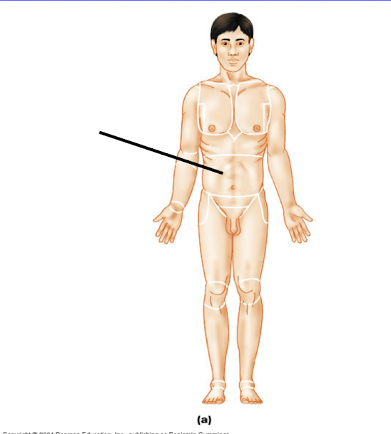
Abdominal
Pertaining to the region of the anterior trunk between the ribs and the upper border of the hips
Acromial
(shoulder)
pertaining to the point of the shoulder

Antebrachial
(forearm)
pertaining to the forearm (the lower portion of the upper limb)
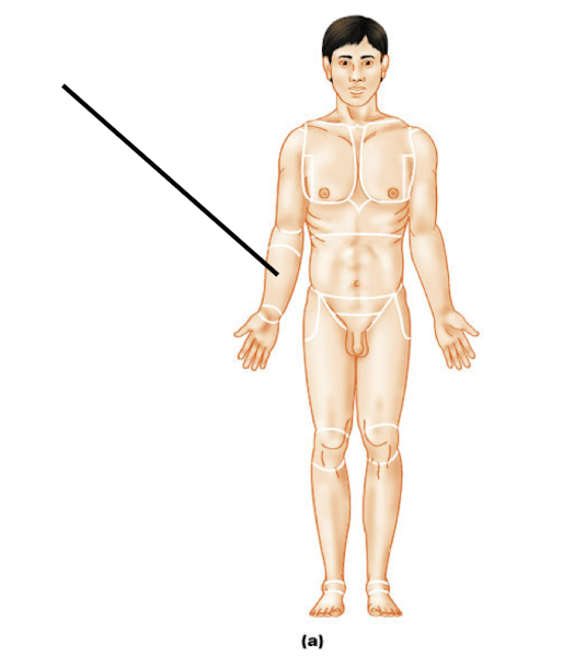
Antecubital
pertaining to the anterior surface of the elbow (the inside of the elbow)

Axillary
pertaining to the armpit

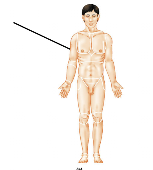
Brachial
pertaining to the arm (upper portion of the upper limb)
Buccal
pertaining to the cheek


Name the regional terminology
carpal
→ pertaining to the wrist
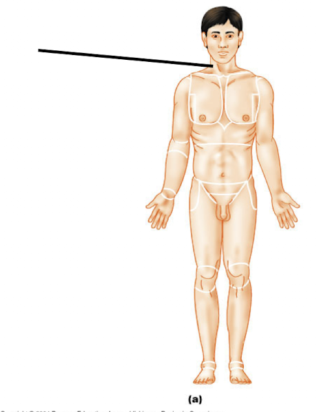
Name the regional terminology
cervical
→ pert. to the neck

Coxal
pert. to the hip

Crural
(shin)
pert. to the leg (lower portion of the lower limb)
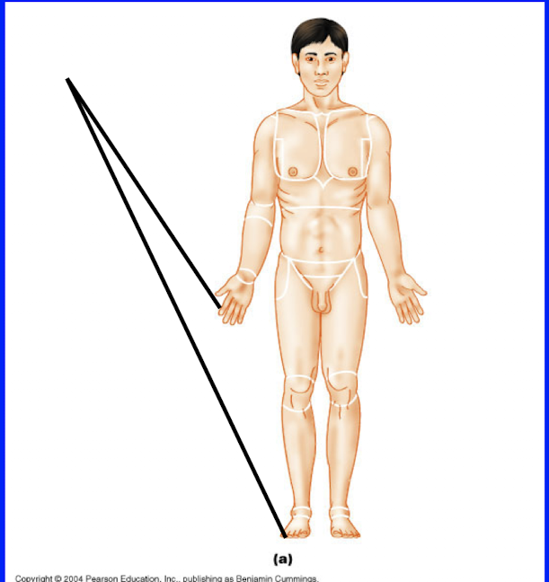
Digital
pert. to the fingers or toes

Femoral
(femur)
pert. to the thigh (the upper portion of the lower limb)

Frontal
pert. to the forehead
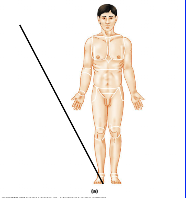
Hallux
pert. to the big toe

inguinal
pert. to the groin

Mammary
pert. to the breast

Mental
pert. to the chin

Nasal
pert. to the nose
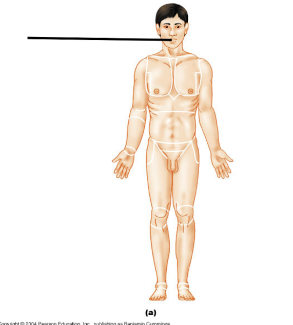
Oral
pert. to the mouth
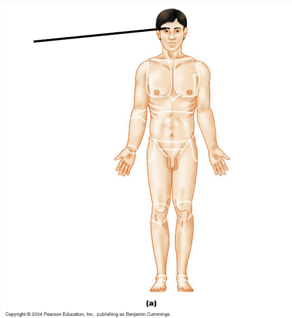
Orbital
pert. to the bony eye socket (orbit)

Palmar
pert. to the palm of the hand

Patellar
(kneecap)
pert. to the anterior knee (kneecap) region

Pedal
pert. to the foot

Pelvic
pert. to the pelvic region (between the upper edge of the hip and groin)

Fibular (peroneal)
pert. to the side of the leg (lower portion)
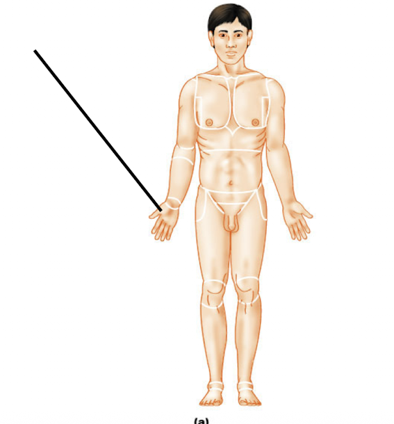
Pollex
pert. to thumb

Pubic
pert. to the genital region
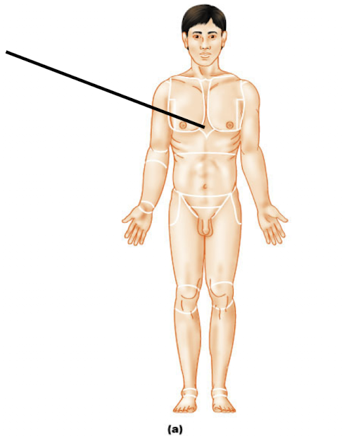
Sternal (sternum)
pert. to the region of breastbone (center of the chest)

Tarsal
pert. to the ankle

Thoracic
pert. to the chest (from the collar bone to the bottoms of the ribs)

Umbilical
(belly button)
pert. to the navel
Regional terminology: Posterior
...
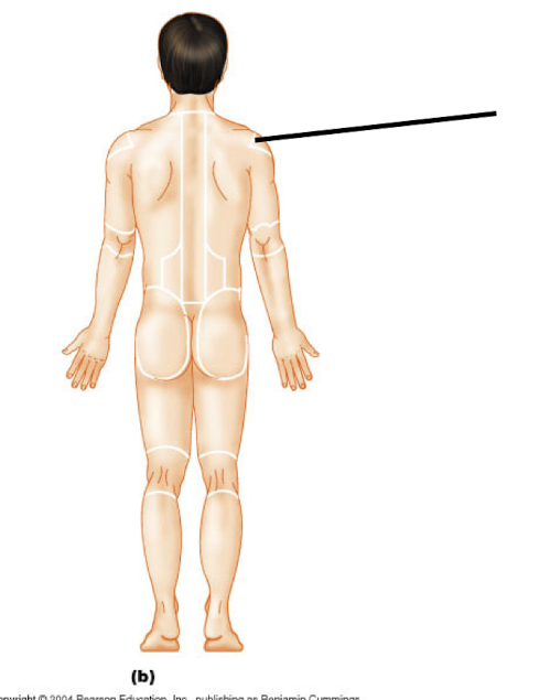
Acromial (P)
pert. to the point of the shoulder
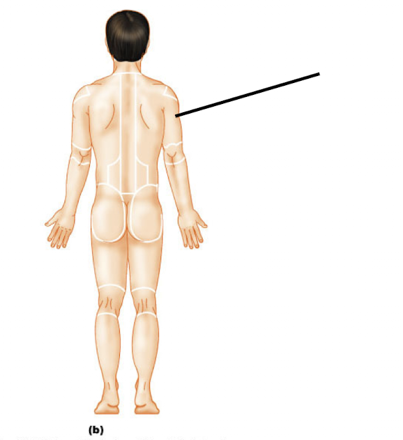
Brachial (P)
pert. to the arm (upper portion)

Calcaneal (P)
pert. to the heel of the feet
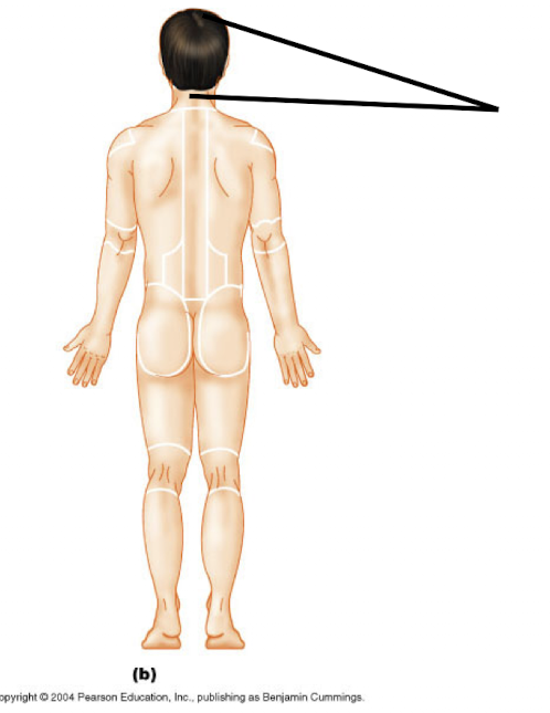
Cephalic (P)
pert. to the head

Dorsum (P)
(whole back)
pert. to the back (trunk only)
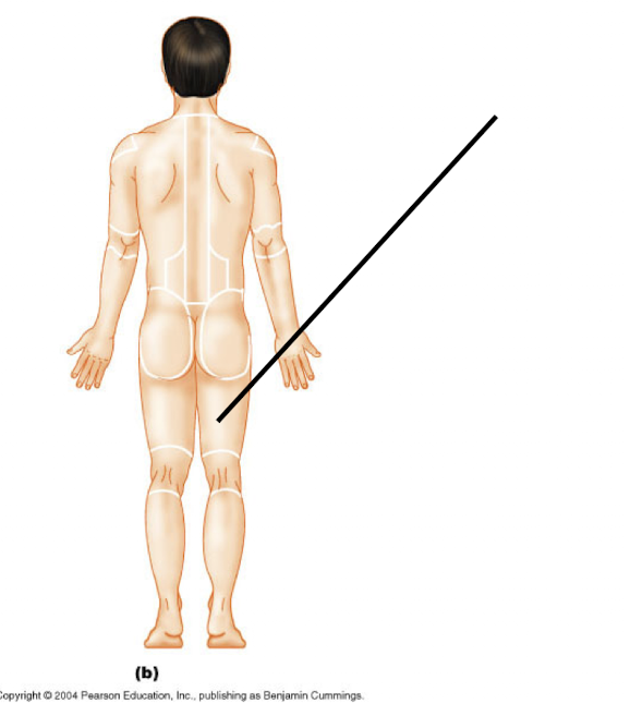
Femoral (P)
pert. to the thigh
(upper portion of the lower limb)

Gluteal (P)
pert. to the buttocks or rump

Lumbar (P)
(lower back)
pert. to the region of the back between the lower edge of the ribs and the hips (the loin)

Manus (P)
pert. to the hand

Occipital (P)
pert. to the posterior aspect of the head or base of the skull
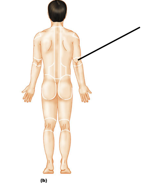
Olecranal (P)
pert. to the posterior aspect of the elbow

Otic (P)
pert. to the ear

Perineal (P)
pert. to the region between the anus and the external genitalia
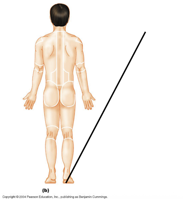
Plantar (P)
pert. to the sole of the foot

Popliteal (P)
Pert. to the back of the knee

Sacral (P)
pert. to the region between the hips
→ (looks like the top of your buttcrack)

Scapular (P)
(shoulder blades)
pert. to the region overlying the shoulder blades

Sural (P)
pert. to the calf or the posterior surface of the leg (the lower portion of the lower limb)
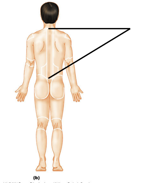
Vertebral (P)
pert. to the region overlying the spinal column
Directional terminology
- superior/inferior
- medial/lateral
- proximal/distal
- anterior/posterior
- superficial/deep
Superior/inferior
placement relative to the long axis of the body
equivalent to cephalad (toward head) and caudal (toward the tail) in humans
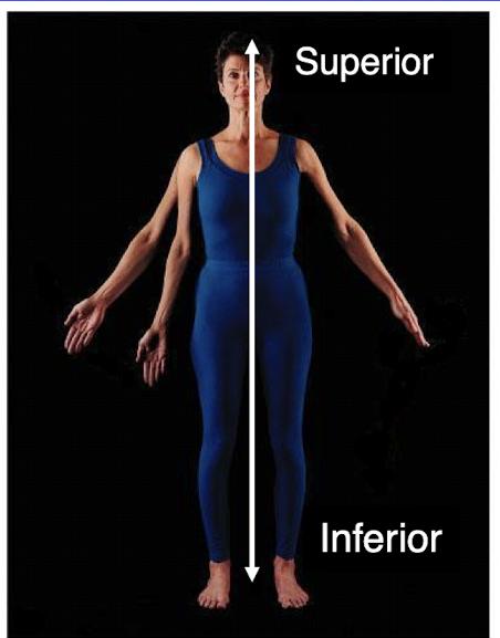
Medial/Lateral
Placement relative to the midline along the width of the body

Proximal/Distal
Placement along a limb relative to the point of attachment of that limb to the rest of the body

Anterior/Posterior
Placement along the axis running from front to back
Equivalent to ventral (front / toward the belly) and dorsal (back / toward the back) in humans

Superficial/Deep
Superficial = toward skin
Placement within the body from the surface toward the interior of the body
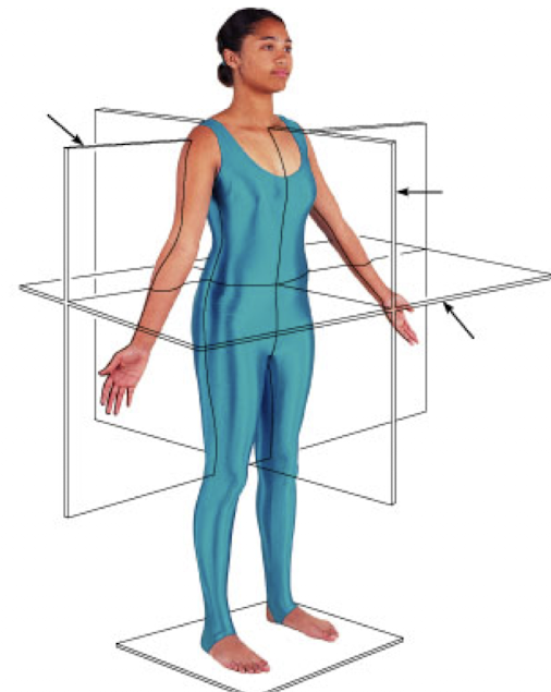
Planes of Section
A cut, or section, is often a useful way of visualizing anatomical structures in relation to other structures
Sections are made along planes of section, which are oriented relative to the axes of the body
Planes of Section (longitudinal defintions)
Longitudinal sections are oriented parallel to the long axis of the body
Longitudinal sections include frontal (front to back) sections and sagittal sections (side to side)


Frontal section
Divide the body into an anterior (front) portion and a posterior (back) portion

Sagittal section
divide the body into left and right portions
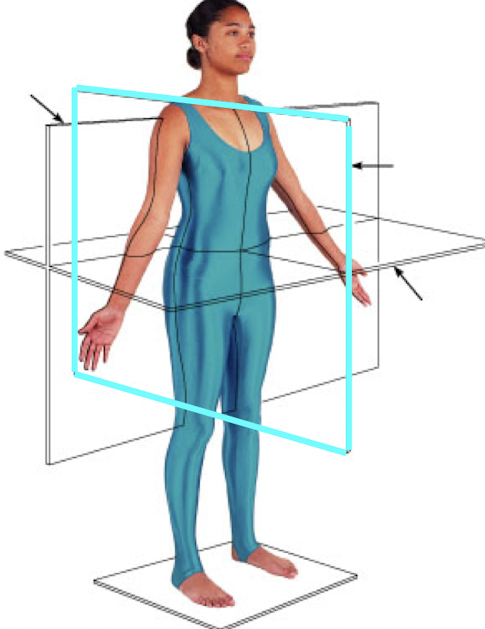
Midsagittal (median) section
Lies along the midline of the body and divides the body into EQUAL right and left halves

Parasagittal section
lies parallel to the midline and divides the body into UNEQUAL right and left portions

Cross section/Transverse section
cross sections lie perpendicular to the long axis of the body
→ (cuts top from body)

Oblique section
lies at an angle (not 90 degrees or 180 degrees) to the long axis of the body
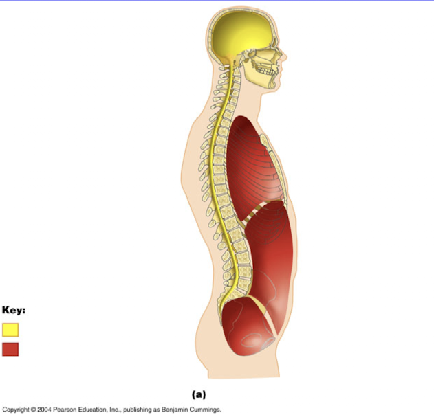
Body cavities
the axial portion of the body contains two main cavities that house the internal organs
these cavities are the DORSAL and VENTRAL cavities (front and back)
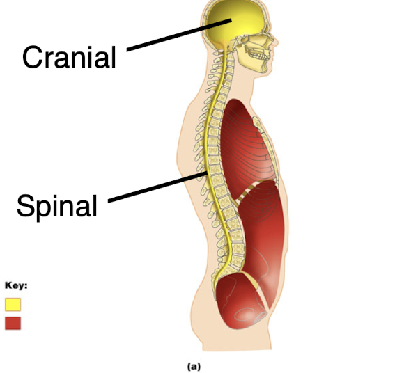
Dorsal cavity
the dorsal cavity lies along the dorsal side of the body and includes the cranial and spinal cavites
the cranial cavity contains the brain, and the spinal cavity contains the spinal cord
Ventral cavity
the ventral cavity lies along the ventral side of the body and contains a number of sub-cavities
these sub-cavities include the thoracic and abdominopelvic cavities

Thoracic cavity (chest)
The thoracic cavity is superior and is bounded below by a muscular wall called the diaphragm
The thoracic cavity includes two pleural cavities and the mediastinum

Pleural cavities
the pleural cavities enclose the lungs and are bounded by serous membranes called pleura
→ pleural cavities enclose in PLEURA

Mediastinum
the mediastinum lies between the pleural cavities and includes the superior mediastinum and the pericardium
the superior mediastinum encloses the esophagus and trachea

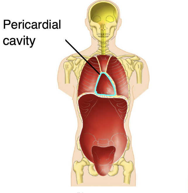
Pericardial cavity
The pericardial cavity encloses the heart and is bounded by a serous membrane called the pericardium
→ pericardial cavity → heart → serous membrane → pericardium
Abdominopelvic cavity
the abdominopelvic cavity includes the abdominal cavity and the pelvic cavity
the abdominopelvic cavity is bounded above the diaphragm
the abdominal cavity encloses most of the digestive organs
the abdominal cavity is bounded by a serous membrane called the peritoneum
abdominal cavity → peritoneum

Pelvic cavity
the pelvic cavity encloses the reproductive organs
the pelvic cavity is bounded by the bony pelvis

Body cavities
serous membranes cover and lubricate most of the organs found within the ventral cavities
serous membranes are continuous membranes that fold to produce two portions, the parietal serosa and the visceral serosa
parietal serosa lines the walls of a cavity
visceral serosa lines the organs within a cavity
serosa includes the pleura, pericardium, and peritoneum
Resolution
the ability to distinguish two closely spaced objects as separate is known as resolution (or resolving power)
Virtual image
the optics of the microscrope produce an image of the specimen projected onto the retina of your eye
→ this is called a virtual image
the image is reversed and upside down relative to the specimen
Field of view
the circular area visible through the oculars is known as the field of view
the field of view decreases in actual size as the magnification increases
if you know the actual size of the field of view given at a magnification, you can use this to estimate the size of an object within this field of view
Stage micrometer
use this to measure the field of view at each magnification
Working distance
the distance between the bottom of the objective lens and the specimen
Depth of field
the depth through which the specimen is clearly in focus is called the depth of field
depth of field decreases with increasing magnification
Histology
study of tissues
The 4 primary tissue types
Epithelial'
Connective
Muscle
Nervous
E.C.M.N
Epithelial tissue
tissue sheets of cells that line the body surfaces, either external or internal
Muscle tissue
tissues are excitable tissues where a specific cell type is embedded within a non-living (acellular) matrix
Nervous tissue
tissues are excitable tissues (“told to do something”) that are capable of transferring and integrating signals from one part of the body to another
Epithelial body tissues line ________
body surfaces (inside or out)
Epithelial tissues function in ________
protection
absorption
filtration
secretion
excretion
and sensory reception
→ epithelial tissues are embryologically derived from either ectoderm or endoderm
What is polarity in epithelial tissue?
(has 2 sides → apical =top and basal = bottom)
Epithelial has two different sides
Apical surface: faces an external or internal open space
Basal surface: faces the underlying connective tissue
What are specialized contacts in epithelial tissue?
cells are joined together via specialized junctions to form sheets
What forms the basement membrane?
Basal lamina: formed by epithelial cells
Reticular lamina: formed by the underlying connective tissue
→ forms from a basal lamina (formed by epithelial cells) and a reticular lamina (formed by underlying connective tissue)
Basal = bottom
reticular = connective
What is the difference between the basal lamina and reticular lamina?
Basal lamina = epithelial cells (bottom layer)
Reticular lamina = connective tissue (formed by the underlying connective tissue)
Characteristics of epithelial tissues
Avascular (NO BLOOD vessels in the tissue layers): epithelia do NOT have a direct blood supply, but are supplied with nutrients via blood supply of the underlying connective tissue
Regenerative: epithelial cells (at least in a portion of the epithelium) will readily divide and regenerate the epithelium. This is especially important for epithelia that form protective layers, since they are often damaged through friction or chemical damage
Classification of epithelial tissues
→ 2 criteria are used to classify epithelial tissue into specific tissue types : number of layers and individual cell shape
Number of layers
→ SIMPLE epithelia are composed of a single layer of cells. (ONE)
→ STRATIFIED epithelia are composed of multiple layers of cells, from two layers to many layers (MORE THAN 1 LAYER)
Cell shape:
→ SQUAMOUS cells are flat. The individual cells are wider than they are tall.
→ CUBOIDAL cells are cube-shaped. The individual cells are the same width as their height
→ COLUMNAR cells are column shaped. The individual cells are taller than they are wide
Specific epithelial tissues are named by giving the layer type and then the cell type
ex: Simple squamous epithelium