West Chapter 2: Ventilation - How Gas Gets to the Alveoli
1/27
There's no tags or description
Looks like no tags are added yet.
Name | Mastery | Learn | Test | Matching | Spaced | Call with Kai |
|---|
No study sessions yet.
28 Terms
How does inspired gas get to the alveoli?
Ventilation
How do oxygen and carbon dioxide cross the blood-gas interface?
Diffusion
How do oxygen and carbon dioxide moved to and from the lung by the blood?
Blood flow
Draw the Lung Volumes
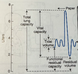
Tidal Volume
Normal breathing
Functional Residual Capacity (FRC)
Volume of gas in the lung after a normal (or tidal) expiration
Vital Capacity
Maximal inspiration followed by maximal expiration
Residual Volume
Gas remaining in the lung after maximal expiration
What can’t be measured with a simple spirometer?
Functional residual capacity
Residual volume
Total lung capacity
Total Ventilation or Minute Ventilation
Total volume leaving the lung each minute
The volume of air entering the lung is very slightly greater because more oxygen is taken in than carbon dioxide is given out
Alveolar Ventilation
Volume of fresh gas entering the respiratory zone each minute, the amount of fresh inspired air available for gas exchange
Dead Space Ventilation Equation
Dead space ventilation = dead space volume x respiratory frequency
Tidal Volume Equation
VT = VD + VA
VT - tidal volume
VD - anatomic dead space
VA - alveolar gas
How can alveolar ventilation be increased?
By raising either the tidal volume or respiratory frequency (or both)
Increasing tidal volume is often more effective because this reduces the fraction of each breath occupied by anatomic dead space (dead space fraction)
Alveolar Ventilation Equation
VA = (VCO2/PCO2) x K
VCO2 - CO2 output or production
PCO2 - partial pressure of CO2
K - constant, total pressure
What can be used to determine alveolar ventilation in healthy individuals?
In healthy individuals, the PCO2 of alveolar gas and arterial blood are virtually identical so arterial PCO2 ca be used to determine alveolar ventilation
What factors increase CO2 production?
Exercise, fever, infection, nutritional intake, and seizures
What factors decrease CO2 production?
Hypothermia and fasting
Anatomic Dead Space
Volume of the conducting areas
What increases anatomic dead space?
Large inhalations because of the traction or pull exerted on the bronchi by the surrounding lung parenchyma
Ratio of Dead Space Volume to the Tidal Volume Equation
VD/VT = (PaCO2 - PECO2)/PaCO2
PaCO2 - partial pressure of arterial CO2
PECO2 - partial pressure of mixed expired CO2
Basic Alveolar Ventilation Equation
VA = VT - VD
VT - tidal volume
VD - dead space volume
What does Fowler’s method measure?
Anatomic dead space
What does Bohr’s method measure?
Physiologic dead space
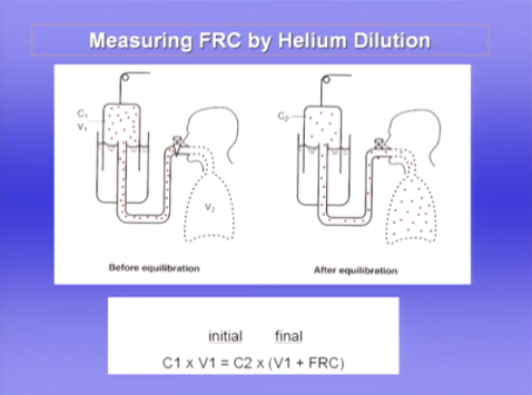
Measuring FRC Using the Helium Dilution Technique
Helium is a very insoluble gas, when the subject breathes it almost none of it is taken up by the blood
C1 - given concentration of spirometer
V1 - volume of spirometer
Subject breathes from the spirometer until there is equilibration of the He between the spirometer and the alveolar gas
Starts to do this at FRC
No helium was lost so the concentration x the volume that you started with will be the concentration that you finished with plus the volume of the lung (FRC)
Difference Between Anatomic and Physiologic Dead Space
In normal subjects, the volumes are nearly the same, but in patients with either acute or chronic lung disease, the physiologic dead space may be considerably larger because of inequality of blood flow and ventilation within the lung
The larger the physiologic dead space, the greater the total ventilation an individual must generate to ensure an adequate amount of air enters the alveoli to participate in gas exchange
Plethysmograph measures the total volume of gas in the lung
Helium dilution only measures the volume of gas that communicates with the mouth
Sometimes smaller if there's lung disease
Regional Difference in Ventilation
The lower regions of the lung ventilate better than the upper zones
When the subject is in the supine position, this difference disappears and apical and basal ventilations are the same
In this posture, the ventilation of the lowermost (posterior) lung, exceeds that of the uppermost (anterior) lung
In the lateral position, the dependent lung is best ventilated
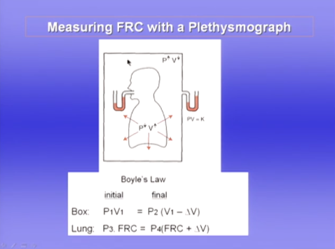
Measuring FRC with a Plethysmograph
In an airtight box
Measure pressure in both with a manometer
Subject has airway occluded and tries to inhale against the closed airway
Small increase in volume of the lung
Volume of gas in the box decreases and pressure increases
Boyle's law - pressure x volume is a constant at a constant temperature